Topological domains have been imaged in superfluid helium-3 using magnetic resonance imaging (MRI). A team of physicists led by Yutaka Sasaki at Kyoto University used the technique to view regions of common chirality as they emerge spontaneously in thin superfluid films. The technique promises to boost our understanding of the complex behaviour of the superfluid state of matter as well as the nature of related types of superconductivity.
When helium-3 is cooled to millikelvin temperatures it becomes a superfluid – a fluid with zero viscosity that can flow forever without any of its kinetic energy being dissipated as heat. Helium-3 atoms are fermions, which normally do not form a superfluid, but in this case the atoms pair-up to behave like bosons – which can form a superfluid. This pairing makes the physics of superfluid helium-3 very rich indeed, and of great interest to physicists studying the topological nature of matter.
Theoretical studies suggest that as helium-3 is cooled to create a superfluid, it forms macroscopic topological domains. Within individual domains, superfluid atoms share the same angular momentum, which gives each domain a certain chirality. However, no previous studies have managed to confirm these ideas experimentally and imaging these structures would reveal the shapes of macroscopic wave functions in the superfluid.
Excited nuclei
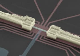
Now, Sasaki and colleagues have used MRI to take direct images of these superfluid domains. Widely used in medical and industrial imaging, MRI works by exciting nuclei in a sample using oscillating magnetic fields and then observing the radio-frequency signals emitted by the nuclei as they decay back to their ground states. The nature of the signals are very sensitive to the immediate surroundings of the nuclei, so MRI is very good at mapping the composition of an object.
This effect was useful to the researchers, as they predicted that the nuclei of superfluid atoms in different domains – therefore with different chiralities – would emit their own characteristic radio waves, helping them to distinguish topological structures. The team tested their prediction using thin films of helium-3 superfluid cooled to 2 mK, obtaining images of the quantum condensates at a spatial resolution of 10 µm.
Unexpected friction found in superfluid helium-3

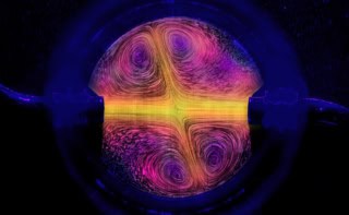
As they predicted, the team saw chiral domains as large as 1 mm across in the MRI signal. The domains were separated by walls, characterized by dark lines where the signal fell away. To test how the texture emerged, the physicists cooled the helium-3 film below its transition temperature several times. Every time, the number of domains and their locations on the film were different – meaning the superfluid’s topology arises spontaneously. This distinguishes superfluids from other fluids, where domains form either because of impurities in the material or from conditions in the surrounding environment.
Writing in Physical Review Letters, the team also reports that when they cooled the film far below the superfluid transition temperature, the domains were relatively stable over longer periods of time.