The Elekta Unity is the first high-field MR-guided radiotherapy system. The device integrates a diagnostic quality 1.5 T MR scanner with a state-of-the-art linear accelerator, and in the second half of last year received both the European CE mark and FDA 510(k) clearance.
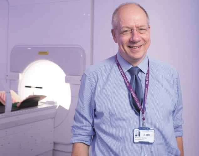
The Royal Marsden and the Institute of Cancer Research (ICR) was among the first sites to acquire an Elekta Unity. And last September, it announced the first patient treatment in the UK using an MR-linac. Tami Freeman spoke to Uwe Oelfke, head of the joint department of physics at The Royal Marsden and the ICR, to find out more about the hospital’s initial clinical experience of MR-guided radiotherapy.
TF: Why did The Royal Marsden/ICR decided to install an Elekta Unity MR-linac?
UO: It can provide us with the opportunity to see what we want to treat, and with exceptional soft-tissue contrast. With MRI, we can now see the complete relevant anatomy prior to and during treatment, and that provides a tremendous advantage.
How did the initial set-up and implementation go?
The commissioning process was very straightforward; it took some time but we had extremely good support from Elekta. We had to train physicists and radiographers to work in the environment of a magnetic field, but we started these efforts way before our first treatment. We also hired some new staff and now have two radiographers and a physicist assigned to the operation of this machine.
Your first treatments were on patients with prostate cancer, why was this particular cancer type chosen?
One reason for choosing the prostate was that Alison Tree [consultant clinical oncologist at The Royal Marsden] is the lead of the prostate tumour site group in Elekta’s MR-linac consortium. She wrote the protocol that is used for all prostate treatments in the consortium.
For us, it was also the easiest site to adopt. On a purely practical level, there’s no large organ motion expected and there are many patients available. These prostate treatments are part of the PRISM clinical trial, which states that we should include 30 patients. We have finished treatments on 11 patients now.
Are you working closely with the other sites in the MR-linac consortium?
Yes, for the founding members of the consortium there is a common data sharing protocol called the MOMENTUM framework. All seven international centres have signed up to this and we are sharing the data, which are stored in a central database at UMC in Utrecht.
The aim is that all MR-linac treatments will be part of trials. But — with the exception of UMC Utrecht, which has two MR-linacs — each institution only has one. In order to get sufficient patient numbers for the clinical studies we have to use the same protocols, share the study data and analyse all the data together. That’s the spirit of this consortium.
How has having MR guidance impacted the treatments?
Even for a case like the prostate, we have observed anatomical changes that we have never seen before — simply because we couldn’t. For instance, the placement of the bowel with respect to the prostate target completely varied daily for some patients; this was a big surprise to us. We can also see, for example, the bladder filling in detail over the time of treatment. It is very nice to have the ability to see what is really happening in the patient.
MR images of the patient are recorded prior to every fraction, do you use these for treatment replanning?
Every day, with no exceptions. For each fraction delivered on our linac, we adapt the whole treatment plan to the new anatomy. Our philosophy is that if we have this adaptation workflow integrated, and we can do it quickly, then we don’t have to deal with decisions to adapt or not to adapt. It’s much easier to have one strategy and do the optimum that you can for each patient.
Does plan adaptation add much time to the treatment?
When we started the whole procedure, one patient took on the order of 45 minutes, while a normal treatment takes around 15 to 20 minutes. Now we are achieving, depending a little on how difficult the case is, time slots of about 35 minutes. But there’s still quite a bit of room for improvement, with improved software components for instance.
The Royal Marsden/ICR recently begun treating rectal cancer with the Unity, will any other indications be treated too?
We have now treated two rectal cancer patients. The next site will definitely be the bladder, the patient is already selected. Then head-and-neck and breast cancer are the next indications. Everything is going very well, the machine has basically no downtime, so it’s our task to ramp up the patient numbers. At the moment we treat around four to five patients per day, but I think along the road between 10 and 15 should be possible.
What else is planned for the future?
More patients, more indications, and there are also some technical aspects where we are working with Elekta on improving the performance of the machine, for instance in motion management and tumour tracking. We are also developing a way to perform functional MRI on this machine — it’s a great platform for applied research.
How would functional MR be employed?
For prostate cancer, for instance, we could use it to identify the dominant inter-prostate lesion, using diffusion weighted imaging. But it also gives us ample opportunity to run functional imaging while we are treating the patient. Then we can analyse these data for differential changes and see whether we can see early indications of treatment response.
We estimate that in one year we could perform up to 500 functional MRI prostate scans, in only one centre. Then if other centres join us, we could maybe record 2000 scans in a year — how else could you collect this amount of data? The patients are in place and while we are optimizing the new plan, we can do functional imaging. It’s something we call “biomarker discovery for free”.


