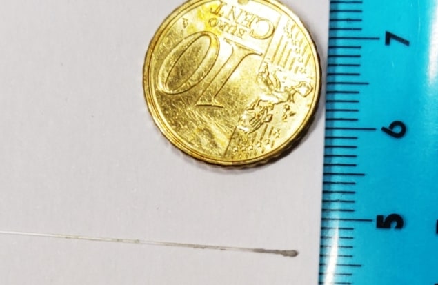
An optical fibre tipped with an inorganic scintillator makes an effective real-time dosimeter for small-field radiotherapy. Researchers at Aix-Marseille University and the Paoli-Calmettes Institute in France created such a device and compared its performance with a pair of commercial small-field dosimeters. The team found that the new device has a much smaller sensitive volume and is less susceptible to detector noise from Cherenkov radiation. It also exhibits excellent dose–response linearity and its output is stable over time (Med. Phys. 10.1002/mp.14002).
Using radiotherapy to treat early-stage tumours or tumours surrounded by critical organs requires small, sharp-edged radiation fields. Verifying the dose delivered in these cases means using a dosimeter with a correspondingly small sensitive volume: larger radiation detectors lack the spatial resolution needed to capture the high lateral dose gradients at the field margins.
To produce a dosimeter suitable for the job, Sree Bash Chandra Debnath and colleagues used silver-doped zinc sulphide (ZnS:Ag) as a scintillator – a compound long known to emit visible light when irradiated by X-rays. The team chose an inorganic scintillator because organic compounds generate more charged particles under ionizing radiation, contaminating the signal with high levels of Cherenkov radiation.
The researchers fabricated the dosimeter by dipping the end of an optical fibre into a mixture of powdered ZnS:Ag and poly(methyl methacrylate) (PMMA) dissolved in an organic solvent. After drying, the result was a ZnS:Ag-filled PMMA sphere about 200 µm across, which they coated in silver to keep out ambient light. At the other end of the optical fibre, the researchers fitted the photon counter and read-out electronics, which translated the scintillation signal into equivalent dose in real time.
To test their inorganic scintillator detector (ISD), the researchers used it to measure the dose inside a water phantom, which they exposed to X-ray fields as small as 0.25 cm2. They compared the ISD’s performance to that of two small-field dosimeters currently used in the clinic, one employing an ion chamber, the other a synthetic diamond.
Debnath and colleagues estimate the sensitive volume of the ISD to be a disc whose diameter and thickness correspond, respectively, to the width of the fibre core (100 µm) and the distance that light travels between emission and reabsorption by the scintillator (1.5 µm). This corresponds to a volume of about 1.2 × 10⁻⁵ mm3 – far smaller than that of the ion-chamber (0.016 cm3) and diamond (0.004 mm3) dosimeters.
Another area where the ISD could offer an advantage is in its high signal-to-noise ratio. This comes about because of the low levels of Cherenkov radiation produced in the small inorganic scintillator and the narrow optical fibre. The researchers think that they can reduce the Cherenkov noise even further in a future version of the device by using an entirely plastic-free fibre.
In all other respects, the ISD performed as well as required for a clinical dosimeter, and comparably to the two benchmark devices. Its dose response was linear over a wide range of dose rates and was consistent over multiple irradiations.
As inorganic scintillators are not tissue-equivalent in terms of radiation absorption, the team’s new device might be best suited to external dosimetry; used internally it would itself risk altering the dose distribution. The actual effect is likely to be small, however, and Debnath is optimistic about its potential in both situations.
“Indeed, we have already tested it for brachytherapy, and probably one of our next articles will focus on this application,” he says. “It might perturb the dose distribution, but negligibly, due to the small volume of the sensor.”
The team also intend to compare the ISD to a range of water-equivalent devices such as plastic scintillator detectors and radiosensitive films. Replacing the scintillator material with a more water-equivalent organic compound is a possibility but would, they think, come at the cost of increasing the detector’s size.


