
Rhodopsin, the protein that enables humans and other vertebrates to sense light, belongs to the family of light-sensitive G protein-coupled receptors (GPCRs). It comes first in the signal transduction pathway for vision to begin. Once it absorbs a photon, an immediate (within 200 fs) conformational change occurs in the retinal, a chromophore located inside rhodopsin. This early structural change initiates the cellular signal transduction processes that set early stages of vision. However, details of the real-time intramolecular events through which the photoactivated retinal induces the activation events inside rhodopsin remain unclear.
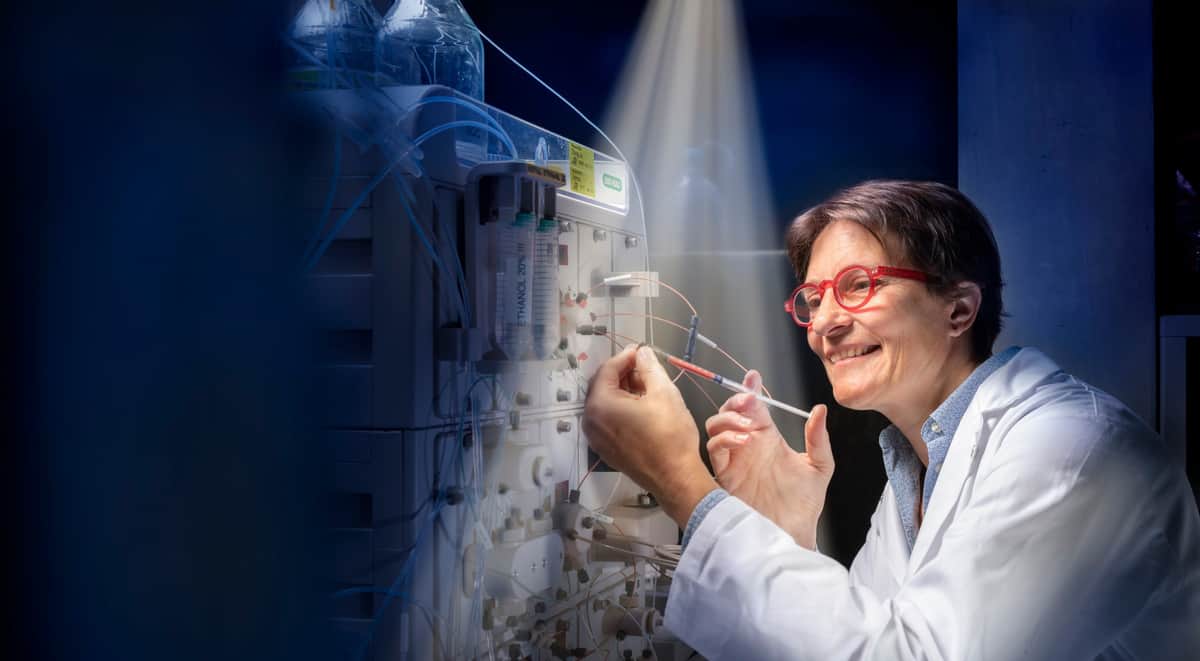
To fill this knowledge gap, researchers at the Paul Scherrer Institute (PSI) in Switzerland used ultrafast time-resolved crystallography to study conformational changes in rhodopsin after it absorbs a photon. Their findings, reported in Nature, explain how the retinal only absorbs part of the photon energy, storing the remaining energy to fuel the conformational changes associated with the formation of the G protein-binding signalling state.
To record and analyse the activation mechanism of the retinal chromophore at the atomic scale, and with ultrafast (picosecond) temporal resolution, the team used time-resolved serial femtosecond crystallography (TR-SFX) at room temperature.
For their experiments, the researchers first grew high-quality rhodopsin microcrystals, and then used TR-SFX to generate series of diffraction pattern images of the crystals. More precisely, they used an optical laser pulse to photoactivate the protein molecules in the crystal and then – after a specified time delay – probed the structure with an X-ray pulse from an X-ray free electron laser (XFEL). Recording with the XFEL, effectively a very high-speed camera, the researchers collected serial frames from tens of thousands of crystals oriented in random manner.
The analyses performed by the team included modelling the rhodopsin structure for electron density changes together with structural refinement against crystallography observations. This revealed that light-induced isomerization (in which a molecule switches between two distinct conformations) with a bend in the retinal chromophore persists for 1 ps, given that the first metastable intermediate of rhodopsin appears 200 fs after photoactivation. Then 100 ps later, the rhodopsin structure adopts a more relaxed conformation. Thereby the results suggest that the protein utilizes active (or functional) zones of the GPCR structural pathways for energy dissipation.
Connecting the dots to artificially restore vision

One highlight of this new study is that the room-temperature structure reveals electron density for all previously described functional and structural water molecules, including those that have a role later in the photoactivation process. The researchers note that previous structures resolved under cryogenic conditions failed to achieve this. Consequently, the new high-resolution SFX structure of rhodopsin showcases the entirety of the water-mediated hydrogen bond network within the protein.
The investigation sheds light on the earliest stages of vision, revealing that ultrafast energy dissipation in rhodopsin occurs through conserved residues of GPCR activation pathways, paving the way to study the early activation events in this largest family of GPCRs (class A).


