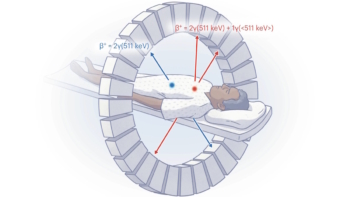
Dynamic PET imaging is an important preclinical research tool used to visualize real-time functional information in a living animal. Currently, however, the temporal resolution of small-animal PET scanners is on the order of seconds, which is too slow to image blood flow in the heart or track the brain’s neuronal activity. To remedy this, the Imaging Physics Group at the National Institutes for Quantum Science and Technology (QST) in Japan has developed an ultrasensitive small-animal PET scanner that enables sub-second dynamic imaging of a rat.
The limited temporal resolution of conventional preclinical PET scanners stems from their low sensitivity (around 10%), caused by relatively thin detection crystals (10 mm) and a short axial field-of-view (FOV). Thus the QST team built a system based on four-layer, depth-encoding detectors with a total thickness of 30 mm. The scanner has a 325.6 mm-long axial FOV, providing total-body coverage without any bed movement, while a small inner diameter of 155 mm further increases detection efficiency.
“The main application of the total-body small-animal PET (TBS-PET) scanner will be assessment of new radiopharmaceuticals, especially for cardiovascular and neurodegenerative diseases, by providing total-body rodent PET images with sub-second temporal resolution,” first author Han Gyu Kang tells Physics World. “In addition, the scanner will be used for in-beam PET imaging, and single-cell tracking, where ultrahigh sensitivity is required.”
Performance evaluation
The TBS-PET scanner contains six detector rings, each incorporating 10 depth-of-interaction (DOI) detectors. Each DOI detector comprises a four-layer zirconium-doped gadolinium oxyorthosilicate (GSOZ) crystal array (16×16 crystals per layer) and an array of multi-anode photomultiplier tubes. The team selected GSOZ crystals because they have no intrinsic radiation signal, thus enabling low activity PET imaging.
The researchers performed a series of tests to characterize the scanner performance. Measurements of a 68Ge line source at the centre of the FOV showed that the TBS-PET had an energy resolution of 18.4% and a coincidence timing resolution of 7.9 ns.
Imaging a NEMA 22Na point source revealed a peak sensitivity of 45.0% in the 250–750 keV energy window – more than four times that of commercial or laboratory small-animal PET scanners. The system exhibited a uniform spatial resolution of around 2.6 mm across the FOV, thanks to the four-layer DOI information, which effectively reduced the parallax error.
In vivo imaging
Kang and colleagues next obtained in vivo total-body PET images of healthy rats using a single bed position. Static imaging using Na18F and 18F-FDG tracers clearly visualized bone structures and glucose metabolism, respectively, of the entire rat body.
Moving to dynamic imaging, the researchers injected an 18F-FDG bolus into the tail vein of an anesthetized rat for 15 s, followed by a saline injection 15 s after injection. They acquired early-phase dynamic PET data every second until 27 s after injection. To enable sub-second PET imaging, they used custom-written software to subdivide the list-mode data (1 s time frame) into time frames of 0.5 s, 0.25 s and 0.1 s.
Dynamic PET images with a 0.5 s time frame clearly visualized the blood stream from the tail to the heart through the iliac vein and inferior vena cava for the first 2 s, after which the tracer reached the right atrium and right ventricle. At 4.0 s after injection, blood flowed from the left ventricle into the brain via the carotid arteries. The cortex and kidneys were identified 5.5 s after injection. After roughly 17.5 s, the saline peak could be identified in the time-activity curves (TACs).
At 0.25 s temporal resolution, the early-phase images visualized the first pass blood circulation of the rat heart, showing the 18F-FDG bolus flowing from the inferior vena cava to the right ventricle from 2.25 s. The tracer next circulated to the lungs via the pulmonary artery from 2.5 s, and then flowed to the left ventricle from 3.75 s.
The TACs clearly visualized the time dispersion between the right and left ventricles (1.25 s). This value can change for animals with cardiac disease, and the team plans to explore the benefit of fast temporal resolution PET for diagnosing cardiovascular and neurodegenerative diseases.
Walk-through PET scanner made for high-throughput imaging at lower cost

The researchers conclude that the TBS-PET scanner enables dynamic imaging with a nearly real-time frame rate, visualizing cardiac function and pulmonary circulation of a rat with 0.25 s temporal resolution, a feat that is not possible with conventional small-animal PET scanners.
“One drawback of the TBS-PET scanner is the relatively low spatial resolution of around 2.6 mm, which is limited by the relatively large crystal pitch of 2.85 mm,” says Kang. “To solve this issue, we are now developing a new small-animal PET scanner employing three-layer depth-encoding detectors with 0.8 mm crystal pitch, towards our final goal of sub-millimetre and sub-second temporal resolution PET imaging in rodent models.”
The TBS-PET scanner is described in Physics in Medicine & Biology.