
The cerebellum, a small region of the brain located at the back of the head, is largely responsible for motor control, as well as being involved in behaviour and cognition. It also plays a role in various disease processes, such as multiple sclerosis (MS), for example, which causes extensive demyelination in the cerebellar cortex. But despite its importance, the structure of the cerebellum has not been fully investigated due to the inadequate resolution of current in vivo imaging techniques.
The key obstacle is that the cortex covering the cerebellum comprises extremely tightly folded layers of tissue and requires high-resolution imaging to fully visualize and study its anatomy. Now, researchers at the Spinoza Centre for Neuroimaging in the Netherlands have developed a method to image the cerebellar cortical layers using a powerful 7 T MRI scanner, describing the technique in Radiology.
First author Nikos Priovoulos and colleagues modified two MRI pulse sequences that image the cortical surface and intracortical layers, to translate the high signal-to-noise ratio of 7 T MRI into high spatial resolution. By also compensating for motion, they generated images of up to 200 μm resolution, with a clinically applicable scan time of less than 20 min.
For their study, the researchers imaged healthy participants in a 7.0 T MRI scanner. To image the layers within the cerebellar cortex, they used a T2*-weighted fast low-angle shot (FLASH) sequence with a 210 × 210 × 15 mm field-of-view (FOV) and a voxel size of 0.19 × 0.19 × 0.5 mm. They used this scan, which covers only part of the cerebellar cortex, to image nine subjects.
With such a small voxel size, involuntary motion can limit the effective spatial resolution. To combat this, the researchers interleaved the FLASH sequence with whole-head fat images, which they used to estimate and correct for motion. In four participants who underwent scans both with and without this step, prospective motion correction improved image sharpness and preserved high-resolution features.
The motion-corrected FLASH scans visualized inner- and outer-layer structures in the cerebellar cortex for all participants. The researchers suggest that these represent the deep, iron-rich granular layer and the less neuronally dense superficial molecular layer, which exhibit differences in magnetic susceptibility at 7.0 T. They note that cerebellar layers are differentially affected in diseases such as MS, thus the ability to observe individual layers could provide valuable diagnostic markers.
“In MS the cerebellum plays an important role,” explains Priovoulos in a press statement. “MS patients have motor lesions, which means that they have damage to the nerve cells involved in movement. Based on previous findings, we know for MS specifically that we could benefit from high-resolution imaging in the cerebellum.”
Unfolding the cerebellum
The researchers also used 7 T MRI to visualize the entire cerebellum in nine healthy participants. Here, they employed a magnetization-prepared 2 rapid gradient-echo (MP2RAGE) sequence with a 210 × 120 × 60 mm FOV and a voxel size of 0.4 mm3. They used the same fat navigator for motion correction.
The motion-corrected MP2RAGE scans resolved cerebellar anatomic features down to individual folia – the tiny folds in the cortical surface. The team, led by Wietske van der Zwaag, note that downsampling the data to match current state-of-the-art MRI acquisitions reduced the visibility of these features.
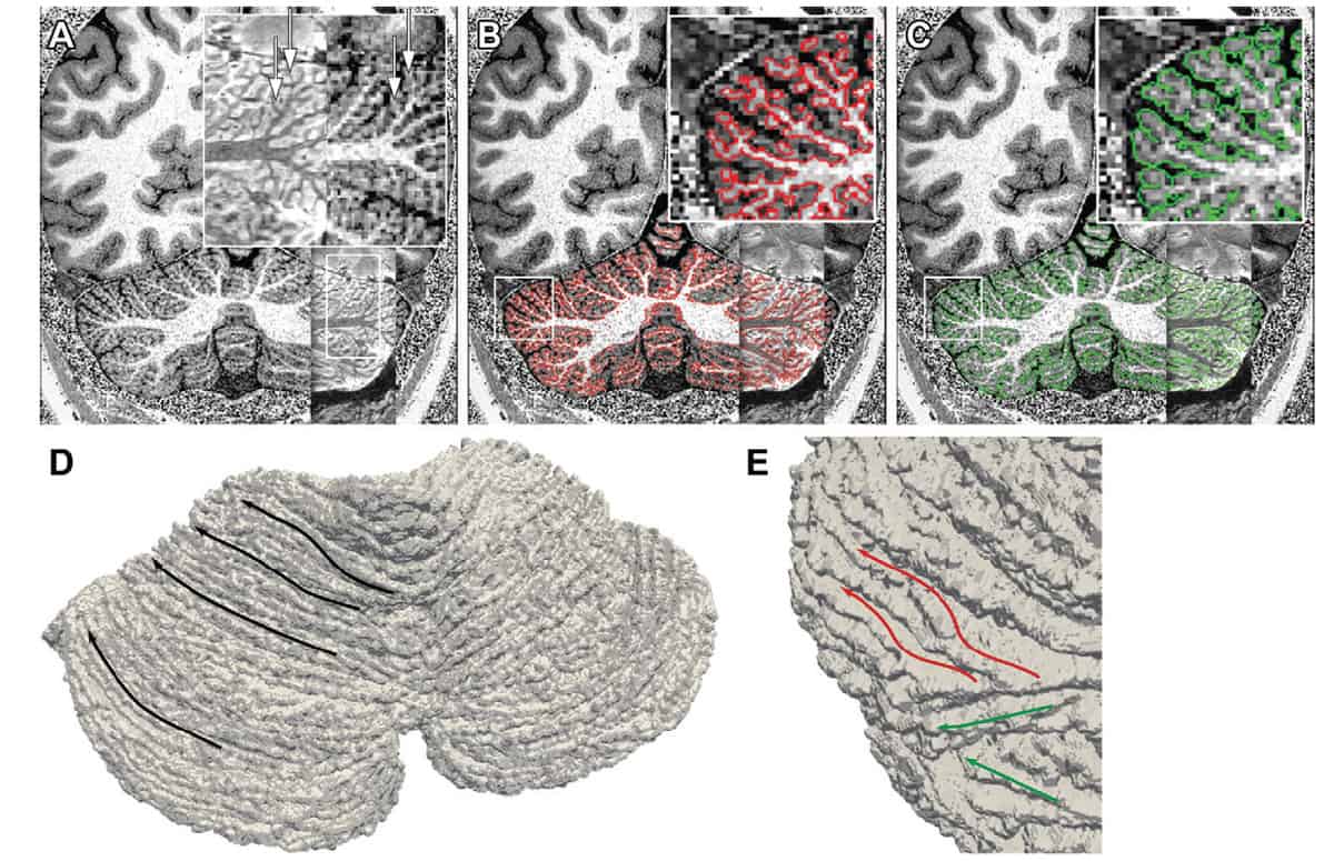
The high spatial resolution of the images allowed the researchers to computationally unfold the cerebellar cortical surface into a continuous sheet. This enabled them to calculate clinical measures such as the cortical surface area and thickness, and examine disease-related factors such as myelin-sensitive T1 values.
The estimated median cerebellar cortical surface area was 949 cm2 (176%–759% larger than previous imaging-based in vivo estimates) and the median cerebellar cortical thickness was 0.88 mm, in agreement with ex vivo reports and four to five times thinner than current imaging-based in vivo estimates.
While most participants in the study were young (a median age of 36), the team included two older subjects (aged 57 and 62). MR images of these participants showed visible cortical thinning in the cerebellum at visual inspection and lower cerebellar cortical thickness and grey matter T1 values than in the younger cohort.
“This is the first time that we can view the human cerebellum directly in living humans, with this much detail,” says Priovoulos. “We can do this specifically because we have a very high-field magnet (which is expensive and hard to build) and also motion correction, because people tend to move during the scans.”
Ultrahigh-field MRI tracks multiple sclerosis progression
Priovoulos, van der Zwaag and PhD student Emma Brouwer are now working to make the MRI signal in the cerebellum more reliable. “The wavelength of the MRI signal at 7 T is comparable to the human head size and this frequently makes the signal in the cerebellum inhomogeneous,” Priovoulos tells Physics World. “To tackle this, we are trying to combine our setup with multiple radiofrequency producing coils to optimize signal generation. The challenge is to do so while still keeping the scan length short and the setup translatable to the clinic.”
The researchers are already applying the 7 T MRI approach to scan patients with MS. They also want to use it to better understand cerebellar ataxia, a muscle-control disease. Alongside, they are using functional 7 T imaging, along with the cerebellar anatomical reconstruction, to examine cerebellar functional responses in detail and explore the role of the cerebellum in human health and disease.


