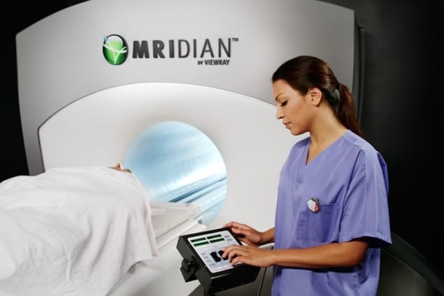
A team of clinicians at the University of Wisconsin have used a ViewRay MRI-guided radiotherapy system to perform brachytherapy planning for cervical cancer. So, what motivated the team to use the ViewRay system to perform brachytherapy planning? And how does use of the system fit time-wise alongside its use for treatments?
Time saving
Writing in the journal Brachytherapy, the authors present the results of some 142 fractions of intracavitary brachytherapy, performed between April 2015 and January 2017 on 29 cervical cancer patients – the first ever clinical use of ViewRay-guided brachytherapy (Brachytherapy 10.1016/j.brachy.2018.04.005). They conclude that “time to treatment using this approach was shorter compared to diagnostic MRI” and that the ViewRay system also provided “significant advantage in visualizing the tumour and cervix compared to CT”.
As co-author Cindy Ko, a radiation oncology resident at the University of Wisconsin School of Medicine and Public Health, explains, the motivation to undertake this research came from necessity. This is because, although Ko and her team typically employ a diagnostic quality 1.5-3T MRI at diagnosis and for “at least the first fraction” of brachytherapy – a quality of imaging that they would ideally use for every fraction of brachytherapy planning – a combination of time constraints and limited resources means that they elect to use such MRI levels for only the first and third treatments.
“We used the imaging component of the ViewRay treatment system, which is in and of itself an independent treatment machine that can be used to directly visualize and treat cancers of the lung, gastrointestinal system [and others] with MRI guidance,” says Ko.
“The ViewRay system is within our department, unlike the diagnostic MRI machines, and thus it can save some travel time between the procedure suite, imaging, and back to the procedure suite for treatment,” she adds.
In addition to the benefits of proximity, Ko reveals that the ViewRay MR image acquisition is also faster, “on account of not requiring multiple sequences”. This is because there is only one proprietary sequence officially available for use with the system. This fast sequence, known as TRUFI, is a hybrid of T2 and T1 sequences that the team uses both with and without contrast for its treatments on the ViewRay system.
Scheduling
Since beginning to use the system in the department, Ko and her team found that scheduling a time to use the machine for brachytherapy planning between treatments worked well. Given the unpredictability of procedures, Ko reveals that they tend to “pink slip” individual brachytherapy cases into a block of time on the ViewRay system’s schedule – and continue to treat or simulate scheduled patients until the brachytherapy case is nearly ready. They will then “image [the case] briefly before going back to the other treatments or simulations on the schedule”.
According to Ko, this requires the therapists to develop a familiarity with how the logistics of a ViewRay imaging session with a brachytherapy patient works, as well as a working knowledge of how long the imaging session will take.
“We don’t find that one use of ViewRay system hinders the other – meaning that the schedule is mostly on time using this approach – and we are happy to find multiple uses for the same machine. The ViewRay schedule is busy, but no busier than our other treatment machines,” she says.
Ko also points out that the brachytherapy instrument placement itself “does not require much more than an ultrasound” for the first fraction, although for some patients, placement can take an hour or more.
In view of the fact that performing instrument placement within the ViewRay suite would probably interfere with the normal ViewRay treatment schedule – during which time the imaging function would not be used – the team found that transferring to the ViewRay system after instrument placement, and then adjusting if needed based on the MR image, was sufficient.
“Very seldom do we have to adjust instrument placement after viewing the ViewRay, MRI [or] CT images, but it can happen and, if so, we image again after adjusting to ensure good geometry,” adds Ko.


