Computed tomography (CT) and X-ray images can be used to determine whether bullets embedded in patients are ferromagnetic or nonferromagnetic, researchers in the US claim. This means doctors can now identify whether it is safe for a patient with gunshot wounds to undergo MRI scans, they say.
According to the Small Arms Survey, 40% of firearms in the world are owned by Americans. In 2017 a survey by the Pew Research Centre found that 44% of Americans know someone that has been shot, either accidentally or intentionally, with 3% of adults saying they have been shot.
Being shot can have important implications for medical diagnostics, even years later, as people with gunshot wounds are frequently denied MRI scans. This is because the composition of embedded bullet fragments cannot be identified to determine whether they are nonferromagnetic, or not.
Ferromagnetic materials are not safe in MRI scanners because the high-powered magnets that the machines use can heat or move them. This could burn the patient. And if the material is near a critical structure, such as the spinal cord, even small movements could cause significant damage.
Jason Allen, a neuroradiologist at Emory University in Atlanta, says that issues arise more frequently with patients that have been shot in the past. “They survive that injury and then they come along later, and they have a new problem,” he explains. “It is particularly problematic at that point because there is really zero chance of us knowing what kind of bullet they were shot with.”
MRI is important, Allen says, because there are clinical questions that doctors are unable to answer with other imaging techniques, particularly for issues related to the brain and spinal cord. “In terms of traumas, if we need to look for damage to those tissues, or someone may have had a stroke or a suspected stroke, these are things that we really need to define with MRI,” he explains.
Allen and his colleagues wondered whether bullets made from ferromagnetic and nonferromagnetic metals deformed and broke up in different ways on impact, leaving identifiable differences in the debris embedded in those who had been shot. This could allow the composition of the bullets to be determined from images taken using non-magnetic radiological techniques, to check MRI safety.
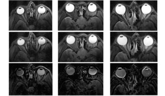
To test this, the researchers fired handgun and shotgun ammunition commercially available in the US into blocks of ballistic gelatin – a material that is an analogue for human tissue and is used to simulate the effect of bullet impact. They then took CT and X-ray images of the gelatin blocks.
The team were able to distinguish between ferromagnetic and nonferromagnetic fired bullets, they report in the American Journal of Roentgenology. They found that a bullet that leaves a metallic debris trail from entry to final position or has been appreciably deformed is of copper, copper-alloy or lead composition, or represents lead shotgun shot. These nonferromagnetic materials deform more because they are softer than ferromagnetic metals. Based on this, the researchers created a simple triage algorithm for patients with retained ballistic fragments that doctors can follow to determine MRI safety. “This can be done by any radiologist of any background,” Allen says.

Allen and his colleagues also took MRI scans of unfired bullets suspended in ballistic gelatin blocks, and assessed magnetic attractive force, rotational torque and heating effects. Although ferromagnetic bullets showed evidence of having moved or rotated during the scans, nonferromagnetic bullets did not. None of the bullets tested showed heating above the US Food and Drug Administration limit of 2 °C. This shows that nonferromagnetic bullets do not pose a significant risk for MRI scans, regardless of when the injury occurred, the researchers say.
“We’ve scanned hundreds of these patients over the years, we’ve not had any cases where the patient has gone into the scanner based on this algorithm and had an injury of some sort, had a heating of the bullet or had a movement of the bullet,” Allen tells Physics World.