Proton arc therapy eliminates hard-to-treat cancer with minimal side effects

Head-and-neck cancers are difficult to treat with radiation therapy because they are often located close to organs that are vital for patients to maintain a high quality-of-life. Radiation therapy can also alter a person’s shape, through weight loss or swelling, making it essential to monitor such changes throughout the treatment to ensure effective tumour targeting.

Researchers from Corewell Health William Beaumont University Hospital have now used a new proton therapy technique called step-and-shoot proton arc therapy (a spot-scanning proton arc method) to treat head-and-neck cancer in a human patient – the first person in the US to receive this highly accurate treatment.

“We envisioned that this technology could significantly improve the quality of treatment plans for patients and the treatment efficiency compared with the current state-of-the-art technique of intensity-modulated proton therapy (IMPT),” states senior author Xuanfeng Ding.

Progression towards dynamic proton arc therapy

“The first paper on spot-scanning proton arc therapy was published in 2016 and the first prototype for it was built in 2018,” says Ding. However, step-and-shoot proton arc therapy is an interim solution towards a more advanced technique known as dynamic proton arc therapy – which delivered its first pre-clinical treatment in 2024. Dynamic proton arc therapy is still undergoing development and regulatory approval clearance, so researchers have chosen to use step-and-shoot proton arc therapy clinically in the meantime.

Other proton therapies are more manual in nature and require a lot of monitoring, but the step-and-shoot technology delivers radiation directly to a tumour in a more continuous and automated fashion, with less lag time between radiation dosages. “Step-and-shoot proton arc therapy uses more beam angles per plan compared to the current clinical practice using IMPT and optimizes the spot and energy layers sparsity level,” explains Ding.

The extra beam angles provide a greater degree-of-freedom to optimize the treatment plan and provide a better dose conformity, robustness and linear energy transfer (LET, the energy deposited by ionizing radiation) through a more automated approach. During treatment delivery, the gantry rotates to each beam angle and stops to deliver the treatment irradiation.

In the dynamic proton arc technique that is also being developed, the gantry rotates continuously while irradiating the proton spot or switching energy layer. The step-and-shoot proton arc therapy therefore acts as an interim stage that is allowing more clinical data to be acquired to help dynamic proton arc therapy become clinically approved. The pinpointing ability of these proton therapies enables tumours to be targeted more precisely without damaging surrounding healthy tissue and organs.

The first clinical treatment

The team trialled the new technique on a patient with adenoid cystic carcinoma in her salivary gland – a rare and highly invasive cancer that’s difficult to treat as it targets the nerves in the body. This tendency to target nerves also means that fighting such tumours typically causes a lot of side effects. Using the new step-and-shoot proton arc therapy, however, the patient experienced minimal side effects and no radiation toxicity to other areas of her body (including the brain) after 33 treatments. Since finishing her treatment in August 2024, she continues to be cancer-free.

Tiffiney Beard and Rohan Deraniyagala

“Radiation to the head-and-neck typically results in dryness of the mouth, pain and difficulty swallowing, abnormal taste, fatigue and difficulty with concentration,” says Rohan Deraniyagala, a Corewell Health radiation oncologist involved with this research. “Our patient had minor skin irritation but did not have any issues with eating or performing at her job during treatment and for the last year since she was diagnosed.”

Describing the therapeutic process, Ding tells Physics World that “we developed an in-house planning optimization algorithm to select spot and energy per beam angle so the treatment irradiation time could be reduced to four minutes. However, because the gantry still needs to stop at each beam angle, the total treatment time is about 16 minutes per fraction.”

On monitoring the progression of the tumour over time and developing treatment plans, Ding confirms that the team “implemented a machine-learning-based synthetic CT platform which allows us to track the daily dosage of radiation using cone-beam computed tomography (CBCT) so that we can schedule an adaptive treatment plan for the patient.”

On the back of this research, Ding says that the next step is to help further develop the dynamic proton arc technique – known as DynamicARC – in collaboration with industry partner IBA.

The research was published in the International Journal of Particle Therapy.

Superconducting microwires detect high-energy particles

Arrays of superconducting wires have been used to detect beams of high-energy charged particles. Much thinner wires are already used to detect single photons, but this latest incarnation uses thicker wires that can absorb the large amounts of energy carried by fast-moving protons, electrons, and pions. The new detector was created by an international team led by Cristián Peña at Fermilab.

In a single-photon detector, an array of superconducting nanowires is operated below the critical temperature for superconductivity – with current flowing freely through the nanowires. When a nanowire absorbs a photon it creates a hotspot that temporarily destroys superconductivity and boosts the electrical resistance. This creates a voltage spike across the nanowire, allowing the location and time of the photon detection to be determined very precisely.

“These detectors have emerged as the most advanced time-resolved single-photon sensors in a wide range of wavelengths,” Peña explains. “Applications of these photon detectors include quantum networking and computing, space-to-ground communication, exoplanet exploration and fundamental probes for new physics such as dark matter.”

A similar hotspot is created when a superconducting wire is impacted by a high-energy charged particle. In principle, this could be used to create particle detectors that could be used in experiments at labs such as Fermilab and CERN.

New detection paradigm

“As with photons, the ability to detect charged particles with high spatial and temporal precision, beyond what traditional sensing technologies can offer, has the potential to propel the field of high-energy physics towards a new detection paradigm,” Peña explains.

However, the nanowire single-photon detector design is not appropriate for detecting charged particles. Unlike photons, charged particles do not deposit all of their energy at a single point in a wire. Instead, the energy can be spread out along a track, which becomes longer as particle energy increases. Also, at the relativistic energies reached at particle accelerators, the nanowires used in single-photon detectors are too thin to collect the energy required to trigger a particle detection.

To create their new particle detector, Peña’s team used the latest advances in superconductor fabrication. On a thin film of tungsten silicide, they deposited an 8×8, 2 mm2 array of micron-thick superconducting wires.

Tested at Fermilab

To test out their superconducting microwire single-photon detector (SMSPD), they used it to detect high-energy particle beams generated at the Fermilab Test Beam Facility. These included a 12 GeV beam of protons and 8 GeV beams of electrons and pions.

“Our study shows for the first time that SMSPDs are sensitive to protons, electrons, and pions,” Peña explains. “In fact, they behave very similarly when exposed to different particle types. We measured almost the same detection efficiency, as well as spatial and temporal properties.”

The team now aims to develop a deeper understanding of the physics that unfolds as a charged particle passes through a superconducting microwire. “That will allow us to begin optimizing and engineering the properties of the superconducting material and sensor geometry to boost the detection efficiency, the position and timing precision, as well as optimize for the operating temperature of the sensor,” Peña says. With further improvements SMSPDs to become an integral part of high-energy physics experiments – perhaps paving the way for a deeper understanding of fundamental physics.

The research is described in the Journal of Instrumentation.

What is meant by neuromorphic computing – a webinar debate

Want to learn more on this subject?

AI circuit board

Want to learn more on this subject?

There are two main approaches to what we consider neuromorphic computing. The first involves emulating biological neural processing systems through the physics of computation of computational substrates that have similar properties and constraints as real neural systems, with potential for denser structures and advantages in energy cost. The other simulates neural processing systems on scalable architectures that allow the simulation of large neural networks, with higher degree of abstraction, arbitrary precision, high resolution, and no constraints imposed by the physics of the computing medium.

Both may be required to advance the field, but is either approach ‘better’? Hosted by Neuromorphic Computing and Engineering, this webinar will see teams of leading experts in the field of neuromorphic computing argue the case for either approach, overseen by an impartial moderator.

Speakers image. Left to right: Elisa Donati, Jennifer Hasler, Catherine (Katie) Schuman, Emre Neftci, Giulia D’Angelo

Team emulation:
Elisa Donati. Elisa’s research interests aim at designing neuromorphic circuits that are ideally suited for interfacing with the nervous system and show how they can be used to build closed-loop hybrid artificial and biological neural processing systems.  She is also involved in the development of neuromorphic hardware and software systems able to mimic the functions of biological brains to apply for medical and robotics applications.

Jennifer Hasler received her BSE and MS degrees in electrical engineering from Arizona State University in August 1991. She received her PhD in computation and neural systems from California Institute of Technology in February 1997. Jennifer is a professor at the Georgia Institute of Technology in the School of Electrical and Computer Engineering; Atlanta is the coldest climate in which she has lived. Jennifer founded the Integrated Computational Electronics (ICE) laboratory at Georgia Tech, a laboratory affiliated with the Laboratories for Neural Engineering. She is a member of Tau Beta P, Eta Kappa Nu, and the IEEE.

Team simulation:
Catherine (Katie) Schuman is an assistant professor in the Department of Electrical Engineering and Computer Science at the University of Tennessee (UT). She received her PhD in computer science from UT in 2015, where she completed her dissertation on the use of evolutionary algorithms to train spiking neural networks for neuromorphic systems. Katie previously served as a research scientist at Oak Ridge National Laboratory, where her research focused on algorithms and applications of neuromorphic systems. Katie co-leads the TENNLab Neuromorphic Computing Research Group at UT. She has written for more than 70 publications as well as seven patents in the field of neuromorphic computing. She received the Department of Energy Early Career Award in 2019. Katie is a senior member of the Association of Computing Machinery and the IEEE.

Emre Neftci received his MSc degree in physics from EPFL in Switzerland, and his PhD in 2010 at the Institute of Neuroinformatics at the University of Zurich and ETH Zurich. He is currently an institute director at the Jülich Research Centre and professor at RWTH Aachen. His current research explores the bridges between neuroscience and machine learning, with a focus on the theoretical and computational modelling of learning algorithms that are best suited to neuromorphic hardware and non-von Neumann computing architectures.

Discussion chair:
Giulia D’Angelo is currently a Marie Skłodowska-Curie postdoctoral fellow at the Czech Technical University in Prague, where she focuses on neuromorphic algorithms for active vision. She obtained a bachelor’s degree in biomedical engineering from the University of Genoa and a master’s degree in neuroengineering with honours. During her master’s, she developed a neuromorphic system for the egocentric representation of peripersonal visual space at King’s College London. She earned her PhD in neuromorphic algorithms at the University of Manchester, receiving the President’s Doctoral Scholar Award, in collaboration with the Event-Driven Perception for Robotics Laboratory at the Italian Institute of Technology. There, she proposed a biologically plausible model for event-driven, saliency-based visual attention. She was recently awarded the Marie Skłodowska-Curie Fellowship to explore sensorimotor contingency theories in the context of neuromorphic active vision algorithms.

About this journal
Neuromorphic Computing and Engineering journal cover

Neuromorphic Computing and Engineering is a multidisciplinary, open access journal publishing cutting-edge research on the design, development and application of artificial neural networks and systems from both a hardware and computational perspective.

Editor-in-chief: Giacomo Indiveri, University of Zurich, Switzerland


A Martian aurora, how the universe fades away, Heisenberg on holiday, physics of fake coins

In this episode of the Physics World Weekly podcast I look at what’s new in the world of physics with the help of my colleagues Margaret Harris and Matin Durrani.

We begin on Mars, where NASA’s Perseverance Rover has made the first observation of an aurora from the surface of the Red Planet. Next, we look deep into the future of the universe and ponder the physics that will govern how the last stars will fade away.

Then, we run time in reverse and go back to the German island of Helgoland, where in 1925 Werner Heisenberg laid the foundations of modern quantum mechanics. The island will soon host an event celebrating the centenary and Physics World will be there.

Finally, we explore how neutrons are being used to differentiate between real and fake antique coins and chat about the Physics World Quantum Briefing 2025.

Ultrasound-activated structures clear biofilms from medical implants

When implanted medical devices like urinary stents and catheters get clogged with biofilms, the usual solution is to take them out and replace them with new ones. Now, however, researchers at the University of Bern and ETH Zurich, Switzerland have developed an alternative. By incorporating ultrasound-activated moving structures into their prototype “stent-on-a-chip” device, they showed it is possible to remove biofilms without removing the device itself. If translated into clinical practice, the technology could increase the safe lifespan of implants, saving money and avoiding operations that are uncomfortable and sometimes hazardous for patients.

Biofilms are communities of bacterial cells that adhere to natural surfaces in the body as well as artificial structures such as catheters, stents and other implants. Because they are encapsulated by a protective, self-produced extracellular matrix made from polymeric substances, they are mechanically robust and resistant to standard antibacterial measures. If not removed, they can cause infections, obstructions and other complications.

Intense, steady flows push away impurities

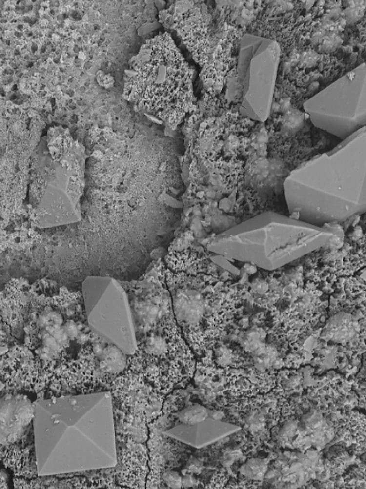
The new technology, which was co-developed by Cornel Dillinger, Pedro Amado and other members of Francesco Clavica and Daniel Ahmed’s research teams, takes advantage of recent advances in the fields of robotics and microfluidics. Its main feature is a coating made from microscopic hair-like structures known as cilia. Under the influence of an acoustic field, which is applied externally via a piezoelectric transducer, these cilia begin to move. This movement produces intense, steady fluid flows with velocities of up to 10 mm/s – enough to break apart encrusted deposits (made from calcium carbonate, for example) and flush away biofilms from the inner and outer surfaces of implanted urological devices.

Microscope image showing square and diamond shapes in various shades of grey

“This is a major advance compared to existing stents and catheters, which require regular replacements to avoid obstruction and infections,” Clavica says.

The technology is also an improvement on previous efforts to clear implants by mechanical means, Ahmed adds. “Our polymeric cilia in fact amplify the effects of ultrasound by allowing for an effect known as acoustic streaming at frequencies of 20 to 100 kHz,” he explains. “This frequency is lower than that possible with previous microresonator devices developed to work in a similar way that had to operate in the MHz-frequency range.”

The lower frequency achieves the desired therapeutic effects while prioritizing patient safety and minimizing the risk of tissue damage, he adds.

Wider applications

In creating their technology, the researchers were inspired by biological cilia, which are a natural feature of physiological systems such as the reproductive and respiratory tracts and the central nervous system. Future versions, they say, could apply the ultrasound probe directly to a patient’s skin, much as handheld probes of ultrasound scanners are currently used for imaging. “This technology has potential applications beyond urology, including fields like visceral surgery and veterinary medicine, where keeping implanted medical devices clean is also essential,” Clavica says.

The researchers now plan to test new coatings that would reduce contact reactions (such as inflammation) in the body. They will also explore ways of improving the device’s responsiveness to ultrasound – for example by depositing thin metal layers. “These modifications could not only improve acoustic streaming performance but could also provide additional antibacterial benefits,” Clavica tells Physics World.

In the longer term, the team hope to translate their technology into clinical applications. Initial tests that used a custom-built ultrasonic probe coupled to artificial tissue have already demonstrated promising results in generating cilia-induced acoustic streaming, Clavica notes. “In vivo animal studies will then be critical to validate safety and efficacy prior to clinical adoption,” he says.

The present study is detailed in PNAS.

Former IOP president Cyril Hilsum celebrates 100th birthday

Cyril Hilsum, a former president of the Institute of Physics (IOP), celebrated his 100th birthday last week at a special event held at the Royal Society of Chemistry.

Born on 17 May 1925, Hilsum completed a degree in physics at University College London in 1945. During his career he worked at the Services Electronics Research Laboratory and the Royal Radar Establishment and in 1983 was appointed chief scientist of GEC Hirst Research Centre, where he later became research director before retiring aged 70.

Hilsum helped develop commercial applications for the semiconductor gallium arsenide and is responsible for creating the UK’s first semiconductor laser as well as developments that led to modern liquid crystal display technologies.

Between 1988 and 1990 he was president of the IOP, which publishes Physics World, and in 1990 was appointed a Commander of the Order of the British Empire (CBE) for “services to the electrical and electronics industry”.

Hilsum was honoured by many prizes during his career including IOP awards such as the Max Born Prize in 1987, the Faraday Medal in 1988 as well as the Richard Glazebrook Medal and Prize in 1998. In 2007 he was awarded the Royal Society’s Royal Medal “for his many outstanding contributions and for continuing to use his prodigious talents on behalf of industry, government and academe to this day”.

Cyril Hilsum at an event to mark his 100th birthday

Despite now being a centenarian, Hilsum still works part-time as chief science officer for Infi-tex Ltd, which produces force sensors for use in textiles.

“My birthday event was an amazing opportunity for me to greet old colleagues and friends,” Hilsum told Physics World. “Many had not seen each other since they had worked together in the distant past. It gave me a rare opportunity to acknowledge the immense contributions they had made to my career.”

Hilsum says that while the IOP gives much support to applied physics, there is still a great need for physicists “to give critical contributions to the lives of society as a whole”.

“As scientists, we may welcome progress in the subject, but all can get pleasure in seeing the results in their home, on their iPhone, or especially in their hospital!” he adds.

Bacteria-killing paint could dramatically improve hospital hygiene

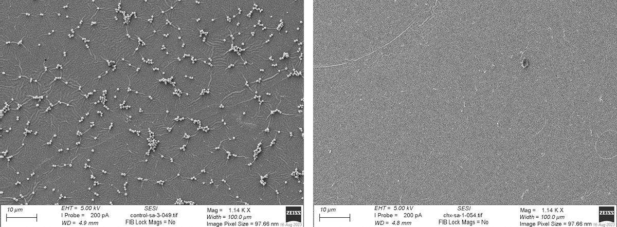
Antimicrobial efficacy of chlorhexidine epoxy resin

Scientists have created a novel antimicrobial coating that, when mixed with paint, can be applied to a range of surfaces to destroy bacteria and viruses – including particularly persistent and difficult to kill strains like MRSA, flu virus and SARS-CoV-2. The development potentially paves the way for substantial improvements in scientific, commercial and clinical hygiene.

The University of Nottingham-led team made the material by combining chlorhexidine digluconate (CHX) – a disinfectant commonly used by dentists to treat mouth infections and by clinicians for cleaning before surgery – with everyday paint-on epoxy resin. Using this material, the team worked with staff at Birmingham-based specialist coating company Indestructible Paint to create a prototype antimicrobial paint. They found that, when dried, the coating can kill a wide range of pathogens.

The findings of the study, which was funded by the Royal Academy of Engineering Industrial Fellowship Scheme, were published in Scientific Reports.

Persistent antimicrobial protection

As part of the project, the researchers painted the antimicrobial coating onto a surface and used a range of scientific techniques to analyse the distribution of the biocide in the paint, to confirm that it remained uniformly distributed at a molecular level.

According to project leader Felicity de Cogan, the new paint can be used to provide antimicrobial protection on a wide array of plastic and hard non-porous surfaces. Crucially, it could be effective in a range of clinical environments, where surfaces like hospital beds and toilet seats can act as a breeding ground for bacteria for extended periods of time – even after the introduction of stringent cleaning regimes.

The team, based at the University’s School of Pharmacy, is also investigating the material’s use in the transport and aerospace industries, especially on frequently touched surfaces in public spaces such as aeroplane seats and tray tables.

“The antimicrobial in the paint is chlorhexidine – a biocide commonly used in products like mouthwash. Once it is added, the paint works in exactly the same way as all other paint and the addition of the antimicrobial doesn’t affect its application or durability on the surface,” says de Cogan.

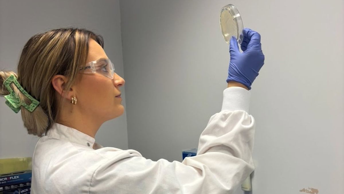
Madeline Berrow from the University of Nottingham

The researchers also note that adding CHX to the epoxy resin did not affect its optical transparency.

According to de Cogan, the novel concoction has a range of potential scientific, clinical and commercial applications.

“We have shown that it is highly effective against a range of different pathogens like E. coli and MRSA. We have also shown that it is effective against bacteria even when they are already resistant to antibiotics and biocides,” she says. “This means the technology could be a useful tool to circumvent the global problem of antimicrobial resistance.”

In de Cogan’s view, there are also number of major advantages to using the new coating to tackle bacterial infection – especially when compared to existing approaches – further boosting the prospects of future applications.

The key advantage of the technology is that the paint is “self-cleaning” – meaning that it would no longer be necessary to carry out the arduous task of repeatedly cleaning a surface to remove harmful microbes. Instead, after a single application, the simple presence of the paint on the surface would actively and continuously kill bacteria and viruses whenever they come into contact with it.

“This means that you can be sure a surface won’t pass on infections when you touch it,” says de Cogan.

“We are looking at more extensive testing in harsher environments and long-term durability testing over months and years. This work is ongoing and we will be following up with another publication shortly,” she adds.

Why I stopped submitting my work to for-profit publishers

Peer review is a cornerstone of academic publishing. It is how we ensure that published science is valid. Peer review, by which researchers judge the quality of papers submitted to journals, stops pseudoscience from being peddled as equivalent to rigorous research. At the same time, the peer-review system is under considerable strain as the number of journal articles published each year increases, jumping from 1.9 million in 2016 to 2.8 million in 2022, according to Scopus and Web of Science.

All these articles require experienced peer reviewers, with papers typically taking months to go through peer review. This cannot be blamed alone on the time taken to post manuscripts and reviews back and forth between editors and reviewers, but instead is a result of high workloads and, fundamentally, how busy everyone is. Given peer reviewers need to be expert in their field, the pool of potential reviewers is inherently limited. A bottleneck is emerging as the number of papers grows quicker than the number of researchers in academia.

Scientific publishers have long been central to managing the process of peer review. For anyone outside academia, the concept of peer review may seem illogical given that researchers spend their time on it without much acknowledgement. While initiatives are in place to change this such as outstanding-reviewer awards and the Web of Science recording reviewer data, there is no promise that such recognition will be considered when looking for permanent positions or applying for promotion.

The impact of open access

Why, then, do we agree to review? As an active researcher myself in quantum physics, I peer-reviewed more than 40 papers last year and I’ve always viewed it as a duty. It’s a necessary time-sink to make our academic system function, to ensure that published research is valid and to challenge questionable claims. However, like anything people do out of a sense of duty, inevitably there are those who will seek to exploit it for profit.

Many journals today are open access, in which fees, known as article-processing charges, are levied to make the published work freely available online. It makes sense that costs need to be imposed – staff working at publishing companies need paying; articles need editing and typesetting; servers need be maintained and web-hosting fees have to be paid. Recently, publishers have invested heavily in digital technology and developed new ways to disseminate research to a wider audience.

Open access, however, has encouraged some publishers to boost revenues by simply publishing as many papers as possible. At the same time, there has been an increase in retractions, especially of fabricated or manipulated manuscripts sold by “paper mills”. The rise of retractions isn’t directly linked to the emergence of open access, but it’s not a good sign, especially when the academic publishing industry reports profit margins of roughly 40% – higher than many other industries. Elsevier, for instance, publishes nearly 3000 journals and in 2023 its parent company, Relx, recorded a profit of £1.79bn. This is all money that was either paid in open-access fees or by libraries (or private users) for journal subscriptions but ends up going to shareholders rather than science.

It’s important to add that not all academic publishers are for-profit. Some, like the American Physical Society (APS), IOP Publishing, Optica, AIP Publishing and the American Association for the Advancement of Science – as well as university presses – are wings of academic societies and universities. Any profit they make is reinvested into research, education or the academic community. Indeed, IOP Publishing, AIP Publishing and the APS have formed a new “purpose-led publishing” coalition, in which the three publishers confirm that they will continue to reinvest the funds generated from publishing back into research and “never” have shareholders that result in putting “profit above purpose”.

But many of the largest publishers – the likes of Springer Nature, Elsevier, Taylor and Francis, MDPI and Wiley – are for-profit companies and are making massive sums for their shareholders. Should we just accept that this is how the system is? If not, what can we do about it and what impact can we as individuals have on a multi-billion-dollar industry? I have decided that I will no longer review for, nor submit my articles (when corresponding author) to, any for-profit publishers.

I’m lucky in my field that I have many good alternatives such as the arXiv overlay journal Quantum, IOP Publishing’s Quantum Science and Technology, APS’s Physical Review X Quantum and Optica Quantum. If your field doesn’t, then why not push for them to be created? We may not be able to dismantle the entire for-profit publishing industry, but we can stop contributing to it (especially those who have a permanent job in academia and are not as tied down by the need to publish in high impact factor journals). Such actions may seem small, but together can have an effect and push to make academia the environment we want to be contributing to. It may sound radical to take change into your own hands, but it’s worth a try. You never know, but it could help more money make its way back into science.

Visual assistance system helps blind people navigate

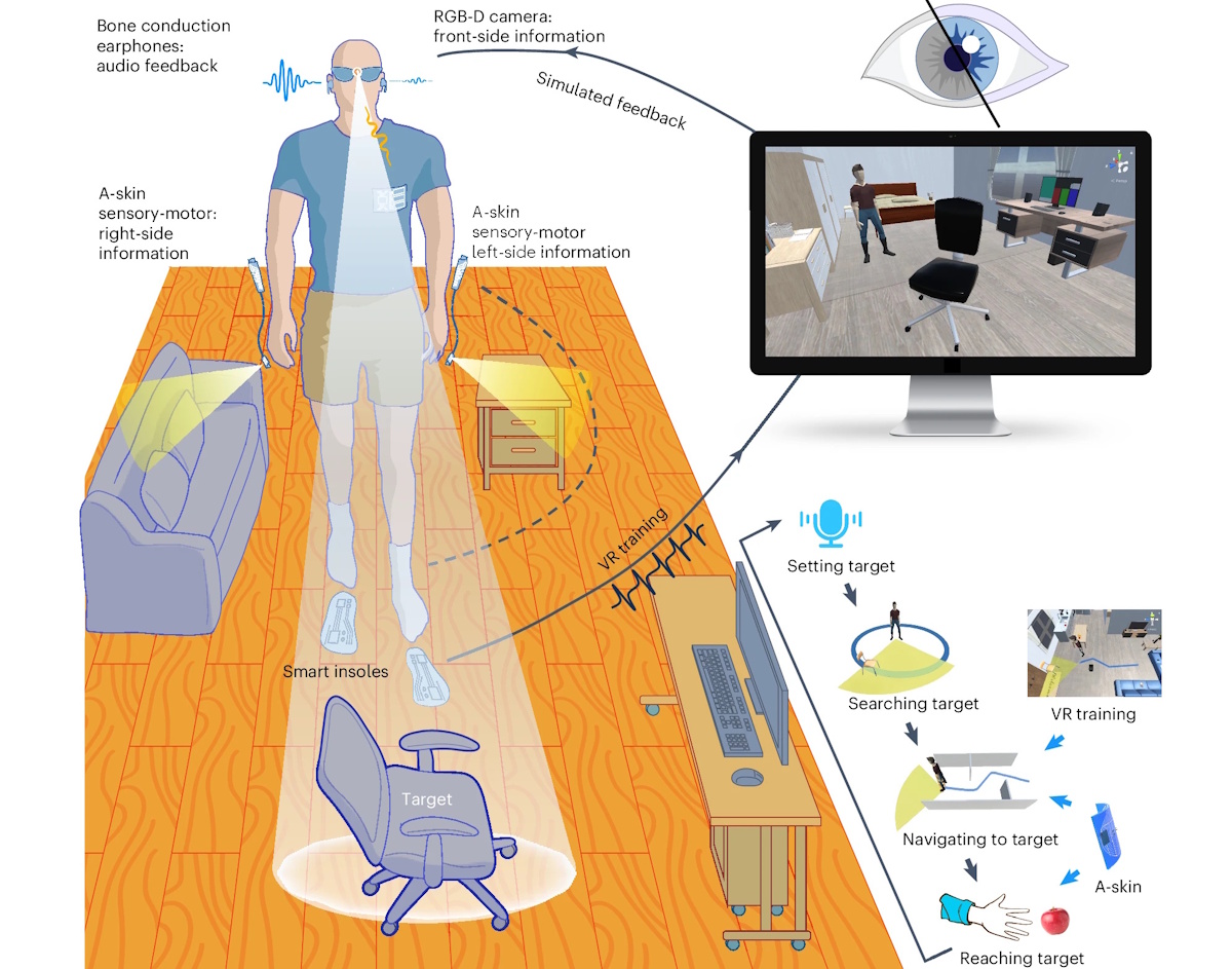
Structure and workflow of a wearable visual assistance system

Researchers from four universities in Shanghai, China, are developing a practical visual assistance system to help blind and partially sighted people navigate. The prototype system combines lightweight camera headgear, rapid-response AI-facilitated software and artificial “skins” worn on the wrists and finger that provide physiological sensing. Functionality testing suggests that the integration of visual, audio and haptic senses can create a wearable navigation system that overcomes current designs’ adoptability and usability concerns.

Worldwide, 43 million people are blind, according to 2021 estimates by the International Agency for the Prevention of Blindness. Millions more are so severely visually impaired that they require the use of a cane to navigate.

Visual assistance systems offer huge potential as navigation tools, but current designs have many drawbacks and challenges for potential users. These include limited functionality with respect to the size and weight of headgear, battery life and charging issues, slow real-time processing speeds, audio command overload, high system latency that can create safety concerns, and extensive and sometimes complex learning requirements.

Innovations in miniaturized computer hardware, battery charge longevity, AI-trained software to decrease latency in auditory commands, and the addition of lightweight wearable sensory augmentation material providing near-real-time haptic feedback are expected to make visual navigation assistance viable.

The team’s prototype visual assistance system, described in Nature Machine Intelligence, incorporates an RGB-D (red, green, blue, depth) camera mounted on a 3D-printed glasses frame, ultrathin artificial skins, a commercial lithium-ion battery, a wireless bone-conducting earphone and a virtual reality training platform interfaced via triboelectric smart insoles. The camera is connected to a microcontroller via USB, enabling all computations to be performed locally without the need for a remote server.

When a user sets a target using a voice command, AI algorithms process the RGB-D data to estimate the target’s orientation and determine an obstacle-free direction in real time. As the user begins to walk to the target, bone conduction earphones deliver spatialized cues to guide them, and the system updates the 3D scene in real time.

The system’s real-time visual recognition incorporates changes in distance and perspective, and can compensate for low ambient light and motion blur. To provide robust obstacle avoidance, it combines a global threshold method with a ground interval approach to accurately detect overhead hanging, ground-level and sunken obstacles, as well as sloping or irregular ground surfaces.

First author Jian Tang of Shanghai Jiao Tong University and colleagues tested three audio feedback approaches: spatialized cues, 3D sounds and verbal instructions. They determined that spatialized cues are the most rapid to convey and be understood and provide precise direction perception.

Real-world testing A visually impaired person navigates through a cluttered conference room. (Courtesy: Tang et al. Nature Machine Intelligence)

To complement the audio feedback, the researchers developed stretchable artificial skin – an integrated sensory-motor device that provides near-distance alerting. The core component is a compact time-of-flight sensor that vibrates to stimulate the skin when the distance to an obstacle or object is smaller than a predefined threshold. The actuator is designed as a slim, lightweight polyethylene terephthalate cantilever. A gap between the driving circuit and the skin promotes air circulation to improve skin comfort, breathability and long-term wearability, as well as facilitating actuator vibration.

Users wear the sensor on the back of an index or middle finger, while the actuator and driving circuit are worn on the wrist. When the artificial skin detects a lateral obstacle, it provides haptic feedback in just 18 ms.

The researchers tested the trained system in virtual and real-world environments, with both humanoid robots and 20 visually impaired individuals who had no prior experience of using visual assistance systems. Testing scenarios included walking to a target while avoiding a variety of obstacles and navigating through a maze. Participants’ navigation speed increased with training and proved comparable to walking with a cane. Users were also able to turn more smoothly and were more efficient at pathfinding when using the navigation system than when using a cane.

“The proficient completion of tasks mirroring real-world challenges underscores the system’s effectiveness in meeting real-life challenges,” the researchers write. “Overall, the system stands as a promising research prototype, setting the stage for the future advancement of wearable visual assistance.”

Universe may end much sooner than predicted, say theorists

The universe’s maximum lifespan may be considerably shorter than was previously thought, but don’t worry: there’s still plenty of time to finish streaming your favourite TV series.

According to new calculations by black hole expert Heino Falcke, quantum physicist Michael Wondrak, and mathematician Walter van Suijlekom of Radboud University in the Netherlands, the most persistent stellar objects in the universe – white dwarf stars – will decay away to nothingness in around 1078 years. This, Falcke admits, is “a very long time”, but it’s a far cry from previous predictions, which suggested that white dwarfs could persist for at least 101100 years. “The ultimate end of the universe comes much sooner than expected,” he says.

Writing in the Journal of Cosmology and Astroparticle Physics, Falcke and colleagues explain that the discrepancy stems from different assumptions about how white dwarfs decay. Previous calculations of their lifetime assumed that, in the absence of proton decay (which has never been observed experimentally), their main decay process would be something called pyconuclear fusion. This form of fusion occurs when nuclei in a crystalline lattice essentially vibrate their way into becoming fused with their nearest neighbours.

If that sounds a little unlikely, that’s because it is. However, in the dense, cold cores of white dwarf stars, and over stupendously long time periods, pyconuclear fusion happens often enough to gradually (very, very gradually) turn the white dwarf’s carbon into nickel, which then transmutes into iron by emitting a positron. The resulting iron-cored stars are known as black dwarfs, and some theories predict that they will eventually (very, very eventually) collapse into black holes. Depending on how massive they were to start with, the whole process takes between 101100‒1032 000 years.

An alternative mechanism

Those estimates, however, do not take into account an alternative decay mechanism known as Hawking radiation. First proposed in the early 1970s by Stephen Hawking and Jacob Bekenstein, Hawking radiation arises from fluctuations in the vacuum of spacetime. These fluctuations allow particle-antiparticle pairs to pop into existence by essentially “borrowing” energy from the vacuum for brief periods before the pairs recombine and annihilate.

If this pair production happens in the vicinity of a black hole, one particle in the pair may stray over the black hole’s event horizon before it can recombine. This leaves its partner free to carry away some of the “borrowed” energy as Hawking radiation. After an exceptionally long time – but, crucially, not as long as the time required to disappear a white dwarf via pyconuclear fusion – Hawking radiation will therefore cause black holes to dissipate.

The fate of life, the universe and everything?

But what about objects other than black holes? Well, in a previous work published in 2023, Falcke, Wondrak and van Suijlekom showed that a similar process can occur for any object that curves spacetime with its gravitational field, not just objects that have an event horizon. This means that white dwarfs, neutron stars, the Moon and even human beings can, in principle, evaporate away into nothingness via Hawking radiation – assuming that what the trio delicately call “other astrophysical evolution and decay channels” don’t get there first.

Based on this tongue-in-cheek assumption, the trio calculated that white dwarfs will dissipate in around 1078 years, while denser objects such as black holes and neutron stars will vanish in no more than 1067 years. Less dense objects such as humans, meanwhile, could persist for as long as 1090 years – albeit only in a vast, near-featureless spacetime devoid of anything that would make life worth living, or indeed possible.

While that might sound unrealistic as well as morbid, the trio’s calculations do have a somewhat practical goal. “By asking these kinds of questions and looking at extreme cases, we want to better understand the theory,” van Suijlekom says. “Perhaps one day, we [will] unravel the mystery of Hawking radiation.”

Copyright © 2026 by IOP Publishing Ltd and individual contributors