An international team of astronomers has uncovered the most ancient habitable exoplanet found to date. The discovery is all the more interesting because the planet originated outside of our Milky Way galaxy. At around 11.5 billion years old, the super-Earth is more than twice as old as our own planet and shows that habitable worlds were around much earlier in the universe’s history than previously thought.
The highly unusual find came from a survey of nearby, low-mass stars led by Guillem Anglada-Escudé of Queen Mary University of London. As part of their trawl, the researchers observed Doppler shifts in the light from Kapteyn’s star. Named after the Dutch astronomer who discovered it, it is one of the nearest stars to the Sun at just 13 light-years away. The Doppler shifts observed by the team were caused by two planets gravitationally tugging on their host and causing it to move slightly towards and away from the Earth. The researchers used new data from the HARPS spectrograph at the European Southern Observatory’s La Silla observatory, the Planet Finding Spectrograph at the Magellan/Las Campanas Observatory in Chile, and the HIRES instrument at the W M Keck Observatory in Hawaii to measure tiny periodic changes in the motion of the star.
In the zone
The team was able to infer that the two planets have orbital periods of 48 days and 121 days, respectively. As the star is a cooler red dwarf, its habitable zone is much closer than that of the Sun. This means that, despite its relatively proximity to its parent star, the innermost planet – dubbed Kapteyn b – should be able to support liquid water. It is thought to be a rocky super-Earth that is about five times more massive than our planet.
A super-Earth that lies within the habitable zone of a red-dwarf star has been found before. What makes this discovery unique, however, is the troubled history of Kapteyn’s star. “It has a very high velocity and a peculiar trajectory – it is not following the other stars around the galaxy,” Anglada-Escudé told physicsworld.com. Most of the stars in the Milky Way orbit slowly around the galactic centre, in the same plane. Astronomers believe that Kapetyn’s star bucks this trend because it did not form inside the Milky Way but rather was dragged into our galaxy at an angle at some later point from a dwarf galaxy that has now merged with the Milky Way.
Extragalactic voyage
This means the conditions that allow the formation of rocky planets in the habitable zones of stars were present in the universe long before the Sun was around Guillem Anglada-Escudé, Queen Mary University of London
The origins of Kapteyn’s star have been traced back to the ancient globular cluster Omega Centauri, the largest such object in orbit around our galaxy. That makes the star 11.5 billion years old – it formed just two billion years after the Big Bang. The planets encircling it are just as ancient and survived their host’s capture by the Milky Way. “We believe that these planets formed around the star – it would be almost impossible for the star to capture these planets at a later date,” says Anglada-Escudé. “This means the conditions that allow the formation of rocky planets in the habitable zones of stars were present in the universe long before the Sun was around,” he adds. It was previously thought that there were not sufficiently heavy elements around in the universe’s infancy with which to construct heavy, rocky planets.
According to Carole Haswell of the Open University in the UK, the finding adds to a growing realization that planets with significantly different histories to Earth might still be capable of hosting life. “It’s beginning to look as though habitable environments are plentiful and persistent in the galaxy,” she told physicsworld.com.
The paper has been accepted for publication in the Monthly Notices of the Royal Astronomical Society; a preprint of the work is available on the arXiv server.
One of the beauties of physics, I’m sure you’ll agree, is that it stretches from the very big (cosmology) to the very small (particle physics). In fact, the great questions at the heart of those fields may well have attracted you to physics in the first place. But a lot goes on in-between these extremes, not least at the nanoscale. It might lack the glamour of research into dark energy or the Higgs boson, but nanotechnology has far more of an immediate impact on everyday life than physics at either end of the length scale.
If you want to find out about some of those applications, take a look at the latest Physics World focus issue on nanotechnology, out now in print and digital formats. It covers, for example, the work of the UK firm P2i, which has developed a “dunkable” nano-coating that can keep a mobile phone functioning after being submerged in water for up to half an hour. Could be handy next time you go swimming.
Sean Carroll in full flow at the 2014 Cheltenham Science Festival.
By Matin Durrani in Cheltenham
I made the short journey yesterday from Bristol to the regency spa town of Cheltenham, which this week is hosting its annual science festival. One of the largest such events in the UK, it’s been running since 2002 and has a packed programme of A-list speakers and topics ranging from genetics to geology, from cocktails to cake, and from the human brain to the Higgs boson.
My main reason for attending the festival, though, was to meet Caltech physicist Sean Carroll, whose book about the search for the Higgs boson (called The Particle at the End of the Universe ) was picked by Physics World last year as one of our top 10 books of 2013. Carroll was in the Gloucestershire town to give a one-hour talk about the Higgs, although the festival organizers were clearly working him hard as he also spoke in separate lectures on dark matter and dark energy, and on his role as a science adviser to Hollywood. (Carroll’s worked on films including Thor, Avengers Assemble and TRON: Legacy and even played a tiny role on TV’s The Big Bang Theory – stay tuned for more on that in our upcoming audio interview with him.)
It started innocuously enough with a 2004 study showing that nanoparticles grown in the presence of certain molecules develop stripe-like structures on their surfaces. In recent years, however, “stripy nanoparticles” have become one of the most controversial areas in nanoscience, so much so that the debate over their existence has given rise to allegations of “cyber bullying” (see box). Now the publication of a new critique on the existence of stripes threatens to fire up the debate once more.
The story of stripy nanoparticles began in 2004, when materials scientist Francesco Stellacci, who was then at the Massachusetts Institute of Technology in Cambridge, US, and colleagues investigated the growth of gold nanoparticles in the presence of ligands – molecules that bond to metal atoms. Nanoparticles are often grown in the presence of ligands, because they act to stabilize the nanoparticles and hence prevent them from getting too big. Stellacci and colleagues claimed that when they used a mixture of two types of ligand – octanethiol and mercaptopropionic acid – for the process, the ligands organized themselves into stripes as thin as 5 Å on the nanoparticles’ surfaces.
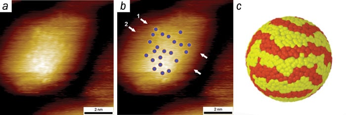
Their principal evidence came from using scanning tunnelling microscopy (STM). In this technique, a fine, electrically conducting tip is passed over a surface, which releases electrons to quantum-mechanically tunnel upwards. By recording the subtle changes in current that ensue through the tip, scientists can reconstruct the structure of the surface. In their STM reconstructions, Stellacci’s group found that the bright circles of their nanoparticles were covered with fuzzy stripes (Nature Materials3 330).
In the same paper, the researchers claimed that the stripy nanoparticles could repel proteins, a property that could be important for certain types of drug delivery. And in the following years Stellacci, working in conjunction with other research groups, has reported various other findings related to the nanoparticles, such as their apparent ability to penetrate biological cell membranes spontaneously. To date, stripy nanoparticles have been the subject of more than 25 papers, some of which have been published in high-impact-factor journals such as Nature, Nature Materials and Science.
Claims and counterclaims
Yet after these papers emerged, biophysicist Raphaël Lévy at the University of Liverpool in the UK became critical of Stellacci’s publications. In 2007 he submitted a technical comment to the journal Science about a paper of Stellacci’s that was about nanoparticle “polarity” – specifically that molecules can easily be placed at either end of a metal nanoparticle (Science315 358). That comment was never published, but it led Lévy to examine in more detail the evidence for nanoparticle stripes.
In 2009 Lévy submitted a manuscript to Nature Materials – the journal in which Stellacci published his original paper in 2004 – entitled “Stripy nanoparticles revisited”, which largely cast doubts on the evidence from Stellacci’s STM images. Nature Materials rejected the manuscript, as did the journal Nano Letters later that year without review; and it was only in 2012, following a lengthy review process taking around three years, that it was finally published as correspondence in the journal Small (8 3714).
Lévy’s criticism for the evidence of stripy nanoparticles derives from the pattern of the stripes themselves. In the STM images, Lévy and colleagues claim that the width of the stripes appeared constant from one pole of a nanoparticle to the other – which is surprising, they say, given that the nanoparticle’s spherical shape must be projected onto the 2D movement of an STM tip.
Just like the surface features of the Earth are distorted when they are projected onto a flat map, say Lévy and colleagues, one would expect the apparent width of the stripes to decrease as the STM tip progressed to the nanoparticle’s edge. They claim that the periodic “stripes” observed were nothing more than a common imaging artefact – the result of oscillatory electrical noise generated by a feedback system that tries to keep the STM tip at a constant distance from the nanoparticle surface.
The real thing? (a) STM image of a nanoparticle; (b) same image with position of molecules showing stripe-like domains (ACS Nano7 8529); (c) a simulation of the stripes (Phys. Rev. Lett.99 226106).
Online discussions
Stellacci, who had by this point moved to the École Polytechnique Fédérale de Lausanne (EPFL) in Switzerland, responded to Lévy and colleagues in the same issue of the journal Small. He countered that the STM tip maintained a constant distance from each particle’s centre of mass, which meant that the nanoparticle features were actually being projected onto a semicircle, for which there would be no distortion. Furthermore, he claimed, it is possible with STM to identify individual molecules on nanoparticles that have no stripes.
If there were distortion, the spacing between such molecules would be greater at the centres than at the edges of the nanoparticles; as it happens, said Stellacci, the spacing in such STM images is constant, meaning that any features were being projected onto a semicircle after all (Small8 3720).
Stellacci’s response did not settle the matter. Frustrated by the three years it had taken him and his colleagues to have their correspondence published, Lévy took to his blog – Rapha-z-lab. Over the next 14 months, he and a few guest contributors wrote more than 30 blog entries pulling apart the evidence for stripy nanoparticles in minute detail. Although most of the commentators sided with Lévy and his colleagues, a few sought to defend Stellacci’s work. At times, the debate became heated.
Stellacci himself was notably absent from the online discussions, but they did prompt him in October last year to publish work in collaboration with two independent groups led by Christoph Renner at the University of Geneva in Switzerland and Steven De Feyter at the University of Leuven in Belgium. The works, which were published in the journals ACS Nano (7 8529) and Langmuir (29 13723), sought to corroborate Stellacci’s original evidence with more advanced STM techniques. Unfortunately, they muddied the water even more: despite the images appearing almost stripe-free at first glance, the authors claimed that their analysis had indeed shown the stripe-like features to be present.
A tirade of comments
A guest post on Lévy’s blog by University of Liverpool chemist Mathias Brust summed up the sceptics’ view: “The authors [of the ACS Nano and Langmuir papers] employ an arsenal of image-analysis techniques to convince presumably themselves and evidently the referees that the now barely discernible ripples at the noise level represent all the features Stellacci et al. had previously reported. The new study thus implicitly admits interpretation errors in the original work, while explicitly aiming to corroborate it.”
This is one of the main criticisms outlined in the most recent paper by Lévy and colleagues, which is currently undergoing review at the journal PLOS One, that also re-examines the body of evidence for stripy nanoparticles to date. The conclusion of the paper states that the STM evidence rests on instrumental artefacts, improper data acquisition and analysis, and “observer bias”.
Already the paper has generated a tirade of comments on PubPeer, an online forum where scientists can review papers freely. The debate looks unlikely to conclude anytime soon, although the central point of contention remains the same. Lévy, like many other sceptics, believes the recognition of feedback artefacts is “elementary” STM science. On the other hand, Stellacci and his supporters consider the data much more difficult to interpret.
“Three groups of the highest standings have done measurements on my particles, and concluded that there are stripe-like domains,” says Stellacci. “Of course they could be wrong, but it is impossible that this is the trivial matter that Lévy portrays.” Stellacci will have a hard time convincing everyone that his nanoparticles are structured as he says they are. If he does, however, he can be assured of one fact: he really will have earned his stripes.
Peer review in the Internet age
Open for discussion Are forums and blogs the best place to discuss controversies in science? (Courtesy: Shutterstock/Maksim Kabakou)
The validity of evidence taken from scanning tunnelling microscopy about ligands organizing themselves into stripes on the surface of nanoparticles is not the only debate surrounding “stripy nanoparticles”. Another area of contention is how scientific discourse should take place in a time when online forums are beginning to displace traditional models of publishing.
Sceptics of Francesco Stellacci’s work, such as Raphaël Lévy, took to the Internet early on because they were frustrated with how long it took for their technical comments to be refereed and published in print journals. Lévy told Physics World it has been a “mistake” for Stellacci not to participate in online discussions too. “The idea that the only legitimate way of discussing scientific data and their interpretation is through the lengthy process of pre-publication peer review is frankly outdated,” he says.
But Stellacci believes the online discussions have not always been professional. He claims to have been accused several times of misconduct and fraud before he has had a chance to respond to the underlying criticisms. This, combined with other “personal attacks” and the “systematic use of lies”, has led him to claim to be a victim of “cyber bullying”.
Stellacci has shown Physics World an open letter that he is planning to publish online (but had not done so as Physics World went to press). “I have nothing against online scrutiny on published data, indeed I believe this is helpful,” he writes in the letter. “I do, however, not wish on any scientist [these] kind of attacks…To be clear what I find bullying is the instantaneous mocking…before the researcher has the physical time to reply to the accusation.”
Stellacci has not been the only one to fall victim to online mockery, however. Lévy’s blog has been copied – allegedly by supporters of Stellacci. The blog, called Fake Rapha-z-lab, is filled with fake posts – some of which are fake “guest posts” from Lévy’s colleagues – that mock the sceptics. The blog seems to have finished, though, as the last post was written last November. The Internet may offer a convenient forum to enter scientific discussion, but it would appear impossible to guard against less professional contributions.
The dust ring around HR 4796A. (Courtesy: ESO/J-L Beuzit et al./SPHERE Consortium)
By Tushna Commissariat
While the fiery shades of the image above may seem familiar to fans of the Lord of the Rings movie franchise, pictured above in exquisite clarity is a ring of dust that surrounds the near-by star HR 4796A. The young, hot star – located a scant 240 light-years from Earth – has been of significant interest to astronomers since the circumstellar ring of debris was detected, thanks to an excess of infrared emissions from the star. While there have been many other images of the HR 4796A system, this particular image has been obtained by the new Spectro-Polarimetric High-contrast Exoplanet REsearch instrument (SPHERE), which was installed in May this year on the European Southern Observatory’s Very Large Telescope (VLT) at the Paranal Observatory in Chile. Thanks to the instrument, not only is the dust-ring clearly outlined, but the glare of the bright star at the centre of the picture has been supressed. This has provided a much clearer view of the whole system, which researchers think also harbours an exoplanet or two.
Sometimes, it feels as if the future has already arrived. That is the case with electronics that can bend and flex, leading to applications such as sensors that can conform to clothing and skin. This short film takes you inside the headquarters of one of the most exciting companies in this emerging technology area: a spin-off firm called MC10 based in Cambridge, Massachusetts.
One of the company co-founders is John Rogers of the University of Illinois at Urbana-Champaign, who is a pioneer in the field of flexible electronics. In the film, Rogers talks about how his interest in the field emerged from the observation that all known forms of biology are soft, elastic and curvilinear, whereas existing forms of electronics are rigid, planar and brittle. “As a result, if you want to integrate electronics with biology – with human skin or tissue – you have severe challenges in a mechanics mismatch and a geometrical form mismatch,” he says.
Rogers describes how MC10 has overcome this limitation by developing a printing process that allows electronic devices to be built on rigid wafers before being removed in thin formats and then printed onto rubber substrates. This innovation enables the company to develop professional and consumer products based on integrated electronics that can flex and reshape in a range of different environments.
The film looks at one of the company’s most high-profile products: the Reebok CHECKLIGHT, which was developed in partnership with the consumer sports giant that also has it global headquarters in Massachusetts. It is essentially a type of skullcap that combines an accelerometer with a gyroscope to measure the magnitude and danger of impacts to the head. One of the problems, particularly with sports such as American football, is that there tends to be something of a hero culture whereby players will respond to head collisions by saying “I’m fine, coach”, even if they are not. The CHECKLIGHT is designed to provide an objective assessment of the impact to the head in that scenario.
Also featuring in the film is Benjamin Schlatka, MC10’s vice-president of business development. He talks about how the company came into existence in 2008 having begun as research in a science laboratory. “[John Rogers] had a connection with an investor, an entrepreneur, here in the Boston area that was a connector to two of the other co-founders.”
A design for what would be the world’s first X-ray frequency comb has been unveiled by physicists in Germany. The team believes that its comb – which would be used to measure the frequencies of X-rays – can be built using existing technologies. A working device could be used to make fundamental measurements in atomic physics with much greater precision than is possible today. The design could even be extended to produce gamma-ray combs, say the physicists.
Conventional frequency combs are short laser pulses comprising light at a number of well-defined frequencies. When plotted as intensity versus frequency, the light is represented by a series of sharp, equally spaced peaks that together resemble the teeth of a comb. Combs are used as a ruler to measure with great accuracy an unknown frequency relative to a precisely defined reference frequency, such as an atomic clock. Combs can therefore detect tiny changes in the frequency of a light signal that is associated with a physical phenomenon. For example, the gravitational tug of an exoplanet causes a periodic change in the Doppler shift of the light from its companion star’s light.
Combs working at optical frequencies were first developed in the 1990s by John Hall at the JILA Lab in Boulder, Colorado, and Theodor Hänsch at Ludwig Maximilians University in Munich, Germany, – who shared a Nobel prize for their efforts. Since then, physicists have been extremely keen to produce combs at higher frequencies. Ultraviolet combs have been produced by high-harmonic generation, in which a lower-frequency laser excites electrons in a gas and causes them to accelerate and emit light at higher harmonics of the original laser frequency. However, the intensity of successive harmonics decreases, so generating pulses in the X-ray region would require an impracticably powerful driving laser.
Subtler approach
Now, Stefano Cavaletto and colleagues at the Max Planck Institute for Nuclear Physics in Heidelberg have come up with a more subtle approach. They propose using three energy levels of the Be2+ ion to create an X-ray comb. The upper excited state lies 123.7 eV above the ground state – a gap that corresponds to low-energy or “soft” X-rays. This state is unstable, and so electrons decay rapidly back to the ground state. The third, metastable state lies just below the main excited state. Electrons in this state remain excited much longer. The group’s idea is to use an X-ray free-electron laser to pump electrons from the bottom state to the top state. Another applied laser pulse then leads the excited electrons to the metastable state, where they remain. If an optical frequency comb irradiates the ion, then with every pulse, some of the photons are promoted from the metastable state to the unstable state, before decaying almost immediately to the ground state with the emission of an X-ray pulse. This produces a series of X-ray pulses modulated at the same rate as the original optical pulses, forming an X-ray frequency comb.
Such a device could have numerous applications in fundamental physics. For example, it would become possible to measure precise values of the transition energies of the inner-shell electrons in highly charged ions. This would allow for stringent checks on the predictions of quantum electrodynamics and whether the fine-structure constant varies over time. “There are papers predicting that such measurements may be more sensitive at higher energies,” explains Cavaletto.
Even higher frequencies
Some of the co-authors of the paper are experimentalists, and Cavaletto says that they are confident it is possible to build the device with available equipment now. He also says that the basic principle could be extended to even higher frequencies. Indeed, gamma-ray frequency combs could be possible, although suitable three-level systems would need to be identified and a free-electron laser would need to be developed that could excite the initial transition.
Jun Ye, who heads the team at JILA that produced the first extreme-ultraviolet frequency comb, is impressed. “This is the first time that a feasible idea for the generation of frequency combs in the X-ray region has been proposed,” he says. “Such work will open up completely new scientific fields and fulfil old dreams. I am very excited about this approach.”
Researchers in the US have for the first time used MRI to follow the dynamics of neurotransmitters with molecular precision. They have demonstrated the technique on dopamine, a neurotransmitter that represents processes of reward and motivation in the brain.
Neurotransmitters are the chemicals released at the end of nerve fibres to communicate signals to other nerve fibres in the vicinity. An understanding of their dynamics is important for a more general understanding of brain function, yet they are hard to study. In the past, scientists have resorted to PET, an imaging technique that relies on a radioactive tracer being inserted into the body so that its path can be monitored by emitted gamma rays. But PET can only supply images that are spatially accurate to within a few millimetres and temporally accurate to within a few minutes.
Contrasting agents
Functional MRI (fMRI) could be more precise. As in standard MRI, fMRI involves the use of a fixed magnetic field to align the spins of protons inside the water molecules of tissue. After radio waves have deflected these spins, their relaxation back to alignment is timed with a radio receiver coil, and this time reveals the tissue’s composition. The difference with fMRI is that paramagnetic molecules known as “contrast agents” interact with some of the water molecules, thereby changing their brightness in scans. Haemoglobin in blood is a natural example: on its own, it acts as a contrast agent, but when it is bound to oxygen, its effect is diminished. In this way, scientists can use fMRI to study the circulation of oxygenated and deoxygenated blood.
Now, medical engineer Alan Jasanoff and colleagues at the Massachusetts Institute of Technology (MIT) in the US have experimented with another contrast agent – the paramagnetic haem protein BM3h. Instead of turning off when bound to oxygen, BM3h turns off when bound to dopamine, and can therefore be used with fMRI to sense the progression of the neurotransmitter between nerve fibres. The team claims that this form of imaging is an order of magnitude more accurate than PET. “Our study is the first to use MRI to study the dynamics of neurotransmitter release and signalling,” says Jasanoff.
The MIT group tested its technique on live rats by injecting BM3h into a region of the brain known as the ventral striatum, which emits dopamine, and electrically stimulating the medial forebrain bundle (a part of the reward system). Each dopamine stimulation lasted 16 s and the researchers took an MR image every 8 s, allowing them to track how dopamine levels changed as the neurotransmitter was released from cells and then disappeared.
Dopamine dynamics
“Neurotransmitters play distinct functional roles, which we can study by imaging their dynamics,” says Jasanoff. “For instance, dopamine is important for the brain’s ‘internal representation’ of reward and motivation, and if we want to figure out how dopamine works to achieve this, we should learn where and when dopamine concentrations are changing in the brain.”
The researchers found that an area known as the nucleus accumbens core (NAcC), which is known to receive dopamine from an area of the brain, showed the highest levels of dopamine release, according to the fMRI scan. They also found that dopamine was released in neighbouring regions such as the ventral pallidum, which regulates motivation and emotions, and parts of the thalamus, which relays sensory and motor signals in the brain.
Jasanoff and colleagues also believe that the technique could help in studies of Parkinson’s disease, which is caused through death of dopamine-generating cells. But Jasanoff is not going to stop with the imaging of dopamine by fMRI. “We are also developing and applying molecular sensors for MRI-based mapping of many other aspects of neural activity,” he says.
Illustration showing a proton (red) confined by magnetic-field lines (green) running down the centre of a Penning trap (yellow). (Courtesy: G Schneider, University of Mainz)
The most precise measurement ever of the proton’s magnetic moment has been made by an international group of physicists. The new result – combined with a similar measurement planned for the proton’s doppelganger, the antiproton – could help explain one of the deepest mysteries of physics – why the universe’s matter seems to vastly outweigh its antimatter.
Every fundamental particle has a nearly identical antiparticle with opposite electric charge. Physicists’ leading theories indicate that particles and their antiparticles were created in equal amounts during the Big Bang and should have annihilated each other long ago. But the universe is full of matter and lacks antimatter, suggesting that an undetected difference might exist between the two.
Minute differences
One possible clue about the difference could lie in tiny discrepancies between the magnetic moments of particles and their corresponding antiparticles. Any difference would be the first-known violation of a fundamental principle that physicists call charge–parity–time (CPT) symmetry. In 2013 researchers working at the Antihydrogen trap (ATRAP) experiment at CERN set the record for the most precise comparison between the magnetic moments of the proton and the antiproton, but the scientists found no difference between the two.
Now, Klaus Blaum, of the Max Planck Institute for Nuclear Physics in Germany, and colleagues are seeking an even more stringent CPT symmetry test. They used a cylindrical device called a Penning trap to confine a single proton using magnetic and electric fields. The trap’s magnetic field causes the proton to circle the cylinder’s axis at a rate known as its cyclotron frequency. The field also makes the direction of the particle’s spin precess like a spinning top, but with a different frequency. From the ratio of these two frequencies, scientists can calculate the particle’s magnetic moment.
Double trap
Measuring the cyclotron frequency is relatively easy, but the precession frequency is harder to pin down. For this reason, Blaum’s group built on a 2008 technique developed by another group of researchers to precisely measure the magnetic moment of the electron. In that work, the researchers applied a second magnetic field that caused the precessing electron to change how it oscillates along the cylinder’s axis. The oscillation frequency then changes slightly again when the particle’s spin flips from pointing up to pointing down, so by forcing a spin flip and measuring the resulting frequency shift, the scientists were able to determine the electron’s precession frequency and thus its magnetic moment.
Researchers have used this cylindrical device, known as a Penning trap, to confine a single proton for more than one year. (Courtesy: C Rodegheri, Max Planck Institute for Nuclear Physics)
To apply this technique to the proton’s much smaller magnetic moment, Blaum’s group developed what it calls a “double Penning trap”. In one trap the researchers determined the proton’s spin state, using a technique they reported in 2011. They then shuttled the proton to a second trap, where they measured the particle’s cyclotron and oscillation frequencies. The researchers repeated the process thousands of times over four months, eventually determining the proton’s magnetic moment to a precision of just over three parts in a billion. This figure is around 760 times more precise than what the ATRAP group achieved in 2012.
“I congratulate this team for showing it could do [this measurement] with the proton,” says Gerald Gabrielse, at Harvard University, who is the ATRAP spokesperson and was also involved in the 2008 research. But he notes that without the antiproton measurement, physicists are no closer to understanding matter’s dominance.
Blaum says his team will soon take that measurement. Team member Stefan Ulmer of RIKEN, a research institution in Tokyo, has already installed a double Penning trap on CERN’s Antiproton Decelerator, which will begin producing particles this summer. Within a year after that, Blaum thinks he and his colleagues should know whether the antiproton’s magnetic moment differs from that of the proton at the precision they have achieved. But Blaum adds that as his “trust in CPT is very high”, he is not betting on a discrepancy.
Big enough already: portrait of Edward Pickering by Sarah Gooll Putnam. (Courtesy: Harvard University Portrait Collection)
By Hamish Johnston
Can you name 10 blunders that have held back the progress of modern astronomy? Avi Loeb of Harvard University can, and he lists them in an essay entitled “On the benefits of promoting diversity of ideas”, which is posted on the arXivpreprint server.
Loeb argues that a common flaw of astronomers is to believe that they know the truth even when data are scarce. This, he argues, “occasionally leads to major blunders by which the scientific community makes the wrong strategic decision in its research plans, causing unnecessary delays in finding the truth”.
The first example he gives is the 1909 pronouncement by Edward Pickering, director of the Harvard College Observatory, that telescopes had reached their optimal size and that there was no point trying to make them any bigger.