Foam result surprises scientists

Benjamin Dollet and François Graner of the Laboratoire de Spectrométrie Physique (LSP) in Grenoble, and Miguel Aubouy of the CEA laboratory in Grenoble, began by filling a tank with a solution made up of 1% washing-up liquid in deionised water, and then blowing nitrogen though the solution to create a foam containing bubbles with an average thickness of 3.5 mm. Next they used a CCD camera to observe how the foam flowed around a small aerofoil-shaped object in the tank. Finally, they measured how the obstacle moved with a force sensor.

The French team found that the flow of foam slowed down beneath the obstacle and accelerated in the region above it. This produces an elastic deformation of the bubbles in the foam. To reduce this deformation, the bubbles below the wing pull the wing down. The bubbles above the wing also push it down, causing the wing to sink into the foam (see figure and movie).

“Our work reveals that complex fluids, like foams, display a completely different physical behaviour to simple fluids, like air or water,” says Dollet. “It could help us better understand, and thus predict, the behaviour of foam in a variety of situations, including flow within porous rock in oil extraction, rinsing in industrial cleaning, and ore separation in the mining industry.”

The results might also have implications for granular materials and polymers, and they might even shed light on how embryonic cells rearrange themselves in a growing foetus.

Secret lives of the solar system

Every academic year, I give one lecture course – on the physics of the solar system – in which I avoid the temptation of simply recycling material from the previous year. Quite simply, so much is happening that it would be impossible to leave the content unchanged. Since last year’s course ended in November, for example, the Huygens probe has transformed our picture of Titan from a fuzzy orange ball into a world replete with methane-carved river valleys, lake beds and shorelines. Deep Impact has given us the first detailed tour of comet Tempel I’s landscape of craters and scarps, and added a new crater of its own. And in the cold underworld beyond Neptune, lonely little Pluto has been displaced as king of the Kuiper-belt objects by a larger, more distant, and as-yet-nameless body simply designated 2003 UB313.

We have all grown accustomed to the “nineness” of the planets in the 75 years since Clyde Tombaugh’s painstaking search revealed the longsought ninth planet. Indeed, the reactions of astronomers and public alike to Pluto’s embattled planetary status have revealed a surprising emotional attachment to this remote little ice-ball. Pluto, like the longer-established planets in our system, is the subject of heroic tales – both mythological and modern. Perhaps we fear that the loss of Pluto’s planethood, like the loss of an ageing relative, will dull the memory of a thread of family history. If so, the timing of Dava Sobel’s new book is fortuitous.

The Planets provides a snapshot of the mythology, the tales of discovery, and the landscapes and physical histories of all the ancient and modern members of the Sun’s family. Like Sobel’s earlier accounts of John Harrison’s chronometers and the life of Galileo, it combines masterful storytelling with clear, engaging explanations of the essential scientific details. Rather than being a single narrative, the book’s chapters form a sequence of essays, one per planet.

In exploring the mythological significance of the planets, Sobel weaves poetry and mysticism seamlessly into the back-plot of each world’s tale. She uses the language of the book of Genesis to illuminate the modern-day creation story from the Big Bang to the violent events in the solar nebula that gave birth first to the planets, then to life on Earth. The history of Mars is recounted from the point of view of one of the red planet’s best-known emissaries to Earth – the meteorite ALH 84001.

Every planet has its unique mythological personality, born of its appearance and the rhythm of its motion across the skies. In the pre-telescopic era, the meticulous record-keeping of Ptolemy, Tycho and many other astronomers revealed inconsistencies in the movements of the planets. Sobel highlights the bits that did not quite fit and the upheavals in human thought that were required to solve the puzzles they presented. She describes, for example, how Copernicus dared to displace the Earth from the centre of the system, and how Kepler and Newton provided the laws needed to predict the planets’ wanderings in the context of the Copernican system.

Sobel also explains how John Adams and Urbain Leverrier used Newton’s laws to predict Neptune’s location from irregularities in the orbital motion of Uranus. This particular tale – replete with heroes and villains – is told in the form of a fictionalized letter from the ageing Caroline Herschel to a young American astronomer Maria Mitchell who had discovered a comet in 1847. Only stubborn Mercury held out, its mischievous perihelion motion spawning searches for a non-existent Vulcan until an elated Einstein found the explanation in general relativity.

The telescopic era brought new wonders, with tantalizing glimpses of the faces of the planets. Today, even from a suburban back garden, anyone with a small telescope can see the moons of Jupiter, the rings of Saturn, and the polar caps of Mars that have entranced astronomers for centuries. Galileo, watching the four “Medicean moons” perform their daily shuffle about giant Jupiter, saw their implications for the unpopular Copernican system. Eugène Antoniadi, Giovanni Schiaparelli and Percival Lowell – patiently awaiting those elusive moments of atmospheric clarity – drew the first, sometimes fanciful, maps of Mercury and Mars. Seasonal changes in the colouring of the Martian deserts suggested tracts of vegetation. For a while, Schiaparelli and Lowell’s fleetingly glimpsed linear features became the canals of Lowell’s doomed Martian civilization. The brilliant cloud tops of Venus, however, yielded no secrets.

In only half a human lifetime – since Sobel made her first model of the solar system for a school project – the planets have one by one been transformed from distant fuzzy blobs into fully fledged alien worlds, with their own landscapes and weather systems that seem at once familiar and bizarre. But new knowledge sometimes carries a price. There was, for Sobel, the bitter childhood disappointment of seeing Mars transformed from a chilly but life-bearing world into a lifeless, black-and-white desert pocked with ancient craters, and Venus from a possible lush tropical paradise into a stifling inferno.

Men walked on the Moon, and brought home images of the Earth that revealed a fragile, lonely jewel the resources of which are finite and should not be taken for granted. But there have been moments of elation and wonder too. We have all seen pictures of the Voyager craft sweeping past the gas giants, and of volcanic Io and ice-bound Europa as coloured billiard balls against the swirling backdrop of Jupiter. We have seen Kepler’s vision of the music of the spheres echoing in the intricate braiding of Saturn’s rings, and unforeseen complexity in the liquid-nitrogen geysers on Triton, their dark plumes pointing downwind in a place once thought too cold to sustain an atmosphere.

As Sobel acknowledges in her final chapter, there is no satisfactory way to end the story of a field where the pace of discovery remains so fast. But the old stories endure as well as the new ones, and to be able to see these wonders for yourself, you need only look up occasionally.


Nanocrystals enhance photovoltaics

The design is based on nanocrystals of cadmium selenide (CdSe) and cadmium telluride (CdTe). The materials were spin cast from a pyridine solution to form 100nm-thick layers on indium tin oxide glass coated with a 0.2nm-thick layer of alumina. The scientists put down a CdTe film first, annealed it to remove residual solvent and then added a CdSe film.

The idea is to combine the solution techniques used to make organic solar cells with the performance and robustness of devices based on inorganic semiconductors. Organic solar cells are attractive due to their potential low cost and ease of manufacture while inorganic devices have broadband absorption and superior charge transport. The researchers found that they could improve the properties of the devices by sintering the nanocrystals. They exposed the films to a solution of cadmium chloride in methanol and then annealed them at 400°C in air. This process increased the photoconductivity of the films by about two orders of magnitude. “We show that by sintering or fusing the nanocrystals together, we can produce cells with efficiencies approaching 3%,” said researcher Ilan Gur. “This is comparable to organic-based technologies, but these cells have the added advantage of being stable in air since they contain no organic components.”

Particles come to life

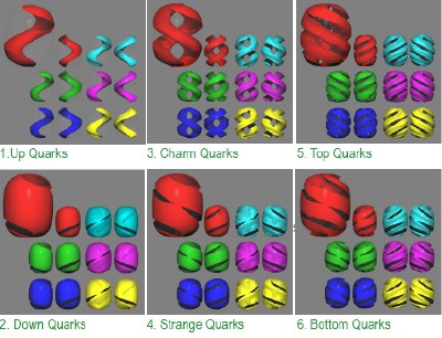
To produce the images Andersen worked with David Gerdes, an experimentalist, Gordon Kane, a theorist, and Sherri Smith, dean of the School of Art and Design at Michigan. The goal of the project was to represent particles in a physically accurate way while being visually appealing and technically feasible.

“Our role was to teach Jan-Henrik enough particle physics to be able to understand the families of elementary particles, their attributes, similarities and differences,” says Gerdes. “He brought his artist’s perception to the project and was able to capture these properties through a set of visual schemes.”

“The particles had to have the same basic form,” Andersen told PhysicsWeb, “yet reflect differences in mass, parity and so on. There also had to be logical coherence between the particles, as is found in the Standard Model, but the scheme also had to be open to ideas beyond the Standard Model such as supersymmetry and string theory.”

Andersen manipulated the equation for the Lamé curve — (x/a)m + (y/b)m = 1, where a and b are the length of the major and minor axes and m is a rational number (figure 1) — to produce images of both the quarks and leptons that make up all the matter in the Standard Model, and the bosons that carry the fundamental forces. Properties like spin, mass, charge and colour were also included.

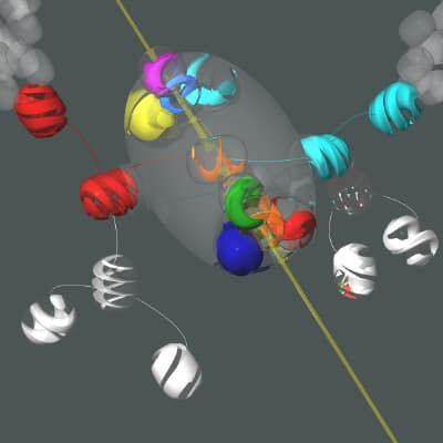
The first generation of quarks, the up and down quarks, are represented by a single curve in space, while images of the heavier, second and third generations are produced by adding these basic shapes together (figure 2). Andersen maintains the conventions of quantum chromodynamics and shows the quarks in three colours: quarks are red, green and blue. Anti-quarks are coloured cyan, magenta and yellow, while electrons and neutrinos are colourless. He also produced images of particles that have not yet been detected, such as the Graviton, the Higgs boson and supersymmetric particles, and interpreted real data from the CDF experiment at Fermilab to produce an image called Top Quark Event (figure 3).

“Our goal from the beginning has been to do more than just create attractive pieces of art,” says Gerdes. “We hope to have developed a pictorial representation of elementary particles that is clear and accurate enough to be understood by lay people and could become a widespread way of imagining the subatomic world, much like the picture of a nucleus surrounded by some elliptical electron orbits is the iconic symbol for an atom.”

STM reveals molecular first

“The Jahn-Teller effect has long been known to play an important role in the relationship between the structure of molecules and their energy levels, but this is the first time anyone has directly imaged it at the single-molecule level,” says Mike Crommie of the University of California at Berkeley and the Lawrence Berkeley Laboratory, leader of the team that saw the effect.

The Jahn-Teller effect occurs in systems that can exist in two or more distinct states that have the same energy. Because such a “degenerate” system is unstable, a molecule will distort itself to split the energy levels. Pure carbon-60 is an insulator because its highest occupied molecular orbital (HOMO) is full of electrons, while the lowest unoccupied molecular orbital (LUMO) is empty. The HOMO state is 5-fold degenerate, while the higher-energy LUMO state is 3-fold degenerate.

If carbon-60 is doped with potassium (K) to produce K3C60, it becomes metallic because the potassium atoms donate three electrons to the carbon-60 molecules, and these electrons go into the LUMO band. However, if an extra potassium atom is added to produce K4C60, the molecule becomes insulating, even though there are electrons in the LUMO band.

To investigate this, Crommie and colleagues studied a gold surface on which monolayers of both materials can exist simultaneously. In particular, they used a low-temperature scanning tunnelling microscope (STM) to both image the monolayer and also map the local density of states for K3C60 and K4C60.

The Berkeley physicists observed different symmetries for the occupied and unoccupied states, which is a telltale sign of a Jahn-Teller distortion. The STM data shows that the K3C60 monolayer exhibits a triangular lattice backbone structure. In contrast, the K4C60 monolayer displays an almost nearly rectangular structure with four molecules per unit cell. If the number of potassium atoms falls between 3 and 4, the system is a mixture of the two phases (see figure).

Moreover, the Berkeley team saw an energy gap of 200 mV in the three-fold degenerate LUMO band of K4C60. This band splits up into a group of two-fold degenerate levels at lower energy and one non-degenerate level at higher energy, which explains why the molecule becomes insulating.

“The beauty of seeing this physics in a monolayer is that it is so accessible: we can image the individual molecules with our STM and look to see where the electrons go at different energies,” says Crommie. “This is a freedom that does not exist for a bulk insulator, which you cannot image with a STM.”

Making the most of doping

Doping generally involves adding impurities to change the electronic properties of semiconducting materials by increasing the number of charge carriers, which can be electrons or “holes”. Although the dopant atoms are randomly dispersed throughout the material, their distribution is assumed to be fairly uniform. However, as the size of semiconductor devices continues to shrink, this uniformity can no longer be taken for granted. Some regions will contain significantly more charge carriers than others, and this will adversely affect the performance of the device.

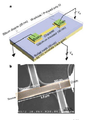
Shinada and co-workers use a single-ion implantation technique to overcome this problem. A small aperture is used to extract ions from a focussed ion beam and they are directed one by one into a nano-sized region of semiconductor until the required number of dopant atoms have been implanted. The team count the number of ions implanted by detecting secondary electrons.

A wide variety of ions – including beryllium, boron, phosphorus, iron and cobalt – can be implanted with a precision of 60 nm. When the Japanese team implanted phosphorus ions at 30 kilovolts into a 100 nm-wide channel in a transistor, they found that the threshold voltage decreased from -0.4 volts, the value for a conventional device with random doping, to just -0.2 volts. The group says that the improvement is a result of the electrostatic potential in the channel being more uniform due to the ordered distribution of dopant atoms (see figure).

Although the technique is too slow for high-volume chip manufacturing, the team plans to modify the ion beam so that single ions can be implanted with an accuracy of better than 10 nm, which should allow it to make single-atom devices.

“Ordered dopant arrays may enhance the prospects of making single-atoms devices whose properties are governed by individual dopant atoms, such as silicon-based solid-state computers,” Shinada told PhysicsWeb. The team also plans to apply the techniques to biomedical materials.

Nanotube switch makes its debut

An electromechanical system is a device in which a mechanical element moves in response to an external force. The new nanoelectromechanical system (NEMS) made by Jae Eun Jang of Cambridge University and co-workers at Cambridge, Sungkyunkwan University and Samsung is driven mainly by electrostatic forces, although Van der Waals forces also come into play.

Unlike conventional electronic devices, it might be possible to routinely manufacture NEMS to high tolerances on the scale of nanometres (10-9 metres). Such devices would rely on carbon nanotubes — rolled up sheets of graphite just nanometres in diameter that have excellent mechanical and electrical properties. However, until now most NEMS have been completely made using complicated lithography and etching techniques.

Jang and co-workers started by defining an underlying three-electrode structure with lithography, and then used electron-beam lithography and a “lift-off” technique to define nickel dots about 100-nm across on the electrodes. Finally, the team grew vertical nanotubes on top of the dots (see figure).

The underlying electrodes are then electrically biased so that one nanotube (the source electrode) is earthed and while the other (the drain) is positively biased. A third electrode — which might or might not have a carbon nanotube on it — close to the drain acts as the gate electrode. As the gate voltage becomes more biased, a threshold voltage is reached where the repulsive electrostatic force felt by the drain nanotube moves it towards the source. When the drain nanotube touches the source, a current passes between the two electrodes and switches on the device.

Depending on the length of the nanotubes, the drain can stay attached to the source through Van der Waals interactions (the “on” position), or spring back to the original “off” position when the gate voltage is reduced below the threshold. Jang and colleagues have shown that the switching behaviour can be controlled by varying the length of the nanotubes so that the balance between electrostatic and Van der Waals forces changes.

“The new switch could be used to replace all electronic switches in certain applications,” says Gehan Amaratunga, leader of the Cambridge team. “Its ability to hold either an ‘on’ or ‘off’ state when the driving voltage is removed also means it can be used in gripper or memory devices.”

Heavy ions damage DNA

Heavy-ion-beam cancer therapy employs protons or ions such as argon and neon that have energies of about 1 MeV per nucleon. One advantage of heavy-ion therapy over other techniques is that most of the energy is deposited in a small region of space, known as the Bragg peak, whereas X-rays, for example, deposit their energy continuously once they enter the body. However, little is known about how heavy-ion radiation damages DNA on the molecular scale, especially in the region beyond the Bragg peak. This damage might be caused by the heavy ions after they have lost most of their energy or by low-energy secondary ions. This is a worry because the tissue beyond the Bragg peak is often healthy.

Previously it was thought that heavy ions caused the same amount of damage as the conventional X-ray or gamma-ray radiation routinely used in medicine. These types of radiation cause damage by simple ionisation of atoms in cells, cleavage of single bonds in molecules, and attack by chemical radicals.

Michael Huels and colleagues at the University of Sherbrooke decided to look into this issue in more detail. They fired low-energy ions onto a film of biomolecules in an ultrahigh vacuum and analysed the ions that desorb from the film with a mass spectrometer. The results show that the initial damage caused by the ions at their track ends is significantly more complex, clustered and lethal than that induced by X- or gamma-rays. Severe damage can be caused by energies as low as 0.25 eV per nucleon — which is very low when compared with the energy of a typical heavy-ion beam.

The new work was prompted by previous experiments by Thomas Schlathölter and colleagues at Gröningen in the Netherlands. In 2003 Schlathölter noticed that low energy (1 to 200 eV) secondary particles could be produced by firing high-energy MeV-range heavy ions at DNA fragments. The latest experiments were made possible by the development of a machine that is capable of producing heavy ions with energies as low as just 1 eV in the Sherbrooke lab.

The team is now investigating how secondary ions, created by the primary ions inside DNA, can also cause damage although they have even lower energies than primary ions. “Our dream is that some day doctors will be able to manipulate the heavy-particle radiation effects at the molecular level – for example, by developing DNA ‘radiosensitisers’ that are specific to the secondary particles created in DNA during ion therapy,” says Huels.

Water in a whirl

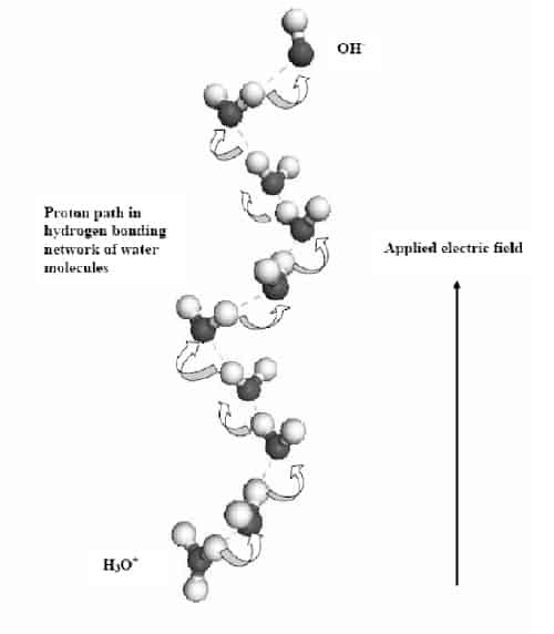
Zackary Chiragwandi and colleagues at Göteburg University and Chalmers University applied an electric field between two gold electrodes covered with pure water and found that the water molecules break up at 3.2 volts. This reaction takes place at both the anode and cathode at the same time. At the anode, the water molecules break up into negative hydroxide (OH–) ions and protons. These protons are released into the bulk of the water, where they create the vortex rings. Meanwhile, the hydroxide ions decompose to form oxygen.

Using an optical microscope, the Swedish team observed that the vortex rings consist of water swirling around in very fine circles with diameters ranging from 10 to 50 microns (figure 1). Moreover, more than one vortex can form at higher voltages. The scientists say that the protons move along a spiral path in solution, which corresponds to the hydrogen-bonding network between water molecules, and that this leads the formation of the vortices (figure 2).

Chiragwandi and co-workers believe that the physics behind the phenomenon is analogous to the transport of electrons in hole-doped semiconductors. The fact that the vortices extend quite deeply into the bulk of the water suggests that they are formed because of a defect at the surface of the anode. The Swedish team has seen such a defect with an electron microscope and also in simulations.

The work could also help improve our understanding of the chemical processes that occur inside living cells and the vortices themselves could be used as a non-invasive way to “stir” aqueous solutions.

Physics goes abstract

The works of the abstract expressionist painter Jackson Pollock have already been analyzed with fractal techniques by Richard Taylor of the University of Oregon and others. This work concentrated on analysing actual patterns or “blobs” formed by a specific colour on a canvas. Now Jonas Mureika of Loyola Marymount University in California and colleagues at the University of Toronto have gone a step further and analysed the “edges” of these blobs as well.

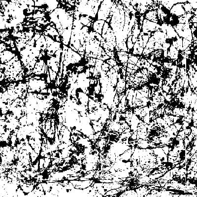
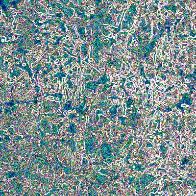
They looked at over 40 abstract works of art by Pollock (figure 1) and members of a movement known as Les Automatistes. To analyse the blobs Mureika and co-workers used digital filtering techniques to isolate specific colours in the painting (figure 2). To analyze the edges they used a Sobel gradient filter: basically, areas of high colour contrast, such as red against green or black against white, appear bright under this filter, while areas of low colour contrast are dull (figure 3).

They then used a standard “box-counting” technique to determine the fractal dimension of these filtered patterns. This involves covering a pattern by a set of squares and counting the number of squares, N, that contain the pattern. Each box is then divided into four smaller boxes and the number of boxes that contain the pattern is counted again. This process is repeated to produce a log-log plot with N on the vertical axis and the size of the boxes on the horizontal axis. The fractal dimension is given by the gradient of this graph.

Mureika and co-workers found that the fractal dimension of the blobs could not distinguish between different artists. However, the edge technique showed that the fractal dimensions of paintings by Pollock were statistically significant higher than those by Les Automatistes.

“Since part of the human visual processing system is based on the detection of such contrast edges, this suggests that there is a perceptually unique quality about paintings by particular artists,” says Mureika. “It also suggests that edge identification is an important contribution to ‘aesthetic’, and might explain why we find one abstract image more artistically appealing than another, even though we have no reference point to judge either as ‘good’ or ‘bad’.”

The technique could also be used to authenticate works of art and identify forgeries.

Copyright © 2026 by IOP Publishing Ltd and individual contributors