Quantum logic gate lights up

Classical computers deal with binary logic and the bits being processed must be either “0” or “1”. Quantum computers, on the other hand, exploit the ability of quantum particles to be in two or more states at the same time. A quantum bit or “qubit” can therefore be “0” or “1” or any combination of the two. This means that a quantum computer could, in principle, outperform a classical computer for certain tasks. However, all the quantum computers demonstrated so far have only contained a handful of qubits.

Although qubits have been made with trapped photons, atoms and ions, it is generally thought that it should be easier to build working devices with solid-state systems. Several teams have made significant progress with the superconducting approach to solid-state quantum computing. Now Steel and co-workers at Michigan, Michigan State, the Naval Research Laboratory and the University of California at San Diego have demonstrated the first all-optical quantum gate in a semiconductor quantum dot.

Steel and co-workers grew a thin gallium arsenide layer 4.2 nm thick between two 25 nm aluminium gallium arsenide barriers to make a quantum dot. Electrons are trapped in the dot because the gallium arsenide layer has a smaller energy band-gap than the surrounding material. When excited by light, electrons from the valence band in the dot move to higher energy levels. The excited electron and the ‘hole’ it leaves behind combine to form an exciton. The system has four states: a ground state containing two unexcited electrons; two states containing one exciton; and a state containing two excitons (see figure). The two single-exciton states can be distinguished from each other because the excitons have different polarizations.

The researchers showed that they can drive Rabi oscillations between the ground state and the one-exciton states, and also between the one-exciton states and the biexciton state, with lasers. In particular they showed that the quantum-dot system behaves like a controlled-NOT gate in which the value of one qubit is reversed (the NOT operation) if – and only if – the value of the other qubit is 1.

Although it will not be possible to scale up the system, the group says that many of the ideas and techniques they have developed could be useful in other approaches to quantum computing based on the optical control of electron-spin qubits in quantum dots.

Where do supernovae come from?

Astronomers believe that a supernova occurs when a star explodes at the end of its life. Supernovae are widely used as “standard candles” in cosmology, and observations of type 1a supernovae provided the first evidence that the expansion of the universe was accelerating – one of the most important advances made in physics in the past decade. However, astronomers disagree about the exact origins of these outbursts.

Supernovae are categorised according to their spectra: type-Ia events are recognized by the presence of elements such as silicon and sulphur, and the absence of hydrogen. However, Hamuy and colleagues have now detected hydrogen emission from SN2002ic, which otherwise has a spectrum that matches those of several other well-known type-Ia supernova.

Hamuy’s group believes that its discovery provides new support for one of the two main theories of the origins of type-Ia supernovas. The ‘single-degenerate’ model says that a white dwarf – the burnt-out remnant of a star – gathers material, including hydrogen and helium, from an ordinary companion star until it is massive enough to explode. Although popular for other reasons, this idea had suffered from the absence of hydrogen in the spectra of any previously observed type-Ia supernova.

But not everyone agrees with their interpretation. “At first glance the observation appears to support a single-degenerate scenario in which the white dwarf accretes from a normal companion”, write Mario Livio and Adam Reiss of the Space Telescope Institute in the US in a preprint just published on arXiv.org (astro-ph/9308018). “However, the opposite may be true, and the observations may support the merger of two white dwarfs as the cause for type-Ia supernovas”.

Although recent calculations have cast doubt on the double-degenerate model preferred by Livio and Riess, it can explain the absence of hydrogen in most type-Ia spectra. Moreover, even though they disagree with the interpretation of Hamuy and co-workers, Livio and Riess add that the detection of hydrogen emission from a type 1a supernova is a “landmark discovery.”

Archaeology turns to superconductivity

Lead is stable in many environments and corrodes only very slowly into lead oxide and lead carbonate. It becomes a superconductor when cooled below 7.2 Kelvin, whereas the corrosion products do not. This means that the magnetization of the lead will be several orders of magnitude higher than that of the corrosion products when the sample is placed in an applied magnetic field at temperatures below 7.2 Kelvin. It is therefore reasonable to assume that the magnetic signal from the sample is coming from the lead only.

Shimon Reich from the Weizmann Institute of Science and two colleagues – Grigori Leitus, a metallurgist, and Sariel Shalev, an archaeologist at the Weizmann Institute and also the University of Haifa – started by measuring the magnetization of small discs of material from different samples. This allowed them to calculate the amount of metallic lead that was present by mass. Next they weighed the samples, which gave them the total mass – the lead plus corrosion products. The difference between these two masses is therefore the mass of the corrosion products alone.

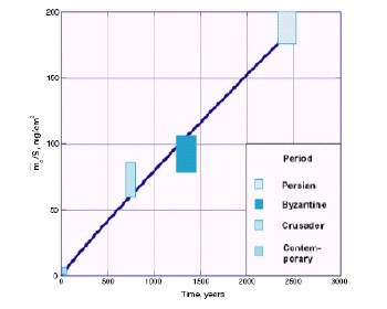
Reich and colleagues then plotted the mass of corrosion product per unit area against ‘archaeological age’ for various well-dated lead samples from the Tel-Dor site in Israel, as well as contemporary data. The Tel-Dor samples include artefacts from the Persian era some 2500 years ago (figure 1) to the Crusader period about 750 years ago. They found that the mass of corrosion product in a sample was directly proportional to its age (figure 2). Therefore, if the mass of corrosion product in a sample can be measured, then its age can be determined from the graph. “Until now, no archaeological method existed that could directly date lead,” said Reich.

The researchers say that their technique is, in principle, non-destructive because the corrosion products need not be mechanically or chemically separated from the lead metal. Moreover, since corrosion increases with time, the relative dating accuracy should improve with the age of the relics being studied.

Organic devices get back on track

Organic field-effect transistors (FETs) are usually thin-film devices. However, structural imperfections in the thin films can adversely affect the performance of the transistor. Organic FETs can also be fabricated on the surface of a single crystal, but this requires a processing method that will not destroy the fragile crystal. Instead the Delft team grew high-quality single crystals of tetracene and relied on electrostatic forces to make them adhere to a silica surface onto which gold source and drain electrodes had already been deposited. Crystals that are very thin – less than a micron – strongly adhere to the substrate without the need for any further processing steps and this minimizes any damage to the crystals.

Klapwijk and co-workers measured the current flowing from source to drain as they varied the drain voltage for different gate voltages. They found that the current had a very weak dependence on the gate voltage, which confirmed the high quality of the devices.

The room temperature mobility of the holes carrying the current was 0.4 centimetres squared per volt per second, which is higher than that of the best tetracene thin-film transistors. The mobility also shows temperature-dependent behaviour typically only seen in high purity organic crystals. Moreover, the mobilities at the surface and in the bulk of the device are similar – further proof that the quality of the crystals remains intact during processing.

Klapwijk and co-workers now hope to optimize their devices by improving the quality of the source and drain contacts, and by further reducing the level of impurities and imperfections in the crystals. They also plan to investigate organic materials other than tetracene.

Research into organic semiconductor devices suffered a setback last year when it emerged that a string of seemingly impressive results reported by Jan Hendrik Schön of Bell Labs – including several based on tetracene crystals – had been fabricated. “It has been very unfortunate that progress in this field appeared to be extremely rapid due to the flurry of results claimed by Schön and co-workers and then, all of a sudden, it dropped to a normal pace,” says Klapwijk. “However, we are now back on the right track, and we are learning step-by-step how to determine the intrinsic properties of these already used but poorly understood ‘messy’ materials.”

The crazy world of quantum computing

Quantum computers could be faster and more powerful than conventional computers. Moreover, they could trigger a revolutionary new approach to information processing. That is the intriguing message conveyed by the science journalist George Johnson in this short book. He explains how this new approach could undermine the security of current cryptography protocols, lead to fast algorithms for searching databases, and offer ways of simulating complex quantum-mechanical systems. However, the full scope of quantum computers is unknown. It might well be beyond our imagination.

Those who wish to obtain a basic understanding of this exciting emerging technology – even those who have never been exposed to the mysteries of the quantum world – will find this book accessible and enjoyable to read. It starts with a gentle introduction to the concept of quantum superposition, explaining how it can open the door to a massive parallel computational power that is far beyond the reach of any classical supercomputer.

Having described this seemingly unlimited computational power, the author takes quite a few chapters before revealing that there is a significant catch. “Reading out” the quantum computer will not provide you with the array of answers calculated in parallel; rather, it will spit out only one, randomly selected answer. This feature of the quantum computer – and of other quantum systems in general – is called the collapse of the quantum wavefunction and has been the centre of philosophical debate since quantum mechanics was founded about a century ago.

Johnson wisely stays out of this debate but explains – without any mathematics – how one can profit from the parallel computation power, despite this wavefunction collapse. He then brings us to the heart of the matter, namely Peter Shor’s algorithm for factoring large numbers, its link to secret code breaking, as well as Lov Grover’s search algorithm. The chapters on these crucial issues help the reader to grasp the underlying ideas rather than understand all the details.

The later chapters explain what it might take to build a fully fledged quantum computer, which is – it is no secret – not yet technologically possible. But given the major efforts in key research centres around the globe, it is not unthinkable that crucial breakthroughs could take place in the near future. The book therefore reviews various approaches to quantum computation based on ion traps and nuclear magnetic resonance, while making clear that others systems also being explored.

Given the technological limitations of controlling quantum systems, quantum computation will unavoidably be prone to errors. If current optimism is to be maintained, it is crucial that we learn how to perform quantum error correction, which would “track” and “repair” faulty operations in a quantum computer. Significant theoretical progress has been made, and Johnson quite rightly dedicates a chapter to this important topic.

There is also an excellent chapter on quantum cryptography, which enables two parties to set up a secret “key” that would let them communicate securely using the quantum properties of photons. The final chapter outlines the hope that quantum computers might solve long-standing unsolved scientific challenges, such as protein folding and the “travelling salesman” problem.

So does the book succeed? “Writing this kind of book is like having two very different people breathing down your neck,” Johnson concludes. “The scientist keeps trying to grab the keyboard to qualify every other sentence with footnotes and equations. Meanwhile, the reader is prodding you to cut to the chase and get on with the story.”

As a scientist myself, I do indeed feel that some explanations are too brief to satisfy my desire to truly understand the issues at hand. However, I have a deep respect for the way in which the author has tried to convey the ideas of quantum computation to people without a background in quantum physics. I know that phrases like quantum cryptography, quantum computation and quantum teleportation can intoxicate the mind of the uninitiated and unleash fantasies in uncontrolled directions. After publishing a paper on experimental quantum teleportation, I once received a message from a religious sect thanking the research team for having proven what they believed in for so many years.

Given the complexity of the topic, the broad target readership, and the decision to keep the book short, A Shortcut Through Time is in my view a remarkable achievement by Johnson. It will help to guide the imagination of the reader into a fascinating future – one in which quantum mechanics might play a prominent role in communication and computation.

Waves and turbulence cause a stir in superfluids

When the pilot gives the order to fasten seat belts during a flight, and the other passengers start to worry about spilling their coffee, physicists might console themselves by recalling that Richard Feynman once described turbulence as the last great unsolved problem of classical physics. Although a complete understanding of turbulence in ordinary classical fluids remains elusive, physicists are making real progress in a different direction – the study of turbulence in quantum fluids.

In an ordinary fluid, such as water or air, the flow can be described by a dimensionless number known as the Reynolds number. This number expresses the ratio of inertial and viscous forces in the fluid, and is defined as Re = UL/ν, where U and L are the typical speed and size of the system, and ν is the kinematic viscosity. Flows with small Re are dominated by viscosity and tend to be smooth and laminar. Instabilities appear in the flow as Re increases, and all flows become turbulent at sufficiently large Reynolds numbers.

In a quantum fluid, however, turbulence acquires new features. According to Landau’s two-fluid theory, a superfluid consists of two components: the viscous normal fluid, which is related to the thermal excitations, and the frictionless superfluid associated with the quantum ground state. And whereas the eddies in the normal fluid can be of all sizes and strengths, quantum mechanics restricts the rotational motion of the superfluid to quantized vortex filaments that are like tiny tornadoes. In particular, the circulation of the superfluid – which is defined in terms of a contour integral of the velocity field, vs, around the vortex core – is quantized in units of h/M, where h is Planck’s constant and M is the mass of a superfluid particle (see figure 1).

Turbulent transitions
Naturally occurring helium is in the main helium-4, but it also contains about one part in 107 of helium-3. The two isotopes behave quite differently near absolute zero because helium-4 atoms are bosons while helium-3 atoms are fermions and must therefore obey the Pauli exclusion principle. For instance, helium-4 becomes a superfluid at a critical temperature, Tc, of 2.2 K, whereas helium-3 only displays superfluidity below 2.7 mK. Moreover, the lighter isotope undergoes a second transition when it is cooled below 2.2 mK to produce the so-called B-phase of helium-3.

While studying this B-phase of helium-3, Antti Finne of the Helsinki University of Technology and colleagues in Japan, Finland, the Netherlands, Russia and the Czech Republic have now observed another transition at 0.6 Tc. This transition from turbulent flow at low temperatures to regular laminar flow at higher temperatures has never been seen before (A P Finne et al. 2003 Nature at press; arXiv.org/abs/cond-mat/0304586).

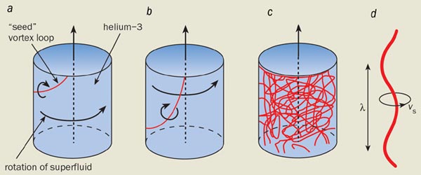
Finne and co-workers start with a rotating sample of helium-3 that does not contain any vortices. Next they inject a few “seed” vortex loops into the sample and then use a non-invasive nuclear magnetic resonance technique to measure how the total length of the vortices – which is proportional to the total energy contained in them – changes as a function of temperature and angular velocity. The results are surprising (see figure).

The total length of the vortices increases with time, with the energy needed to do this coming from the normal fluid (and ultimately from the motor being used to rotate the sample). If the temperature is greater than at 0.6 Tc, the vortices became rectilinear and align themselves along the axis of rotation. However, if the temperature is less than at 0.6 Tc, they evolve into a tangled network that fills the entire superfluid sample (although this eventually relaxes into an ordered array of rectilinear vortices).

What is so special about the temperature at 0.6 Tc? The answer lies in the behaviour of Kelvin waves on the vortex filaments (see part (d) of figure). Finne and co-workers showed that, at high rotations, the superfluid vorticity is determined by the ratio of dissipative and inertial forces in the superfluid. This ratio can be expressed as q = α/(1 – α´), where α and α´ are related to the strength of the interaction between the vortex and the normal fluid. For a single vortex filament, q = 1 represents the crossover from waves that propagate (i.e. Kelvin waves) to waves that are overdamped (q > 1).

Numerical simulations show that an injected vortex loop will expand and become rectilinear when q > 1. However, if q q depends on the temperature, and q is almost equal to one (1.3) when the temperature is 0.6 Tc. This suggests that Finne and co-workers have discovered a dimensionless quantity, q, which plays a role in superfluid turbulence similar to that played by the Reynolds number in classical hydrodynamics.

Moreover, the experiment suggests that we may be close to a breakthrough in understanding all the individual steps that lead to the formation of a turbulent state in a superfluid, and its subsequent decay. Indeed, we are rapidly approaching a time when we can numerically model these phenomena with reasonable accuracy. This is in marked contrast to our understanding of classical turbulence.

Superfluidity at the double
The superfluid states of helium-3 and helium-4 have both proved to be incredibly rich sources of new physics, so it seems only natural to wonder what would happen if both states could co-exist. It is possible to dissolve small amounts of helium-4 in liquid helium-3, which, in principle, provides us with a starting point for such experiments. Unfortunately this reduces the critical temperature of the helium-3 to a few microkelvin, which is currently beyond the reach of experimenters.

However, Gavin Lawes from Cornell University, and colleagues at Manchester University and the University of Delaware have now overcome this problem by studying the properties of helium-3-helium-4 mixtures in aerogels (Phys. Rev. Lett. 90 195301). An aerogel is a porous network of silica strands with diameters of a few nanometres. The 98% porous aerogel used by Lawes and colleagues increased the Tc of the helium-3 in the mixture to a few millikelvin, which is well within the range of current experimental techniques.

So how did the team prove that the superfluid phases of both helium isotopes were co-existing? Landau’s two-fluid theory predicts the existence of two sound modes in liquid helium: in “first sound” the superfluid and normal fluid components move in phase, while in “second sound” they move in antiphase. First sound corresponds to ordinary sound in a classical fluid, and can therefore be observed both above and below Tc. Second sound, on the other hand, is only found in the superfluid state below Tc. The researchers were able to exploit the link between second sound and superfluidity to prove that superfluid helium-3 and superfluid helium-4 were present in their experiment at the same time.

Quantum fluids can display remarkable behaviour in porous materials and other confined geometries. In a rigid porous material, the normal component is clamped by viscosity and cannot move. But an aerogel is not rigid, so it is possible for both the normal fluid and the aerogel to move together, either in phase with the superfluid in a “fast” mode, or against it in a “slow” mode. This slow mode is the key because it depends on the existence of superfluidity: if two different superfluid components are present in a mixture, we would expect to observe two different slow modes.

Lawes and colleagues first filled the aerogel with pure helium-3 and tracked this slow mode at increasing temperatures until it vanished at Tc = 1.62 mK. Then they repeated the experiment with increasing amounts of helium-4 in the mixture, and when the overall concentration of helium-4 reached 10.5%, they observed not one but two slow modes below the Tc for helium-3. This clearly demonstrated that superfluid phases of both isotopes were co-existing inside the aerogel. Above this temperature one of the slow modes vanished, while the researchers tracked the other up to 337 mK.

Superfluidity was first observed in helium-4 in 1938, and these new experiments show that the subject is still going strong some 65 years later. Moreover, they demonstrate that the barriers between different areas of physics – such as low-temperature physics and fluid dynamics – are slowly coming down.

How particles can be therapeutic

Until a cure for cancer is found, about one in three of us will probably have to undergo surgery, chemotherapy or radiotherapy at some point in our lives. Traditional radiotherapy uses X-rays to target cancerous tissue, but there is growing interest in using particles instead. Beams of hadrons, such as protons, neutrons and ions, offer important advantages over X-ray radiotherapy. Their clinical applications, however, are much less widespread.

Most of the 25 or so hadron-therapy facilities that are currently in operation around the world are sited at large particle-physics laboratories – not hospitals. A major focus of the hadron-therapy session at the World Congress in Medical Physics and Bioengineering, which is taking place in Sydney this month, will be on efforts to transform particle therapy into a practical and affordable treatment option.

Particle therapy works by damaging the DNA of cancerous cells – mostly by ionization – so that they cannot grow and multiply, while minimizing damage to surrounding healthy tissue. This is the same basic principle behind X-ray therapy (see “X-rays pinpoint tumour targets”). However, photons lose a significant amount of their energy before they reach the tumour, which can damage healthy cells and cause unpleasant side effects.

On the other hand, when a beam of charged particles enters the body, it deposits most of its energy at a depth that depends precisely on the energy of the particles. This means that tumours can be targeted more accurately, allowing a larger radiation dose to be delivered and speeding up the treatment programme.

“There is no justification for irradiating disease-free tissue if it is not absolutely necessary,” says Al Smith of the M D Anderson Cancer Center at the University of Texas, who is chairing a session devoted to proton therapy at Sydney. “There is a growing consensus that protons offer the potential for superior clinical results over photons – especially in reducing ‘late’ effects such as secondary tumours, which are caused by the treatment itself.” The trouble is that while X-rays are cheap and easy to produce, particle beams are not.

Proton therapy
The most widely used form of particle therapy is proton therapy, which was proposed by Robert Wilson – the first director of Fermilab in the US – in 1946. The first proton therapy was carried out in 1954 with the Bevatron accelerator at Berkeley, and to date about 35,000 patients around the world have undergone the treatment, mostly for cancers of the retina. Over 9000 of these were treated over the course of 30 years at the Harvard Cyclotron Laboratory in Massachusetts. The programme was transferred to the Massachusetts General Hospital in Boston last year, making it the world’s second hospital-based proton-therapy centre.

Protons, like all charged particles, slow down as they travel through a material as a result of electromagnetic interactions. The slower they move, the more efficient they are at ionizing atoms in their path and the more likely they are to interact with atomic nuclei. This means that the highest radiation dose is delivered at the point in the body at which they stop – the so-called Bragg peak – while the dose elsewhere is low (see figure 1). The ratio of radiation deposited at the tumour site to that in the path of the particle is called the relative biological effectiveness (RBE), which is basically a measure of the benefit versus risk of radiotherapy techniques.

Before treating a patient, radiation oncologists have to identify the precise location of the tumour by imaging it. This is often done using another physics-based technique – positron emission tomography (PET). The energy of the proton beam – which is generally between about 100 and 200 MeV – is then adjusted to match the tumour depth. Moreover, by combining protons with different energies in a single beam, the Bragg peak can be modulated into a plateau that dumps a high dose of radiation throughout the depth of the tumour.

There are currently six proton-therapy facilities under construction worldwide and a further 20 have been proposed, many of which will be in the US. This explosion of interest is partly due to the success of the first hospital-based proton facility at Loma Linda University in California – which has treated over 8000 patients since it opened in 1990 – and the fact that medical-insurance companies now include proton therapy as a reimbursable treatment programme.

But the technique still has some way to go before it becomes a routine service. According to Al Smith, much current research is focused on the use of lasers to accelerate protons, rather than using existing cyclotron and synchrotron sources.

“Laser-based devices can be made small enough so that each treatment room could have its own proton source,” he says. “They also offer the most substantial reduction in cost, but it is unlikely that laser proton accelerators will become a reality in the next 10 years.”

Heavy treatment
Newer than protons on the particle-therapy scene is ion therapy. Heavy ions, such as carbon, have a higher RBE than protons and are thought to provide more effective treatment for certain, deep-seated tumours that are often “radioresistant”.

This is because the rate at which a charged particle loses energy in a material – which is quantified by its linear energy transfer (LET) – increases with the mass of the particle. Helium ions were used to treat over 2000 patients at the Bevelac accelerator at Berkeley from 1957 and 1992, while neon ions have been used to treat a further 430. However, the optimal RBE using ions has since been found to lie in the range between lithium and carbon. There are currently just three heavy-ion treatment facilities in the world – two in Japan and one in Germany – all of which use carbon ions.

“It is easy to use protons, but heavy ions are superior for very resistant cancer cells,” says Kanai Tatsuaki of the Heavy Ion Medical Accelerator (HIMAC) in Japan, which has treated over 1500 patients with carbon ions since 1994. “However, many people think that heavy ions can damage healthy tissue and we are trying to demonstrate its clinical effectiveness.” Significant advances in improving the precision of carbon-ion beam delivery have been made at the GSI laboratory in Germany, where 150 patients have undergone carbon-ion therapy since 1997.

The third carbon-ion facility opened last year in Hyogo, Japan, and further facilities are planned by the TERA foundation in Italy and the University of Heidelberg in Germany. These projects have benefited from the proton ion medical machine study (PIMMS), which was based at the CERN particle-physics lab in Geneva (see Physics World June 2000 p9, print version).

Particles take charge
Charged particles such as protons and ions are not the only way to tackle tumours – neutral particles can be even more effective. Fast neutrons lose energy in the tumour via interactions with nuclei, rather than ionization, which causes cell damage that the body cannot repair. Neutrons therefore have a higher linear energy transfer than charged particles, and can produce a RBE that is up to three times that of protons or ions.

In the 1970s accelerator physicists at Fermilab realized that their linear accelerator could produce more protons than they needed, which could be used to generate a neutron beam. Funds were later secured to investigate a dedicated facility for fast-neutron therapy, which has since treated more than 3000 patients. Several other neutron facilities are currently in operation worldwide, but Arlene Lennox of the Fermilab facility thinks that neutron therapy needs more resources for outreach and marketing activities. “Fundamental science is not the issue here,” she says. “Most physicians and patients know nothing about the advantages of fast-neutron therapy; the main reason why people know about proton therapy is that Loma Linda University has been aggressively marketing it for over 10 years.”

A newer form of particle therapy that gives a high linear energy transfer is internal hadron therapy. In boron neutron-capture therapy, for example, a boron compound is first injected into the patient, which accumulates in the cancerous tissue. A beam of slow (thermal) neutrons from either a reactor or accelerator is then directed towards the tumour, where the boron atoms are split into lithium ions and alpha particles. These charged particles then destroy nearby cells but they only have enough energy to do so in a localized region, thereby sparing the surrounding healthy tissue.

“The World Congress is the first time that external-beam and internal-hadron therapies are being discussed together,” says Barry Allen of George Hospital Cancer Care Centre in Kogarah, Australia, and co-organizer of the Sydney meeting. “Internal hadron therapy offers systemic rather than local treatment, which is the key to tackling cancer.”

While still at a very early stage, a small company in the US is turning to antimatter as a potential form of particle therapy. PBar Medical is currently using antiproton beams at CERN to investigate the effects of antiprotons in biological material (see Physics World March 2003 p12, print version). Antiprotons exhibit the same Bragg peak as protons, but when they meet protons and neutrons at the treatment site, they annihilate to produce gamma rays and high energy particles that cause further damage to cancerous cells. Antiprotons might therefore have a RBE that is twice as great as protons, although this remains to be demonstrated.

The rise and rise of medical imaging

Medical physics has come a long way since Wilhelm Conrad Röntgen first described a “new kind of ray” back in 1895. The discovery that X-rays could be used to display the innermost workings of the human body on a photographic plate was of immediate interest to the medical community at the time. Today, over a century later, the phrase “going for a scan” can refer to any one of a multitude of different medical-imaging techniques that are used for diagnosis and treatment.

The transmission and detection of X-rays still lies at the heart of radiography, angiography, fluoroscopy and conventional mammography examinations. However, traditional film-based scanners are gradually being replaced by digital systems that are based primarily on caesium-iodide scintillators coupled to flat-panel detectors. Some systems rely on charged-coupled devices (CCD) rather than flat panels but the end result is the same: the data can be viewed, moved and stored without a single piece of film ever being exposed.

The humble X-ray also forms the heart of modern computed tomography (CT) systems, which can obtain a series of 2D “slices” through the body. A whole host of other physics-based techniques or “modalities” are also routinely used to look inside the body without the need for a scalpel. Single photon emission CT (SPECT) and positron emission tomography (PET) rely on the properties of radioactive isotopes, while magnetic resonance imaging (MRI) exploits the well known principles of nuclear magnetic resonance (NMR), and is the starting point for functional MRI (fMRI). Last but by no means least, ultrasound uses high-frequency sound waves in a similar manner to submarine sonar to produce images of tissue and blood vessels.

Data explosion
Whatever the modality, a typical scan now yields more information than ever before. This is particularly true for “multislice” CT scanners in which the X-ray source and the detectors are both rotated around the body of the patient. The multislice approach has improved the spatial resolution of CT scans from about 6 mm to better than 1 mm, which is making it increasingly attractive for a wider range of diagnostic and screening tests.

“Lots of new clinical applications have opened up due to the fast-scan capability of state-of-the-art CT,” says John Boone, professor of radiology and biomedical engineering at the University of California Davis Medical Center in Sacramento. “Multislice CT has given rise to the realistic prospect of CT coronary angiography, for instance, and there is also a large clinical trial in the US looking at the feasibility of using it to screen for lung cancer.”

However, the rise in data per CT scan, and the greater use of the machines, means that many hospitals are experiencing a dramatic – often exponential – increase in the amount of data they generate. Computer networks in hospitals – and existing software for the manipulation of images – are struggling to cope with the sheer volume of data now being generated. The end result is that there is often a delay before a doctor can view the results of a scan.

The problem of data overload is only going to get worse according to Sébastien Ourselin, project leader for medical imaging and a member of the e-Health team at CSIRO Telecommunications and Industrial Physics in Epping, Australia. “The issue will not be with the quality of data,” says Ourselin. “Rather, the question will be ‘How can I extract the information that is relevant to me from these hundreds of megabytes of data in just one or two minutes so that I can make a clinical diagnosis?’. Can you expect a radiologist to wait two hours to get a good segmentation of a heart?”

Andrew Todd-Pokropek, head of the medical-physics department at University College London, believes that time would be saved if medical physicists could ensure that all the data leaving the scanner were usable. “There should be ways of controlling data acquisition during the scan to optimize the quality of the data produced,” he says. Todd-Pokropek would like to see “intelligent acquisition” systems that allow for the effects of patient motion. Indeed, even if patients remain absolutely still while being scanned, a beating heart or the movements associated with breathing can sometimes distort the final images.

Combining strengths

Another trend in medical imaging involves combining the power of different imaging techniques. For instance, there is considerable interest in using a combination of multislice CT and PET in cancer imaging. PET is especially adept at mapping areas of metabolic hyperactivity, which is a strong indication that cancer is present, while the sub-millimetre resolution of multislice CT offers a clear picture of anatomical detail. Put the two together and you have an accurate means of locating a tumour and assessing its size and shape. This could prove particularly valuable when characterizing or “staging” tumours when they are first diagnosed, and then monitoring their response to treatment.

There are three options for generating a combined PET/CT scan according to Todd-Pokropek. The first is to collect the PET and CT images independently and then morph them together with powerful image-registration software. This is relatively straightforward for brain scans, but more difficult for whole-body scans. The second option involves fixing the patient to a bed and then wheeling them into the scanners in quick succession. However, it is quite a challenge to design a bed that will restrict the motion of patients to within a millimetre. The third option is to literally bolt the two scanners together in a single system. This last option has found favour with the major medical-imaging manufacturers, all of whom now market such systems.

“Multimodality image fusion is the future of nuclear medicine,” says Todd-Pokropek. “It is absolutely critical that imaging data from nuclear medicine is combined with that from other modalities to extract the most information.” Indeed, Todd-Pokropek predicts that PET/CT scanners will eventually replace single PET systems. This would be part of a general move towards blending physiological information with the anatomical information provided by traditional, diagnostic images.

David Townsend, senior PET physicist at the University of Pittsburgh Medical Center and co-inventor of the first working PET/CT scanner, agrees. “Three years ago we were taking an hour to do a PET study,” says Townsend. “Now we see that the promise of five minute whole-body scans for PET/CT is within reach.”

So what other combinations of techniques might prove useful? The jury is out on the merits of combined PET/MRI – even if the challenges of fitting a PET scanner within the small bore of an MRI unit and then getting it to work in a fairly strong magnetic field could be overcome.

A more promising approach is the merger of fMRI with electroencephalogram (EEG) studies to examine brain activity. This marriage would benefit from MRI’s high spatial resolution and the excellent temporal resolution of EEG studies. “There is a great potential for combined imaging using fMRI and EEG,” says Karl Friston, head of the functional imaging laboratory at University College London. Friston cites studies of epilepsy as a good example. “fMRI is a vital tool in understanding the genesis and nature of epileptic disorders,” he says, “and the complementary spatial and temporal properties of fMRI and EEG should finesse many of our observations about brain responses.”

Animal magic
While many medical physicists are working on ways to combine different imaging modalities, others are busy shrinking their scanners to study an entirely new class of subjects – laboratory mice. This growth of interest in rodent imaging has been triggered by a shift on interest (and research funds) towards gene therapy and other targeted treatments in clinical medicine (see “Watching biology in action”).

Most of the approaches being tested involve attaching a “normal” gene to a virus carrier and injecting it into an infected region in which the cells contain “abnormal” genes that carry disease. The general idea is that the normal genes replace the abnormal ones. The success or otherwise of the therapy can be assessed by attaching another agent to the carrier virus that will show up when a particular imaging technique is used, which allows researchers to monitor the physiological activity at the target location after therapy.

“There is a tremendous amount of activity in relation to small-animal scanners as a result of interest in gene therapy and genetic manipulation,” says Todd-Pokropek. “There is small-animal MRI, small-animal CT, small-animal PET and small-animal PET/CT, as well as systems based on optical fluorescence. Just about everybody is jumping onto this and trying to get small-animal imaging systems because they need them to do their genetic experiments.”

However, designing a scanner for a mouse is not as straightforward as it may seem, according to Ron Price, director of radiological sciences and professor of physics at Vanderbilt University Medical Center in Nashville. For example, miniaturizing a CT scanner means that the individual volume elements used to build up each image become smaller, which tends to reduce the resolution of the scan. This means that the only way to achieve adequate resolution is to increase the duration of the scan and/or the X-ray dose.

“Unlike human studies, where we can now produce a CT image in a fraction of a second, it will take several minutes for an animal,” says Rice. “That is a problem for us right now.”

Surgical solutions
Biologists are not the only group pressing for a new generation of tailor-made medical scanners. Physicists and engineers are also trying to modify the design of traditional, fixed imaging systems so they can be used during certain surgical procedures. The acquisition of clinical images in the operating theatre allows surgeons to operate via thin wire catheters, which is less invasive than other approaches. Surgeons use the images obtained during the operation to ensure that the catheters and other devices are in the correct place, and can also to check that the treatment has been successful.

Ultrasound and X-ray fluoroscopy have long been used in this role, though both have certain drawbacks. The accuracy of the former is highly dependent on the skills of the person who is performing the scan, while the latter exposes patients to ionizing radiation. Now attention is focusing on the potential of MRI-guided procedures, particularly in neurosurgery and cardiac operations where accuracy and the risks from X-ray exposure are of prime importance. “Interventional MRI is going to be an expanding area,” says Price.

The MRI scanners used for surgical applications must have an open magnet structure to give the doctors access to their patient. However, commercially available open MRI scanners tend to have magnetic field strengths of between about 0.2 and 0.6 T, compared with 1-3 T for most closed-bore MRI scanners. The leads to a lower signal-to-noise ratio and correspondingly poor spatial resolution.

One way to improve the image quality would be to increase the field strength, but this is difficult with an open-bore set-up says Price. “In open magnets you are forced to spread things out,” he says. “Think of a typical magnet that you might make at home by wrapping wires around an iron rod or a nail – the field is highest when the windings are close together. But if you spread the wires out to give the surgeon access to the patient, you are going to have to compensate by increasing the current.”

The problem is that the current can become too high for the wires to carry – even if superconducting wires are used. Price is optimistic that clever engineering will eventually solve the problem, but adds that the scanners are going to be “pretty expensive”.

As hospitals wait for the arrival of truly high-field open MRI scanners, so-called XMR scanners might provide a compromise. An XMR device consists of an X-ray scanner and a closed-bore MRI machine. The scanners are configured so that patients can be moved quickly from one to the other, allowing high-field MRI to be used in the middle of surgical procedures. XMR systems are already undergoing trials for intricate heart operations.

Surgeons can also benefit from interactive software tools that allow them to manipulate information from previously acquired images. “It is becoming fairly routine for surgeons to have access a whole series of volumetric images that he or she is able to co-register with the patient while they are on the operating table,” says Price, who is currently involved in a project to develop a “smart image recall device” that will be able to handle multiple sets of data from different scans.

Good vibrations
Ultrasound is another technique that could be used to provide functional data for many clinical applications, according to Price. It is already widely used to monitor changes to blood flow, courtesy of the Doppler effect, and could prove valuable in monitoring so-called anti-angiogenesis cancer therapies, which attack the growth of new blood cells in tumours.

“Blood flow is a very important parameter in cancer imaging,” says Price. “Ultrasound is very sensitive to blood flow, so it has a lot of potential in this area. Unfortunately it is mostly used as a qualitative modality, and a lot of effort needs to be put into making it more quantitative.” Price adds that the potential of ultrasound is sometimes overlooked as attention focuses on the seemingly more hi-tech – and certainly more expensive – approaches such as MRI, PET and CT.

Paul Carson, director of ultrasound research at the University of Michigan, agrees: “Ultrasound lends itself to targeted therapy and imaging.” Carson is also keen to improve the performance of ultrasound by using “contrast agents” to enhance the quality of images. Ultrasound contrast agents are suspensions of tiny gas bubbles that are just a few microns in diameter. Exposure to ultrasonic frequencies causes the microbubbles to resonate, which increases the strength of the reflected signal.

Very intense pulses can cause the bubbles to burst, boosting the signal still further. Data from advanced ultrasound techniques like these could also be fused with CT data in yet another variation on the multimodality imaging theme says Todd-Pokropek.

Other researchers are working on ways to glean more information on tissue behaviour from ultrasound scans, again with an emphasis on measurement rather than qualitative mapping. “Medical physicists and others have developed new methods of quantifying tissue characteristics, which at the very least have led to improvements in image quality and resolution,” says Carson. The bubble-bursting mechanism is also being investigated as a possible method for targeted drug delivery.

Outlook
The past century has seen medical imaging emerge as a powerful method for diagnosing disease, and also for monitoring treatment. Researchers are now striving to enhance the clarity of images, collect more quantitative data, monitor physiological processes, merge existing techniques and find more intelligent ways of displaying and using the information generated. But the greatest challenge could be ensuring that their hi-tech solutions have some real benefit to patients.

“The goal is to make the quality of medical images clinically acceptable,” says David Townsend. “If scanning for three times as long means you get a beautiful image for marketing, it does not mean it has any more diagnostic significance.”

Further information

Wanted: multi-talented physicists
The growing interest in ever-more-sophisticated imaging systems has fuelled job prospects for medically minded physicists, many of whom are now tailoring their training accordingly. David Townsend of the University of Pittsburgh Medical Center notes the growing trend for graduates to enrol on dedicated medical-imaging courses, prior to applying for research posts.

Townsend contrasts this route with his own path to a successful career in scanner design. Having trained as a nuclear physicist in the UK, he spent 10 years at the CERN particle-physics lab before deciding to apply his knowledge of particle detectors to medical scenarios. However, postdoctoral researchers with similar experience now face tougher competition for places in medical-imaging research. “I give talks to high-energy physics groups, and then a line of people ask me how I got into the field,” he says. “But nowadays, if I am looking to recruit a physicist, I can find someone with specific experience in medical imaging. It is harder for high-energy or nuclear physicists to get into the field.”

Entrants to the field should hone up on computer programming, according to John Boone of the University of California Davis Medical Center. Commercially available software tools are not yet sufficiently sophisticated to analyse the quality of digital images, he says, so medical physicists must be prepared to pitch in themselves. “In my opinion, having good computer coding skills is an absolute necessity to becoming a relevant medical physicist in radiology,” he says.

A thorough grounding in biology is also important says Ron Price of Vanderbilt University Medical Center, who used to find it difficult to communicate effectively with colleagues on some collaborative projects before he took a course in molecular biology. “In the main, we lack the vocabulary and background to communicate with some of these investigators right now,” he says.

But prospective candidates should not be put off, because the supply of multi-talented physicists has not yet matched demand and there are jobs aplenty. “There is even more work at the moment than ever before,” adds Andrew Todd-Pokropek, head of the medical-physics department at University College London. “We have trouble recruiting enough people.”


Extraterrestrial plasma crystals

A plasma is typically a gaseous discharge containing electrons and positively charged ions. However, in the surface-processing industry – in which plasmas are used to fabricate integrated circuits, for example – it has long been known that plasmas can contain larger particles too. The electric fields at the plasma boundaries, which are caused by electric sheaths around the discharge electrodes, can trap micron-sized “dust” particles in the plasma volume. These particles can support a high negative charge of up to several thousand electrons, and are therefore characterized by Coulomb electrostatic interactions.

However, our understanding of crystal growth in Earth-bound dusty plasmas has been hampered by the influence of gravity. Now astronauts on board the International Space Station have observed plasma crystals in microgravity for the first time.

These two results – which appear in a focus issue of New Journal of Physics on complex plasmas – represent only a taste of what is to come from further dusty-plasma experiments on board the International Space Station. Many teams in the world are joining this programme not only to gain fundamental insights into complex plasma science, but also with a view to developing applications such as particle coating and surface functionalization.

In the August issue of Physics World André Bouchoule and Laifa Boufendi from GREMI at Orléans University in France explain how the experiments have revealed crystal structures that have never been seen on Earth.

Cooling off with physics

The microstructure, and hence the texture that you experience when you eat ice cream, is created in a freezing process that has remained fundamentally unchanged since the first ice-cream maker was patented in the 1840s. The ingredients – water, milk protein, fat, sugar, emulsifiers, stabilizers, flavours and a lot of air – are mixed together before being pasteurized and homogenized. They are then pumped into a cylinder that is cooled from the outside with a refrigerant. As the mixture touches the cylinder wall it freezes and forms ice crystals, which are quickly scraped off by a rotating blade. The blade is attached to a beater that disperses the ice crystals into the mixture. At the same time, air is injected and broken down into small bubbles by the shear that the beater generates.

As the mixture passes along the cylinder, the number of ice crystals increases and its temperature drops. As a result, the viscosity of the mixture increases, so that more energy input is needed to rotate the beater. This energy is dissipated as heat, and when the ice cream reaches about -6ºC the energy input through the beater equals the energy removed as heat by the refrigerant. The process therefore becomes self-limiting and it is not possible to cool the ice cream any further. However, at -6ºC the microstructure is unstable. The ice cream therefore has to be removed from the freezer and then hardened in a very cold air flow.

In the August issue of Physics World Chris Clarke of Unilever R&D in the UK describes the latest research into ice cream.

Copyright © 2026 by IOP Publishing Ltd and individual contributors