Smarter, wiser, happy or sad?

Long ago, when I was a postdoc at Harvard, I read a book by Herman Kahn, futurologist and all-round guru at the Hudson Institute in New York State. He gave a ranked list of the 50 major problems that science and technology – broadly defined – needed to solve over the coming decades. “A pill to control appetite” ranked something like seventh from the top (Kahn was grossly obese), while “effective methods of controlling world population growth” was down in the high 20s. I have mistrusted futurology ever since.

This mistrust is reinforced by a chrestomathy of predictions put together by some bright sparks in the UK Office of Science and Technology’s Foresight team a few years ago. Their aim was to underline that the main purpose of the Foresight exercise is to forge links between academia and industry by bringing together the two sides to think about likely futures, and not to be deluded that such visions of tomorrow would be accurate in usable detail.

The collection includes such gems as: “heavier-than-air flying machines are not possible” (Lord Kelvin, 1895); “there is a world market for 15 computers” (IBM chairman, 1945); and “space flight is hokum” (Astronomer Royal, 1956). My all-time favourite is “the time has come to close the book on infectious diseases”. This comment, made by the US Surgeon General some 30 years ago, is remarkable not only for its misguided arrogance, but also for its stunning disregard of the high mortality caused (then as now) by infectious diseases in developing countries.

So I may be the wrong person to review this book. The dust jacket promises “a glittering panel of some of the world’s leading scientists brought together to discuss the future of science – and its implications”. And the 25 contributors are indeed a collection of notable science writers, who have thoughtful and interesting things to say; a significant subset are also outstandingly distinguished research scientists.

Not surprisingly, the book defies any crisp summary. Insofar as any overarching message does emerge, it is that anyone who burbles about “the end of science” in the sense that everything will soon be known – as some have done recently – is lamentably out of touch with their purported subject.

The 25 chapters are divided into a first dozen on “The future, in theory”, and a second set on “The future in practice”. The opening three chapters, by Lee Smolin on fundamental physics, Martin Rees on cosmology and Ian Stewart on mathematics, are superb.

Smolin analytically catalogues what he thinks the seven “big questions” were 50 years ago, suggesting that we have answered the first four but are still working on the other three. He then lists today’s “big seven” questions, the first four of which continue and deepen past explorations while the last three are new. Smolin hazards the guess that in 50 years’ time only two will remain unanswered. These are: “what explains the exact values of the parameters that determine the properties of the elementary particles?”; and “what explains the large ratios of scales we observe?”, such as why the length scale of the universe is 1060 times bigger than the Planck scale.

Likewise, Rees gives a lucidly compressed account of current ideas and ignorance about the evolution of the universe (or multiverses), while Stewart surveys the current state of play regarding the 23 great problems that the mathematician David Hilbert set out a century ago. Stewart focuses on the seven remaining unsolved problems, each of which now has a million-dollar prize awaiting the solver. Boldly, he offers a guess at how these seven will stand in 2050.

Seven of these first dozen essays discuss aspects of cognitive science and consciousness, with varying degrees of focus on the authors’ particular interest. I especially liked Mihaly Csikszentmihalyi on “The future of happiness” and Robert Sapolsky’s counterpoised essay “Will we still be sad fifty years from now?”. Steve Strogatz gives a substantial and admirably hype-free account of chaos, complexity theory and “small worlds”, and Stuart Kauffman rounds off the section with a lively – although some would say idiosyncratic – view of “What is life?”.
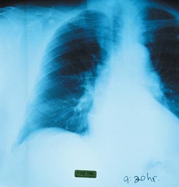
These first essays do a good job of looking to the future of fundamental research in their allotted areas. However, I found the second group of 13 essays, under their banner of practical implications for the future, to be on the whole less satisfactory. They start well enough, with Richard Dawkins noting: “Today, a chest X-ray will tell you whether you have lung cancer or tuberculosis. In 2050, for the price of a chest X-ray, you will be able to know the full text of every one of your genes. The doctor will hand you not the prescription recommended for an average person with your complaint but the prescription that precisely suits your genome.”

Most of the other “future-in-practice” essays either deal with tomorrow’s (excessively?) information-rich wired world and the bionics of “the merger of flesh and machines”, or revisit earlier themes of consciousness, childhood development and the analyst’s couch. A notable exception is John Holland’s essay on the meta-level process of thinking about “what is to come and how to predict it”; I would have placed this essay at either the start or end of the book.

Instead, the book concludes with an essay by the biologist Paul Ewald entitled “Mastering disease”. Harking back to our US Surgeon General, the essay could have been an appropriate finale, because the problem is hugely important and has wide implications for the shape of 2050. Sadly, what we get is a quirky marshalling of selected anecdotes to support the notion that “we are already dying of terrible global pandemics of heart attack, stroke, Alzheimer’s disease, and cancer – pandemics caused by infectious agents that are lethal but overlooked. We have been worrying about a few stray cats [the conventionally recognized infectious diseases, such as malaria, TB, HIV, etc] while we are being stalked by leopards”. Maybe Ewald’s ideas are right; after all, no one denies that infectious agents are implicated in some subspecies of his “leopards”. But I will bet against.

Overall, John Brockman’s book is great fun to read. But as a vision of important questions for science in 2050, it is more than a bit Kahn-like. There is no mention of climate change – neither the basic science of it nor the implications for tomorrow’s world. Ditto for human population growth. Ditto for feeding the world of 2050, and the associated problems of future water supplies. Ditto for alternative energy sources. Ditto for diminishing biological diversity, much less its likely adverse consequences for the delivery of ecosystem services that humans depend on.

Indeed, Ewald’s cat/leopard metaphor appears to be the only substantial reference to animals other than humans in the entire book. Nor is there much discussion of the ethical and practical dilemmas that tomorrow’s genomics will usher in. Abortions following the use of ultrasound to determine a foetus’s sex are already causing local female/male birth ratios to drop to as low as 0.8 in parts of India and China; the consequent social problems are arguably but a shadow of larger ones to come. The closest the book gets to these central ethical issues for 2050 is Roger Schank’s chapter on “Are we going to get smarter?”

The real question – never asked, much less addressed here – is: are we going to get wiser?
The Cavendish Laboratory and structural biology

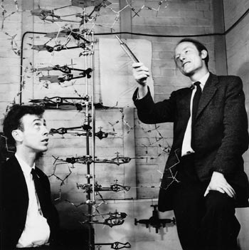
ON 25 April 1953 James Watson and Francis Crick, working in a small Medical Research Council unit in the Cavendish Laboratory, Cambridge, published a short letter in Nature. It described a remarkable two-chain helical structure for DNA, the biological polymer that constitutes the hereditary material in living organisms. The structural details of their model immediately suggested a mechanism by which the genetic material would replicate itself. Furthermore, the model indicated clearly what principle is used by the genetic material to store all the information needed to synthesize the proteins required to build a living organism.

In constructing their model, Watson and Crick were helped by advance knowledge of X-ray data obtained by Rosalind Franklin and Maurice Wilkins at King’s College London. These results – and additional data and reasoning supporting the Cambridge structure – were published in the same issue of Nature. The double-helix model provided the key to a detailed understanding of how living cells can produce two exact copies of themselves. It was a major factor in the enormous revolution in biology that dominated science in the second half of the 20th century, just as the revolution in physics had dominated the first half.

In the same laboratory, and only a few months later, a second major advance was made. Less immediate in its impact, it was no less far-reaching in its effect on the biological sciences. This was the discovery, made by Max Perutz, of a technique that would in principle make it possible to determine the phases of the X-ray reflections from a protein crystal and thence calculate a high-resolution structure for these very large molecules. After a further seven years’ work, John Kendrew and Perutz were able to use this approach to determine the structures of myoglobin and haemoglobin. Since then, X-ray structural analysis of these and many thousands of other protein molecules has helped us to understand the detailed chemistry of biological reactions.

These two foundation stones of modern biology and medicine – DNA structure and protein structure – were recognized in the same year (1962) by the award of a Nobel Prize in Physiology or Medicine to Watson, Crick and Wilkins, and a Nobel Prize in Chemistry to Perutz and Kendrew. Franklin had died of cancer in 1958 at the tragically young age of just 37.

A particularly surprising feature of these discoveries was that they were both made in the Cavendish Laboratory – a physics laboratory. Under J J Thomson and then Ernest Rutherford, the Cavendish had played a dominant role in the development of atomic and nuclear physics in the years before the Second World War. But why had work in the then very new field of X-ray analysis of biomolecular structure reached such an advanced state in a physics laboratory?

The answer lies in two directions. The more obvious is the strength of experimental physics in Cambridge, beginning in the later part of the 19th century with Maxwell, Rayleigh and Thomson. This provided the intellectual environment where William and Lawrence Bragg (father and son) were trained, and where Lawrence Bragg – as an undergraduate and then as a research student – had the first ideas in 1912 that led them to invent the technique of X-ray structural analysis.

Although the diffraction of X-rays by crystals had already been discovered by Max von Laue, Walter Friedrich and Paul Knipping, it was Lawrence Bragg who first realized the simple way in which it could be understood. This was in terms of reflection by “Bragg planes” – sheets of atoms in different crystallographic directions that can diffract strongly at specific angles determined by the separation between sheets. Using this approach, the two Braggs were able to calculate the exact arrangement of sodium and chloride atoms in a crystal of salt. For this work, Lawrence Bragg, aged just 24, shared the Nobel Prize for Physics with his father in 1915. Both went on to play powerful roles in establishing major schools of X-ray analysis in Britain, building on their initial discovery and the many subsequent contributions that they made.

But while these earlier events created the right environment from which the 1953 discoveries could flow, that they actually did so depended on the personalities and decisions of many individuals, on many chance encounters, and on fortuitous sets of circumstances. As so often is the case in life, the outcome could have taken quite different directions on many occasions.

The early years: William Bragg, J D Bernal and Max Perutz

William Bragg graduated in mathematics in Cambridge in 1884, and immediately became professor of physics in Adelaide, Australia. In 1909 he returned to Britain to take up a chair in Leeds, where he continued his work on the nature of X-rays. He became director of the Royal Institution in London in 1923, where he attracted some outstanding young scientists interested in the X-ray field. Among them were William Astbury and John Desmond Bernal – both recent Cambridge graduates. They became interested in the problem of protein structure – Astbury as a result of being asked by Bragg to provide X-ray diagrams of wool and silk for lectures that he was to give.

Bernal moved back to Cambridge as a lecturer in structural crystallography in 1927, working in four dilapidated rooms that were later demolished to make way for the Austin wing of the Cavendish. In 1931 he was promoted to assistant director of research in crystallography, which was by then a sub-department of the Cavendish, although his group remained housed in the same old rooms as before. Bernal’s main scientific interest was initially in the atomic structure of crystals of metals and minerals, then of hormones and sterols, and of some amino acids – the building blocks of proteins.

Astbury, meanwhile, moved to Leeds in 1928, where he also began working on amino acids and proteins. He and Bernal corresponded amicably about Astbury’s unsuccessful attempts to obtain well-ordered X-ray diffraction patterns from crystals of the protein pepsin, and about the possibility of Bernal providing help in obtaining crystals of other proteins. In the event, a friend of Bernal’s in Cambridge called Glenn Millikan – son of Robert Millikan of oil-drop fame – happened to visit a lab in Uppsala, Sweden, where large crystals of pepsin had just been obtained. Millikan, who knew of Bernal’s interest in proteins, brought some of the crystals back to Cambridge, still in their mother-liquor.

Bernal and Dorothy Crowfoot (later Hodgkin) initially obtained patterns of the crystals in the dry state, as Astbury had, with similar disappointing results. But when Bernal observed the crystals in a light microscope, he noticed that on drying they became disordered, as the large amount of water in the crystal lattice evaporated. The X-ray experiment was then repeated, but now with the crystal surrounded by its mother-liquor and sealed in a glass capillary. This time they obtained patterns with large numbers of crystalline reflections from the fully hydrated crystals, revealing a hexagonal lattice with a 67 Å spacing and a third axis that was too long to be measured accurately.

This was the first defining moment in protein crystallography associated with the Cavendish. The results were published as a letter in Nature in 1934 (133 794), together with a related letter by Astbury (133 795). Astbury had deduced the presence of polypeptide chains – indicated by 4.5 Å and 10 Å rings that he had seen in diffraction patterns from dried crystals of pepsin. Subsequently, Astbury continued to pursue his pioneering studies of polypeptide-chain configurations in fibrous proteins. He also obtained the first X-ray patterns of partially oriented samples of DNA, showing the characteristic 3.4 Å axial repeat, which he correctly ascribed to the repeat of certain chemical structures (known as bases) along the polynucleotide chains.

In 1935 Max Perutz, a chemistry graduate from Vienna who wanted to do research on the structure of proteins, arrived in Cambridge to work as a graduate student with Bernal, to whom he had been recommended by his mentor in Austria. Despite the run-down appearance of the laboratory, Perutz found it a magical place to work, largely because of Bernal’s charismatic character. Bernal had a very wide range of interests, making contributions of great originality and force on everything from liquids, minerals, metals and organic compounds to proteins and viruses. He developed apparatus for X-ray data collection and contributed to the International Tables used in crystallographic data analysis. He wrote and lectured widely on science and society, and was well versed in history. During the Second World War he was one of the founders of “operational research”, and, as Louis Mountbatten’s scientific advisor, played a large part in the planning of the Normandy landings. Mountbatten described him as being “one of the most engaging personalities I have ever met…with a clear analytical brain…tireless and outspoken”. He was also a convinced and very active Marxist.

In 1936 Perutz was given excellent crystals of haemoglobin by Gilbert Adair, and soon produced the best X-ray diffraction patterns to date, with reflections extending out to a resolution of 2-3 Å. These were published jointly with Isadore Fankuchen in Nature in 1938, together with similarly promising X-ray patterns from crystals of the enzyme chymotrypsin. However, the observable diffraction pattern – i.e. the intensities and positions of the individual reflections – represents only half of the data needed to deduce the structure of the diffracting object. In mathematical terms, it gives the amplitude – but not the phases – of the terms in the 3D Fourier series that represents the object. Without the phase information, the data could not be deciphered and the atomic positions could not be determined.

With simpler structures made up of small numbers of atoms, where chemistry could provide considerable guidance as to the atomic arrangements, a solution could often be found by an educated trial-and-error process. But proteins, which contain thousands of atoms, were far too complicated for this to work. So despite the enormous amount of excellent data that could be (and was) collected, the solution remained tantalizingly out of reach.

One possibility that was considered at the time involved attaching a strongly scattering atom, such as gold or mercury, to a specific site on a protein in a crystal. The atom might produce changes in the intensities that could then be used to obtain the phase information. However, this solution was not pursued after preliminary experiments proved unpromising. The technical difficulties were great, and the belief grew that proteins had so many atoms that the intensities of the reflections would not be altered measurably by adding a single, heavy-metal atom. But the faith remained that detailed information about protein structure could be obtained from the X-ray patterns in some way, if only it could be discovered.

Radio waves measure body water

Martin Robinson, Darren Stone and Janet Clegg at York University in the UK have used a novel electromagnetic technique to measure changes in the body’s water content. Their method is fast, accurate and cheap, and is now attracting interest from hospitals to help monitor diseases that affect patients’ water levels (M P Robinson et al. 2003 Phys. Med. Biol. 48 113­125).

In the March issue of Physics World, features editor Matthew Chalmers describes the experiment in more detail.

The subtle secrets of exotic helium

Since Niels Bohr presented his revolutionary model of the atom in 1913, physicists have come a long way in understanding the subtle spectroscopy of atoms. In the mid 1920s the quantized energy levels of orbiting electrons were found to be split into sublevels due to effects associated with the spin of the electron. The discovery of this “fine structure” was followed by that of hyperfine structure ­ even smaller shifts in the electronic energy levels caused by the magnetic interaction between the electrons and the nucleus.

Now Eberhard Widmann and co-workers in the ASACUSA (Atomic Spectroscopy And Collisions Using Cold Antiprotons) collaboration, which includes researchers from Japan, Switzerland and Hungary, have observed hyperfine structure in an exotic form of helium (E Widmann et al. 2002 Phys. Rev. Lett. 89 243402).

In the March issue of Physics World, Yasushi Kino from the Department of Chemistry at Tohoku University in Japan explains these developments.

Balancing atoms probe fundamental symmetries

Recently, however, spin-exchange collisions have been put to constructive use. Michael Romalis and Tom Kornack at Princeton University have used them to make significant progress in the technology of magnetometers ­ devices that measure changes in magnetic field. Using a concept that was developed by the same researchers together with Joel Allred and Rob Lyman at Washington University in 2002, Romalis and Kornack have demonstrated a new, ultra-sensitive “co-magnetometer”. The device could be used to image the weak magnetic fields in living systems such as the brain, and to test the fundamental symmetries of nature (T W Kornack and M V Romalis 2002 Phys. Rev. Lett. 89 253 002).

In the March issue of Physics World, Richard Stoner from the Charles Stark Laboratory in Cambridge, Massachusetts and Ronald Walsworth from the Harvard-Smithsonian Center for Astrophysics discuss this magnetometer in more detail.

Biological quantum dots go live

Biologists have long been eager to probe living cells in full colour over extended periods of time. Such a technique could reveal the complex processes that take place in all living organisms in unprecedented detail, such as the development of embryos.

Existing imaging techniques use natural molecules that fluoresce, such as organic dyes and proteins that are found in jellyfish and fireflies. However, each dye emits light over a wide range of wavelengths, which means that their spectra overlap. This makes it difficult to use more than three dyes at a time in order to tag and image different biological molecules simultaneously. The fluorescence of dyes also tends to fade away quickly over time.

Inorganic semiconductor nanocrystals – quantum dots – can get round these problems. As well as being brighter and living longer than organic fluorophores, quantum dots have a broader excitation spectrum. This means that a mixture of quantum dots of different sizes can be excited by a light source with a single wavelength, allowing simultaneous detection and imaging in colour. Turning these ideas from physics into biology, however, has remained a challenge because quantum dots cannot survive in water, and they must remain non-toxic.

Now a team of physicists and biologists led by Albert Libchaber and Ali Brivanlou at Rockefeller University in the US has produced new, biocompatible quantum dots and used them to image a live frog embryo (B Dubertret et al. 2002 Science 298 1759-1762). The researchers were able to “dress” quantum dots in an organic disguise that prevents them from coming into direct contact with the aqueous biological environment.

Molecular beacons

Quantum dots are nanometre-scale crystals that were developed in the mid-1980s for optoelectronic applications. They are composed of hundreds to thousands of atoms of an inorganic semiconductor material in which electron-hole pairs can be created and confined. The size of quantum dots can be tuned with nanometre precision during chemical synthesis, which gives them intriguing optical properties.

When the electron-hole pairs in the core of a quantum dot are excited with a beam of light, they re-emit light (fluoresce) with a narrow and symmetric emission spectrum that depends directly on the size of the crystal. This means that quantum dots can be fine-tuned to emit light at a variety of wavelengths simply by altering the size of the core, and therefore constitute a set of multicoloured molecular beacons for use in imaging. A 3 nm particle made from cadmium selenide, for example, radiates green light at 520 nm, while a slightly larger 5.5 nm particle of the same material radiates red light at 630 nm.

The core of the quantum dot is usually contained within a protective inorganic shell such as zinc sulphite, which has a higher electronic band gap than the core. This improves the confinement of the electron-hole pairs and therefore increases the intensity of the fluorescence. However, it is the hydrophobic nature of this outer shell that prevents quantum dots from being used in aqueous biological environments. In addition, to ensure that they remain non-toxic and that they recognize specific targets in a cellular context, quantum dots need to be modified with biological molecules to make them biocompatible before they can be let loose in a living organism.

Watertight

Since the mid-1990s a great deal of effort has gone into the development of robust, versatile and biocompatible surface chemistries to produce nanocrystals that are both soluble and functional. Various modifications of the outer surfaces of quantum dots have been successful, but at the expense of increased size and a compromise in the fluorescent and colloidal properties of the nanocrystals.

Now the Rockefeller team has produced soluble nanocrystals by a rapid and rather simple one-step procedure that does not require any surface modification of the particles. The breakthrough came in Albert Libchaber’s lab when Vincent Noireaux and Benoit Dubertret realized that an individual hydrophobic quantum dot could be encapsulated in a micelle – a simple chemical aggregate that has a hydrophobic centre surrounded by a hydrophilic shell.

The micelle was made from two kinds of phospholipids – fat-related products that are the major constituent of cell membranes in living organisms. Natural phospholipids were mixed together with polymer-grafted phospholipids. The resulting mixture forms a micelle because the hydrophobic tails of the mixture move away from water, while the polar heads immerse themselves in aqueous environments. Quantum dots are quickly engulfed during this process and stay protected at the centre of the micelles. Aqueous suspensions that consist of cadmium-selenide quantum dots coated with zinc sulphite are stable for months within the micelles.

The crucial question, then, was whether the encapsulated quantum dots retained their optical and colloidal properties? The Rockefeller group used high-resolution transmission electron microscopy to obtain images suggesting that the majority of the micelles contained a single quantum dot. These appeared to be spherical and of the same size, and they did not form any aggregates. The fluorescence quantum yield – the ratio of the amount of light emitted from a sample to that absorbed by the sample – of the quantum-dot micelles in water was 24%, a value very similar to the original quantum dots.

Having successfully kept the quantum dots away from water, the team went on to chemically alter the surface of the micelle to allow it to act as glue onto which biological macromolecules could be attached. These modified micelles were then linked to short single-strand fragments of DNA. The highly specific nature of DNA bonding meant that the DNA-micelle could recognize and pair with its complementary DNA strand. These experiments therefore demonstrated how the quantum-dot micelles could be adapted to locate a specific target, at least in vitro.

Frogs first

The success of these experiments prompted the Rockefeller team to investigate quantum-dot micelles in a live biological system. They injected a billion of the particles into Xenopus embryos (a South African frog) that were in early stages of development, and performed in vivo imaging using time-lapse microscopy.

The researchers made several key observations. Upon cell division, the fluorescent particles appeared to be solely distributed to the offspring of the injected parent cell, and did not diffuse out of the cell. The path taken by a fluorescent cell could therefore be traced back to the cell that was first injected. The researchers also found that all embryonic cell types can be stained, which came as a pleasant surprise since the behaviour of the particles in a cellular context was totally unknown. Crucially, they found that the quantum-dot micelles do not appear to have any detrimental effect on the frog’s development, and that the quantum-dot micelles were stable in vivo. The dots were still producing a detectable fluorescence signal after four days of embryonic development, and fluorescence was visible even in high-background regions such as the embryo gut. Finally, the quantum-dot micelles were found to be more resistant to photobleaching than other fluorophores. (see figure 1).

The Rockefeller results clearly show that fluorescence imaging with micelle-encapsulated quantum dots can be successfully applied to a live organism. But there are still issues that have to be addressed. The team used 4 nm (green) particles only, and we need to know if quantum dots of other sizes can sustain similar micelle encapsulation. The optical properties of the particles that were observed in vitro also need to be validated experimentally in a biological system. For example, can they be excited and detected simultaneously to form a multicolour image of live cells? While a very large number of quantum dots were used to image these embryos, it would also be interesting to know the lower detection limit of the nanocrystals within a cell.

Furthermore, although the targeting of quantum dots to specific biological objects has been demonstrated in principle using DNA, this has not yet been verified in a living biological system. It remains to be seen if the surface of the micelle can be used to target macromolecules or ligands of interest, in order to penetrate cells and pinpoint their constituents.

But quantum-dot imaging is itself at an embryonic stage. After fascinating physicists for over a decade, fluorescent semiconductor nanocrystals are finally fulfilling their promise in the biology lab. Fluorescence-based techniques are already widely used in biology, so we can expect that quantum dots will rapidly expand the biologist’s toolbox.

The Rosalind Franklin question

I once attended a series of lectures at the Brookhaven National Laboratory delivered by James Watson. In his initial talk he spoke of the historical events that surrounded the discovery of the structure of DNA. Afterwards, there was time for one question. A person raised her hand. “Dr Watson,” she asked, “I wonder if you could comment a little bit more on Rosalind Franklin’s contribution to the discovery of DNA.” Whereupon some in the audience applauded.

The applause puzzled me. Those people evidently felt that the mere act of asking that question was praiseworthy. I’ve heard since that this scene – the asking of the Rosalind Franklin question, to a smattering of audience applause – often takes place at Watson’s public lectures and has for years. Why?

Recognition

Is the question motivated by the feeling that Franklin’s contribution to that Nobel-prize-winning discovery is insufficiently recognized today? This cannot be. Franklin, who died in 1958 aged just 37, is the subject of two biographies: Anne Sayre’s 1975 book Rosalind Franklin and DNA and Brenda Maddox’s Rosalind Franklin: The Dark Lady of DNA (reviewed in Physics World in December 2002; p42, print version).

The National Portrait Gallery in London hangs her photograph alongside those of Watson and the two physicists with whom he shared the 1962 Nobel Prize for Physiology or Medicine – Francis Crick and Maurice Wilkins. The Franklin-Wilkins Building at King’s College London is co-named after her, while the Royal Society recently announced a £30,000 prize in her name. The Institute of Physics even has a new “Rosalind Franklin room” in its expanded headquarters in London. It is impossible to think about the discovery of DNA today without bringing to mind Rosalind Franklin.

Ironically, Watson is largely responsible. But for his coarse caricature of her in his 1968 book The Double Helix, Franklin’s collaborator Aaron Klug would not have written an article for Nature forcefully countering Watson’s account. Nor would the outraged community of crystallographers have sought to prevail on the initially reluctant Sayre (whose husband David is a crystallographer) to undertake a book about Franklin. Watson’s account was thus the triggering event that catapulted Rosalind Franklin to fame as one of the key players in the DNA discovery – even though this episode was only one of her contributions to science.

Justice

Is the Rosalind Franklin question motivated by the need to acknowledge a past injustice? This is more plausible. Maddox and Sayre describe the hurdles Franklin faced as a Jewish woman in science in British scientific institutions of the 1950s. For Sayre, the hurdles were almost entirely related to gender, while Maddox more accurately and persuasively attributes them to class and religion. And in his new biography, Watson and DNA, Victor McElheny notes how relentless Crick and Watson were, quoting Watson as saying: ” ‘Nice’ is what you do when you have nothing else to offer.”

Crick and Watson relied on two key pieces of information that were due to Franklin but obtained without her knowledge. One was her DNA Photograph 51, which Maurice Wilkins showed to Watson in January 1953. “The instant I saw the picture my mouth fell open and my pulse began to race,” Watson writes in The Double Helix, for he recognized immediately its tell-tale helical signature. It was psychologically the key event that inspired him to drop everything to search for the DNA structure.

The other piece of information used was Franklin’s measurements of a DNA unit cell, which she included in a report to the Medical Research Council. When Max Perutz passed this non-confidential but not really public report to Crick in February 1953, Crick realized that the two strands of the helix run in opposite directions.

Lacking these clues, Crick and Watson would not have been able to piece together the DNA structure as fast as they did. However, Crick and Watson also obtained other important clues from Astbury, Chargaff, Donohue, Furberg, Wilkins and others. Did Crick and Watson steal something from Franklin and present it as their own? No. She was close to figuring out the structure of DNA, but did not do it. The title of “discoverer” goes to those who first fit the pieces together. In rejecting Sayre’s claim that Franklin was the victim of “robbery”, the physicist Jeremy Bernstein has expressed the logic quite forcefully: “They made the double-helix scheme work. It is as simple as that.”

The critical point

The Rosalind Franklin question, however, is motivated by the feeling that “it” – not the priority claim, but something else – is not simple.

In the Nature paper of April 1953 in which Crick and Watson announced their discovery, they acknowledged being “stimulated by a knowledge of the general nature of the unpublished experimental results and ideas of Dr M H F Wilkins, Dr R E Franklin and their co-workers at King’s College”. This sentence was carefully crafted so that Franklin would not realize just how effectively Crick and Watson had used her data. She would die five years later without ever knowing that Watson and Crick had seen Photograph 51 and her unit-cell measurements, although Maddox says that she must have suspected. Neither Crick nor Watson mentioned her in their Nobel speeches.

Such stingy behaviour may not be unknown, or even uncommon, among scientists. However, our sensitivity to the norms of science and to violations of fair play heighten in proportion to the importance of the discovery – the DNA structure probably ranks as the single most important scientific discovery of the past 50 years – and in situations involving marginalized individuals.

The Rosalind Franklin question – and the applause it receives – is, I think, motivated less by the feeling that her contributions should be more widely recognized. Instead, it is more to do with the scientific community recalling and reasserting norms of fairness when they find these violated. Some day it may no longer be necessary to ask the Rosalind Franklin question. When this happens, it would also be worth applauding.

Physics and DNA

It will not have escaped readers’ notice that the 50th anniversary of the discovery of the double-helix structure of DNA will soon be upon us. Countless articles, books, conferences and the like have been commissioned, written and organized to celebrate what is widely agreed to have been the single biggest advance in biology since Darwin proposed the theory of evolution through natural selection.


However, two aspects of the DNA story seem to have been overlooked amid all the discussions about the past, present and future of genetics. First, the double-helix structure emerged from a physics laboratory. Second, DNA is still the focus of intense study by a small band of physicists around the world. The aim of this special issue of Physics World is to add a physics perspective to the celebrations.

It would be churlish, of course, to claim that physicists discovered the double helix all on their own – to claim the “Bragging rights” as it were. After all, James Watson was a biologist. However, as Hugh Huxley describes in this issue in The Cavendish Laboratory and structural biology (p29, print version), a unique set of events and characters conspired to make the Cavendish Laboratory in Cambridge the home of the double helix.

In the 1930s the Cavendish was dominated by Ernest Rutherford and nuclear physics. However, when the legendary New Zealander died unexpectedly in 1937, the electors to the Cavendish chair of physics decided that nuclear physics had become too expensive for a university laboratory, and they opted to replace Rutherford with the crystallographer Lawrence Bragg. The Austrian-born chemist Max Perutz was already there, Francis Crick arrived in 1949 – having graduated with a degree in physics from University College London in 1937 – and Watson followed two years later. Whereas Watson was a mere 22 years old when he arrived in Cambridge, Crick was in his mid-thirties when he joined the Cavendish. Progress was slow to begin with and Crick was told to work on other projects. However, the pair persevered and the double-helix paper was published in Nature on 25 April 1953. The Nobel prize followed in 1962 and the rest, as they say, is history – except for the Rosalind Franklin question.

It does seem odd and unfair that neither Crick nor Watson mentioned Franklin, who died in 1958 aged just 37, in their Nobel speeches. However, that was more than 40 years ago and today Franklin is almost as well known as Crick and Watson. Indeed, as Robert Crease writes in The Rosalind Franklin question (p17, print version): “It is impossible to think about the discovery of DNA today without bringing to mind Rosalind Franklin.” It is time to move on.

Fast forward to 2003 and DNA is still fascinating from a physics point of view. John Marko and Simona Cocco describe experiments in which physicists measure the mechanical properties of single molecules of DNA in The micromechanics of DNA (p37, print version). Genetics is primarily interested in the order of the four bases – adenine (A), cytosine (C), guanine (G) and thymine (T) – on the double helix. However, the processes by which cells replicate and repair DNA are highly mechanical and their study requires a variety of tools and techniques from physics. And as attention turns to more complex structures such as enzyme-DNA interactions, the experiments will become even more demanding.

The double helix has a diameter of 2 nm, so it is not surprising that DNA is of considerable interest to researchers in nanotechnology. The fact that adenine always binds with thymine, and cytosine with guanine, also gives DNA built-in “intelligence” when used as an engineering material. As Andrew Turberfield explains in DNA as an engineering material (p43, print version), DNA has already been used as a molecular glue and as fuel for molecule tweezers. Moreover, a “DNA computer” recently solved what is probably the largest mathematical problem ever tackled using non-electronics means.

Other physicists have explored the electronic properties of DNA and its use as a scaffold for solid-state dye lasers. DNA might even be a superconductor. There are sure to be a few more twists and turns in the story of physics and DNA.

Energetic star becomes a cool supergiant

Novae and supernovae usually undergo explosive outbursts that eject stellar material into space. When V838 Mon erupted, it brightened by a factor of 10 000 and so led astronomers to believe that it was a classical nova. However, the star did not eject its outer layers and expose a hot core – unlike a conventional nova – but simply expanded to become a cool, luminous supergiant instead. This transformation defies the conventional understanding of the life cycle of stars.

Bond and co-workers found that the star underwent rapid and complex changes in brightness between January and April 2002. Hubble Space Telescope images show “light echoes”, which are a series of nearly circular arcs and rings, centred on the star (see figure). These echoes are created by light propagating into the surrounding stellar dust.

Using these measurements, the researchers calculated that the star is about 20 000 light years away. It appears to be a new type of outburst in which the star expands rapidly to supergiant dimensions in a hitherto unseen mechanism.

“At this point, we can only say that we know of two sources that could release so much energy so quickly: gravitational energy, or thermonuclear energy,” Bond told Physics Web. “Gravitational energy, such as in a stellar collision or merger, seems unlikely because the surrounding circumstellar dust suggests that V838 Mon has undergone previous outbursts – a stellar interaction would be a one-time event. We may be seeing the release of energy through nuclear fusion, but in a region of parameter space that we have not seen before.”

The team now hopes to continue with the Hubble Telescope observations and create a three-dimensional map of the circumstellar dust. It also wants to refine the distance calculations and determine the exact nature of the underlying stellar system.

Trapped ions make logic gates

In conventional electronics, a NOT gate inverts the value of a bit of information from 1 to 0 or from 0 to 1. Such binary information could also be stored in two-state quantum systems, such as the horizontal and vertical polarization states of photons, or the spin-up and spin-down states of electrons.

One of the new two qubit logic gates was made using a pair of trapped beryllium ions, by David Winefield from the National Institute of Standards and Technology in Colorado and colleagues from Universities of Colorado, Oxford and Belgrade. The hyperfine structure of their spin-up and spin-down electronic energy states allows them to store extremely stable qubits.

The researchers applied an oscillating force that only acts when the two ions are in a mixture of spin-up and spin-down. The forces on the ions are then unbalanced and the ions oscillate slightly towards and away from each other. This changes the Coulomb forces between the charged ions and, while the force is acting, makes the energy of the mixed states different from that of half the energy of a completely spin-up or spin-down state. This is called a “phase gate” and is the logic equivalent to the controlled NOT operation in conventional electronics.

Meanwhile, Rainer Blatt and colleagues at the University of Innsbruck have created a similar logic gate between two trapped calcium ions. The added advantage of this system is that the ions can be addressed individually by focused laser beams.

In both experiments the ions are “entangled”, which means that a measurement of the internal state of one ion reveals the internal state of the other. The results show that considerable progress has been made in the controlled manipulation of entanglement, which is essential for quantum computing.

Copyright © 2026 by IOP Publishing Ltd and individual contributors