Campbell left his position last Friday after a series of anonymous faxes was sent to officials at the Department of Energy (DOE) saying that he did not have a doctorate degree. Although Campbell finished his PhD coursework at Princeton University, he did not complete his dissertation. Campbell joined Livermore in 1977, and has won a series of awards including the Weapons Program Award of Excellence (for developing the X-ray laser), the American Physical Society Award for Excellence in Plasma Physics Research, the Lawrence Award and the Edward Teller Award.
Campbell is officially ‘on leave’. His departure comes at a time when DOE employees are facing increasing scrutiny by security officials following leaks of nuclear secrets from the US’s other nuclear weapons design lab in Los Alamos.
When the European Physical Society (EPS) holds its general conference in London this month, there is likely to be much talk over coffee about the need for Europe to match the United States in a variety of activities. These will include journal publishing, the organization of conferences, funding and, maybe, even physics. The scientific communities in Europe and the US are of comparable sizes so why, delegates will ask, does Europe have nothing to rival the Physical Review? Why is the organization of conferences dominated by the US? Why has the European Union not pledged to double spending on science, as the US Senate recently did? And why has the US so thoroughly dominated the Nobel Prize for Physics in the 1990s? Of the 20 physics laureates decorated in the 1990s, 15 were born in the US.
Some of these questions are easy to answer. Physical Review Letters and its siblings are so prestigious because European physicists are just as keen to publish in them as their North American counterparts. American scientific societies are much larger than those in Europe, and this gives them an enormous advantage when it comes to organizing conferences. Promises to double funding for research over the next decade are fine in principle, but this will not happen in practice if the US Congress continues to cut R&D budgets.
The question about Nobel prizes is trickier. Roughly speaking, the US and Europe shared the honours in the 1980s. Moreover, four of the European laureates carried out their research at a lab owned by an American company (IBM), while three of the American laureates were actually born in Europe. Germany and Switzerland supplied most of the Nobelists in Europe, but there were also prizes for physicists from Italy, the Netherlands and Sweden. In the 1990s, however, only France has challenged the US’s domination of the prizes.
Of course counting Nobel prizes is not a very representative way of comparing the scientific excellence of nations, and various studies have shown that Europe is competitive with the US in many areas, including physics (Science275 793). Not surprisingly US researchers publish more of the world’s scientific papers (35% of the total) than researchers from any other country, although Europe as a whole is not far behind. (Exactly how far behind depends on how you define Europe.)
Moreover, papers from the US receive 49% of the citations in the scientific literature, which places it at the top of the list when countries are ranked by “relative citation impact” (the number of citations for papers published by scientists from nation X, divided by the number of papers published by scientists from nation X). There is, however, a great deal of variation among different subjects, and in physics the top five nations ranked by the relative citation impact are: Switzerland, Denmark, US, Netherlands and Israel.
Apart from its sheer size, do any other factors explain why the US is such a dominant force in world science? A recent study by two American sociologists used a variety of measures, including election to the National Academy of Sciences and publication of highly cited papers and patents, to identify scientists who had made “exceptional contributions” to US science and engineering (Science285 1213). The study found that a “disproportionate” number of these scientists were born and educated outside the US. The paper concludes that “the US has benefited from the educational investment made by other countries, presumably to their own detriment”. The authors do not discuss what attracts these exceptional scientists to the US, but the size of the US science community, and the facilities and funds available there, must surely be a factor.
But, as the EPS meeting in London will make clear, many areas of physics are thriving in Europe. The Large Hadron Collider at CERN will define the high-energy frontier in physics in the next decade, the neutron and synchrotron radiation sources in Grenoble are the envy of the US, and from Helsinki in the north to Catania in the Mediterranean, Europe has numerous excellent physics laboratories. Journals, conferences and prizes are important but, at the end of the day, the quality of the physics is what matters most.
It is 10 years since the Large Electron Positron collider (LEP) smashed its first particles together at CERN, the European centre for particle physics near Geneva. Since then it has made valuable contributions to particle physics by confirming many of the predictions of the Standard Model. But to explore new science beyond this, physicists will need to build a successor to LEP, which at 200 GeV has just about reached its maximum energy.
LEP is one of a handful of electron-positron colliders in the world. Others include Tristan at the KEK lab in Japan, CESR at Cornell University in the US, and the Stanford Linear Collider (SLC), also in the US. (The HERA machine at the DESY lab in Germany collides electrons or positrons with protons.) With the exception of the SLC, the charged particles in these machines all radiate synchrotron radiation as they travel in a circle and this causes them to lose energy. The lighter the particle being accelerated, the more synchrotron radiation it generates.
New physics is most likely to occur in proton colliders, such as the Tevatron at Fermilab near Chicago, or the Large Hadron Collider (LHC), due to come on line at CERN in 2005. Protons are far heavier than electrons and therefore produce higher energies when they collide. But protons are composite particles, containing point-like quarks bound together by gluons. Their collisions are therefore messy and have no well-defined energy. To tease out the precise properties of any new particles produced in proton-proton collisions, physicists need to collide together point-like particles such as electrons and positrons.
So what kind of machine can physicists build that will collide point-like particles at the energies needed to observe new physics? One possibility is to build a muon collider. Muons are much heavier than electrons so the synchrotron-radiation losses are lower. They can consequently be collided at high energies. But muons decay into lighter particles, having a half-life of only about 63 µs, which would make it difficult to generate a sufficient collision flux. It would therefore be decades before a muon collider could be built, and a full feasibility study has still to be carried out (Physics World May 1997 p8).
Indeed, for more than a decade there has been a consensus that the next major particle accelerator should be a linear collider in which synchrotron-radiation losses would not be a problem. The SLC, at the Stanford Linear Accelerator Center (SLAC) in California, is the world’s only linear collider. It is about 3 km long and accelerates electrons and positrons to energies of 50 GeV in the same tunnel, before separating the beams and bringing them back together in order to collide at 100 GeV. In contrast, a future linear collider would accelerate electrons and positrons along separate tunnels that faced each other head on. Such a machine would measure some 30 km in length and produce centre-of-mass collision energies of the order of 1 TeV (1012eV).
According to current estimates, this energy would be high enough to create the Higgs boson, the so-far elusive particle that is thought by many physicists to endow all fundamental particles with mass. The Higgs is predicted to have a mass of about 100 GeV (see Net tightens round the Higgs boson). If LEP or the Tevatron do not see it first, the LHC will get the first glimpse of the Higgs, but an electron-positron machine will be needed to measure the most basic properties of the Higgs particle in detail, such as its decay width.
Finding the Higgs would plug a gap in the Standard Model and might point to new physics such as supersymmetry. In this theory, every known fundamental particle and force carrier would have a much heavier supersymmetric partner. Quarks would be partnered by “squarks” for instance, while W and Z bosons, the particles that carry the weak force, would be partnered by winos and zinos. Again the LHC and a linear collider could work hand-in-hand to put this theory to the test, between them having the potential to identify the supersymmetric partners of all the particles within the Standard Model.
There are currently three linear collider designs on the drawing board: one from a partnership between SLAC and KEK called the Next Linear Collider / Japanese Linear Collider (NLC/JLC); one from the TESLA collaboration, which is based at DESY; and one based at CERN called CLIC. Each of these has been, or is about to be, bench tested. However, since each proposal is likely to run to billions of dollars, the world is likely to fund only one design, if any. Like all major physics projects, the criteria for the successful design will be science and money. To do good science the machine will have to produce lots of high-energy collisions. And to be cheap the machine will have to be as short as possible.
All the designs rely on radio waves generated in cavities. The NLC/JLC will accelerate its particle beams to 1 TeV in conventional copper cavities. TESLA would operate at a slightly lower energy of 0.8 TeV, but would produce about four times the rate of collisions per unit area (luminosity). Part of the reason for this superior luminosity is TESLA’s use of superconducting cavities, which drain less power from the electric fields used to accelerate the particles within the cavities. But each superconducting cavity will be longer than its copper counterpart because of the longer-wavelength radio waves produced within these cavities. The result: a longer, and therefore more expensive, machine.
“We are convinced that TESLA has the highest performance potential for a future linear collider,” says Reinhard Brinkmann of DESY, who is in charge of TESLA’s collider design. “But there is a challenge too. Based on superconducting technology available about 10 years ago, the machine would be too expensive. The TESLA collaboration has set an ambitious goal of drastically reducing this cost figure.” Brinkmann says that a technical report, including cost and construction schedule, will be presented to the German government in 2001, at which point it will be evaluated by an independent body, the national science council. If the outcome from the council is positive, Brinkmann says that the TESLA collaboration would then seek international contributions to funding. If all goes smoothly construction will begin in 2003 and the machine will be built by around 2010.
The Americans already have a price for the NLC/JLC – and it frightens them. Last month the Department of Energy (DOE), which funds the bulk of high-energy physics research in the US, estimated that the NLC would cost $7.9bn, a figure which alarmed Congress. As a result, the Senate committee that funds the DOE voted to cut the NLC research budget from $12m to $6m next year. The outgoing director of SLAC, Burton Richter, says that if this cut goes ahead this will make at least a two-year hole in the NLC programme.
The memory of a costly failure still lingers – in 1993 the Superconducting Super Collider was cancelled in mid-construction after its costs soared to $11bn (Physics World November 1993 p5). A spokesman for the DOE says the US would be taking a major step backwards if it did not participate in a future linear collider. Senators will meet their opposite numbers from the House of Representatives this month to agree the budget. The House has already passed a bill that did not cut research funds.
Gregory Loew of SLAC says the Americans are working hard to make savings, estimating that costs may fall by 20%. He then hopes that a formal proposal for the NLC/JLC can be submitted to participating governments in about three years’ time. The plan is that construction would begin shortly after this submission and, like TESLA, the NLC/JLC should be completed in about 2010.
CLIC, on the other hand, is not so far down the road – construction would not start until about 2009. Ian Wilson, deputy study leader of the project at CERN, says CLIC takes a more futuristic approach. It would operate at about 3 TeV by extracting the power for the main beam from a secondary beam. “We believe that the CLIC two-beam scheme is the only way to go to multi-TeV linear colliders,” says Wilson. Because the design involves radio frequencies almost three times as high as those in the NLC/JLC, Wilson says that the accelerator would be considerably shorter than the other proposals. “CLIC could end up cheaper than the other designs, but until we have the figures to back this up we cannot say for definite.”
In addition to the design decision, there is also the question of where to site the machine. In the US, the two possible sites are SLAC or Fermilab, says Richter. In Europe, DESY is putting a strong case together, and local authorities are “very supportive”, according to Brinkmann. A central site at DESY could accommodate the experiments of both the collider and the free-electron laser that is part of TESLA. Japan is also certain to offer to host any global linear collider. In the end it may be the country that stumps up most of the cost that will host the machine.
Choice of design and site will involve complicated political negotiations, as Loew fully appreciates, and the state of the world economy will have a significant bearing on decisions. But he hopes that the physics remains at the focus of the debate. “If the Higgs is indeed found in the next two or three years, I believe the motivation for a linear collider somewhere will be very strong,” he says. “My personal hope is that at that point, an international linear collider will then be agreed upon to be built with the best and the ripest technology.”
Results presented at the International Europhysics Conference on High Energy Physics in mid-July provide strong hints that the Higgs boson, one of the holy grails of particle physics, is lurking just around the corner. Over 500 physicists gathered in Tampere, Finland, armed with the latest data and hot on the trail of this elusive particle, the interactions of which are thought to be responsible for giving particles their mass. The Higgs boson is the only unobserved piece in the jigsaw puzzle of elementary particles in the Standard Model. And it now appears to be within tantalizing reach of experiments that will take place over the next few years.
Particle physicists maintain a curiously “Jekyll and Hyde” approach to the Standard Model. On one hand it is an elegant theoretical framework, based on fundamental symmetry properties of nature, which encapsulates our knowledge of elementary-particle interactions via the electromagnetic, weak and strong forces. The Standard Model is hence a cherished icon that has passed every test of comparison with experimental data with flying colours.
On the other hand, the so-called Higgs mechanism was introduced in an ad hoc fashion. This mechanism “breaks” the symmetry between the electromagnetic and weak forces, and leads to the carriers of the weak-force interactions – the W and Z bosons – acquiring mass.
The Standard Model is hence incomplete, and perhaps there is a deeper underlying symmetry principle in nature that, if broken, would naturally give rise to this effect.
One currently favoured idea is the notion of “supersymmetry” – affectionately known as SUSY. This is a symmetry between the interaction properties of elementary fermions (particles with half-integer spin) and bosons (particles with integer spin). When supersymmetry is broken, it gives rise to the particles of the Standard Model as we know them today. The theory also predicts a new set of massive particles containing a fermion partner for each known boson, and vice versa. It is a standing joke that the best evidence for SUSY is that we have already discovered half of the expected particles!
New particles are hence eagerly sought in experiments throughout the world. As reported at Tampere, despite vigorous searches at the world’s particle-physics laboratories, there is no direct evidence for these particles yet. However, the experimental sensitivity continues to improve and eats into the mass range that the new particles are allowed to have in SUSY (and other) theories.
In the absence of direct evidence for new particles, attention has focused on precision tests of the Standard Model itself, in the hope of observing some subtle discrepancy with the data that may provide telltale evidence of new physics.
Many properties of the Z boson have been measured in electron-positron annihilation experiments at the LEP collider at CERN, near Geneva, and at the Stanford Linear Collider (SLC) in California. Some of these properties have been measured with an accuracy of a few parts per thousand and compared with predictions from the Standard Model. Although most of the reported measurements are in good agreement, there are a few that differ at the level of roughly two standard deviations. One of these is the cross-section for producing the Z boson. Another relates to the strength of the coupling between the Z boson and the massive bottom quark. And a third is associated with the “mixing angle”, qW, which relates the electromagnetic charge, e, to the weak charge, g, via e = gsinqW.
Intriguing though these differences are, given the fact that a total of 16 properties of the Z boson have been measured it is natural that several will deviate from their predicted values by a couple of standard deviations. It would require much more significant deviations to shake the foundations of the Standard Model.
In proton-antiproton scattering experiments at the Tevatron collider at Fermilab in the US, the W bosons and the super-massive top quark (175 times heavier than the proton) have been studied in detail. In particular their masses have been measured to precisions of 0.1% and 3%, respectively. The mass of the W boson has also been measured with similar precision at LEP.
These masses are very interesting because in order to predict them in the Standard Model we need to know the mass of the yet-undiscovered Higgs boson. In fact, all the predictions from the Standard Model depend to some extent on the Higgs mass, although the degree of sensitivity is different for different physical quantities.
This apparent weakness can be turned to our advantage by confronting the Standard Model with all of the data – 20 measured quantities in total – and determining the Higgs mass that fits best (). The latest estimate, presented at Tampere, gives a Higgs mass of 92-45+78 GeV. Although the errors are rather large, this value is within the reach of several current experiments. For example, LEP is now producing electron-positron collisions with an energy of 200 GeV and experiments there should be able to detect the Higgs mass within the next two years if its mass is less than about 108 GeV.
Meanwhile, the Tevatron has been upgraded to provide more than ten times as many proton-antiproton collisions per unit time (the “luminosity”) compared with the last run carried out in 1996. Data-taking will commence in 2000, and within a few years the CDF and D0 experiments expect to be able to detect the Higgs boson if its mass is less than 120 GeV. Further improvements in luminosity would increase the mass-reach yet further, perhaps to 180 GeV by 2005.
Exciting though these near-term discovery prospects are, if nature is unkind then the Higgs mass could be great enough to elude both LEP and the Tevatron. However, the CERN Large Hadron Collider (LHC), due to start operation in 2005, will smash protons together with an energy seven times higher than that at the Tevatron. The LHC will be able to detect the Higgs even if it is as massive as 1000 GeV, which is well above the region suggested by the current data.
At the conference it was also reported that plans are advancing in Europe, the US and Japan for a higher-energy, linear electron-positron collider to follow on from the pioneering SLC at Stanford. Such an accelerator would be the ultimate Higgs “factory”, producing tens of thousands of Higgs particles a year. This would allow incisive precision measurements of the Higgs, and any other new particles, perhaps as early as 2010.
Although colliding elementary particles together at the highest possible energies is a good way to uncover new phenomena, experiments performed at lower energies can also be sensitive to important new effects. Good examples were provided by the latest measurements based on the decays of millions of K mesons. The NA48 experiment at CERN and the KTeV experiment at Fermilab have firmly established a tiny asymmetry, at the level of 1 part in 1000, which indicates a violation of the charge-parity (CP) symmetry operation. Such “CP violation” is allowed within the Standard Model at the level of a few parts in 10 000, although a precise calculation of the exact amount is tricky.
Interestingly, the average of the new measurements is roughly four times larger than expected from some calculations. This will stimulate renewed efforts to firm up the theoretical predictions to see if such a “large” effect can be accommodated within the Standard Model, or whether the data provide evidence for new physics.
CP violation will also be pursued in the next decade by collecting and studying the decays of billions of B mesons at the new generation of “B factories.” The BaBar experiment in the US and the Belle experiment in Japan have successfully been turned on. They will soon be joined by the CLEO experiment in the US, and all three collaborations expect to show results at future conferences.
One of last year’s major developments was the announcement by the SuperK collaboration in Japan of evidence for a deficit of muon neutrinos in particle showers produced by cosmic rays striking the Earth’s atmosphere. The SuperK experiment also showed an “up-down asymmetry”, with more muon neutrinos detected entering the experiment from above (“up”) than from below (“down”). By contrast no up-down asymmetry was observed for electron neutrinos. This provided indirect evidence for the “oscillation” of muon neutrinos into a type of neutrino other than the electron neutrino. Such “flavour oscillations” are already known for K mesons and B mesons, and they are a remarkable manifestation of quantum mechanical behaviour.
At Tampere, the SuperK collaboration reported its up-down asymmetry measurement is now an impressive eight standard-deviations effect. The most likely explanation of the SuperK results and other data is that the muon neutrinos can oscillate into tau neutrinos and vice versa.
There is nothing in the Standard Model that forbids neutrino oscillations, but their existence implies that neutrinos have non-zero mass, whereas their mass is usually set to zero in the Standard Model. The measured neutrino oscillations imply that one species has a mass of at least ~ 0.05 eV. As neutrinos are so prevalent throughout the universe, massive neutrinos could provide an important component of “dark” matter. Their mass is, literally, of cosmic significance.
Several new neutrino experiments have just started taking data, and others are in the construction and planning phases, so the study of neutrino oscillations promises to be lively for some time to come.
Back on Earth, a group from the University of Mainz in Germany presented a preliminary, and very precise, direct experimental limit on the mass of the electron neutrino. The Mainz group studied the high-energy tail of the ß-particle energy spectrum in tritium radioactive decay with impressive accuracy. They determined that the mass of the electron neutrino is less than 2.3 eV at a 95% confidence level.
Apart from these selected highlights, an enormous number of beautiful results – both experimental and theoretical – were presented in several hundred talks. Undaunted, the plenary-session speakers summarized admirably the wealth of new information, much of it extremely precise, on all aspects of particle physics. The combination of the magnificent Tampere Hall location and the careful planning of the organizing committee ensured an enjoyable and successful conference.
Biomedical science has undergone a revolution in the past few years. A decade ago biologists studied proteins and genes one at a time. Today they are able to monitor almost 10 000 genes in a single experiment using so-called “labs on a chip”. These devices are made from silicon wafers that have been machined using lithography techniques borrowed from the semiconductor industry. As a result biological data are flooding in at an incredible rate, and biologists are beginning to realize that they have much to learn from physicists and astronomers who have been dealing with large amounts of experimental data for decades.
Present-day biology relies enormously on tools that have been largely developed by physicists. Synchrotron radiation sources are in continual demand for protein-crystallography experiments, while techniques such as electron microscopy, nuclear magnetic resonance and optical tweezers have led to key advances in structural biology.
New challenges for physicists
In his article, Harold Varmus, director of the National Institutes of Health in the US, asserts that physicists do much more than simply develop instruments for biologists. He surveys the past contributions of physicists to biology and outlines three areas where he believes physicists can make a real impact in scientific terms. First, they can develop methods to examine the physical and chemical properties of single macromolecules and single complexes of large molecules. Second, they can interpret complex data sets to understand why cells develop in different ways (i.e. to understand the process of “gene expression”). And third, they can understand the “signalling pathways” that link changes in gene expression within the cell nucleus to molecular interactions at the cell surface. Varmus also addresses some of the problems involved in enabling researchers from different scientific disciplines and cultures to work together on biological problems.
The behaviour of living biological systems, such as cells, cannot be explained by the sum of their components. And the challenge of tackling this complexity in biology is attracting increasing numbers of physicists. Manfred Radmacher for example, describes in his article how experiments with atomic-force microscopes are shedding light on a whole host of biological processes. These versatile instruments can be used to measure the elasticity of single molecules, determine the strength of individual chemical bonds, and monitor how the shape of a single molecule changes as it functions.
The shape of a protein molecule is just as important as its chemical composition, and one of the greatest challenges in biology is to understand how a long, one-dimensional molecule “folds” into a working three-dimensional structure. In their article (see summary), Peter Wolynes and William Eaton explain how in the last decade physical scientists have made real progress in understanding how proteins fold. The theory of protein folding has many parallels with the theory of so-called “spin glasses” in magnetic alloys. If we can understand the physical process of protein folding then we may be able to develop computer algorithms that can predict the structure of a protein from its chemical components. This would allow biomedical scientists to create drugs and therapies much faster than is currently possible.
An equally challenging problem is to understand the ways in which information is stored and processed by the brain. This information is transmitted as electrical pulses, and many of the basic processes are understood at the level of single nerve cells or “neurons”. Physics-based techniques, such as positron-emission tomography and functional magnetic-resonance imaging, are also revealing much about how the different areas of the brain work. However, we cannot simply describe the brain as a vast collection of nerve cells. In their article (see summary), Chris Wilkinson and Adam Curtis describe attempts to grow simple networks of living nerve cells on artificial substrates to better understand the nervous system. This work, which is still at an early stage, requires the combined efforts of many types of biologist as well as biophysicists and electronic engineers. Indeed, funding agencies and academic institutions throughout the world are recognizing the need for interdisciplinary research by promoting new fellowship schemes and building dedicated research centres (see Physics World February 1999).
The importance of physics
Many of today’s high-profile biological advances have their roots in physics-related technologies that were developed decades ago. Now biology is rapidly becoming a science that demands a more intense mathematical approach, and a new way of thinking, if we are to have a more physical explanation of biological behaviour. With physicists who can manipulate single molecules and solve complex problems in a quantitative way, the revolution in the biomedical sciences looks set to continue for some time to come.
1 Tools of the trade An atomic force microscope (AFM) comprises a sharp tip about 10–10 m in diameter mounted at the end of a soft cantilever spring, which can be brought in direct mechanical contact with the sample. The deflection of the spring is measured via a laser beam reflecting from the cantilever and onto a position-sensitive photodiode. The sample can be moved in all three dimensions by a piezoelectric scanner, which allows the contours of its surface to be mapped out. The AFM can also be used to apply or measure forces at very specific points on the sample.
Biological cells, molecules and organisms are complex systems that are increasingly capturing the imagination of physicists. Unlike many objects in the physical domain, biological systems cannot simply be described by the collective behaviour of individual components. The difference is that biological cells are living systems – a property that has less to do with the number of components they contain and more to do with the way these various components are arranged. Many physicists recognize that understanding this complexity is a fundamental challenge of biology.
Yet the components of a living cell obey the same laws of physics as all other systems, from stars and galaxies down to the smallest subatomic particle. Although there are four fundamental forces in nature, the electrostatic force is the most important in biology. (We will overlook some special cases where the gravitational force also plays an important role, such as the stability of trees and the flow of blood in a giraffe’s neck.)
As an example of the way in which biological systems are governed by the laws of physics, consider the concept of “charge screening” in an ionic solution. The electric field around a positively charged ion, for example, will effectively decrease with distance because negatively charged ions cluster around it. Charge screening is important in a whole host of physical systems, from plasma physics to colloids. Although most biological molecules and membranes are charged, the charges only play a role at very small distances.
Unlike chemical reactions in a test tube, the reactions in a biological system cannot simply be started or speeded up by changing the temperature. In biological systems, enzymes are used as catalysts to start and control reactions because the temperature is either fixed by the environment or by the organism itself, as in the case of warm-blooded animals.
Despite their complexity much can be learnt about complex biological systems by studying their individual components. And in recent years new experimental techniques have been developed that allow researchers to study and manipulate individual molecules. These range from single-molecule fluorescence and optical tweezers, where laser beams are used to control biological specimens (see Padgett and Allen in further reading), to scanning-probe techniques, such as atomic force microscopy.
Versatile device
Atomic force microscopes (AFMs) are particularly well suited to studying biological systems thanks to their versatility. These instruments can be operated in many different ways, ranging from simply mapping the surface of a sample in detail to measuring the local forces on its surface. Another advantage of the AFM is that we can also study the properties of living molecules as they undergo various reactions, and thereby learn how they function.
An AFM comprises a very sharp tip, typically 10-10 m in radius, attached to a weak cantilever spring that can be brought into direct, mechanical contact with the sample (figure 1). The sample is typically mounted on a piezoelectric “positioner” that can be moved in all three dimensions. We can obtain a contour map of the surface of the sample by simply moving the tip over the surface in two dimensions and measuring the way the spring bends. Or by pressing the tip into the sample, we can apply a force to a specific position on the sample. By pulling the tip away from the sample we can also measure the force between the two to an accuracy of around 10 pN. Thanks to this high sensitivity, the AFM can probe and study the structure and function of single, intact biological molecules.
Elasticity of single molecules
How far can a polymer molecule stretch? This is one of the most important factors in determining how certain molecules function. For instance, some antibodies have to uncurl and stretch in order to fight disease-producing organisms. And the elasticity of connective tissues, such as collagen, is clearly important.
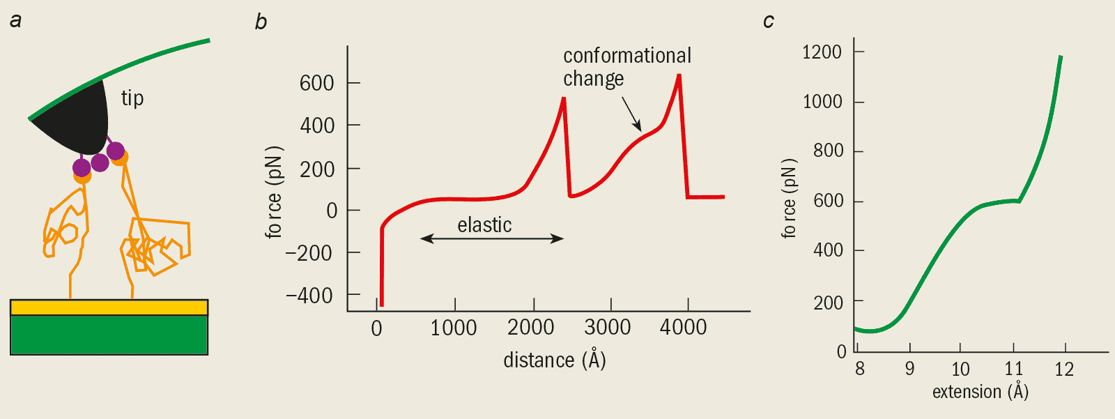
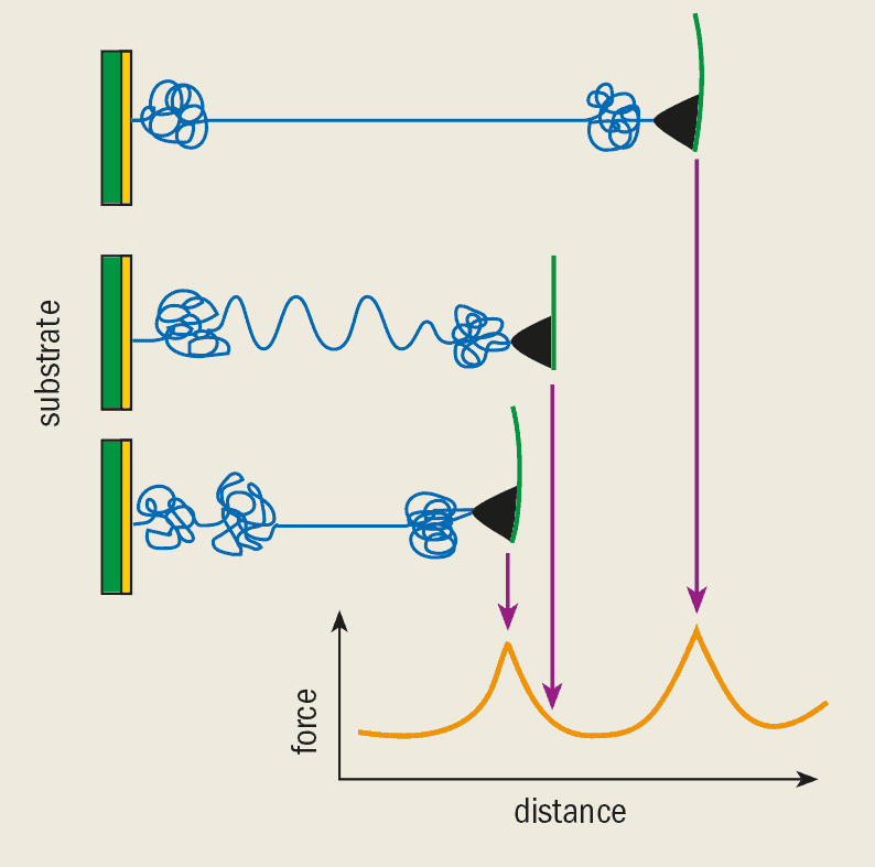
In 1997 Mathias Rief and co-workers at the University of Munich used an AFM to measure the elastic properties of a single dextran molecule – a long sugar molecule that is produced by certain bacteria. The basic idea is to hold one end of the molecule tightly by binding it to a substrate and to pull the other end with the tip of the AFM. The distance between the tip and the substrate is then increased steadily. As the dextran molecule is pulled, it changes shape and the force is measured by monitoring the deflection of the cantilever spring. The variation of the force with this distance is known as a force-extension curve (figure 2).
2 Elastic molecule (a) A single dextran molecule is bound covalently to a gold substrate while part of it is picked up with the tip of the atomic force microscope. The molecule is then pulled at a steady rate and the force is measured from the deflection of the cantilever spring. (b) A plot of the force versus the distance pulled shows how the force varies as the molecule changes shape. For forces below 50 pN the molecule behaves according to the rubber elasticity model. At larger forces, other elastic contributions become important. As the molecule is pulled further, the chemical bonds within the molecule rotate until they cannot rotate any more. At this point the only way the molecule can change shape is through flattening. This effective change in the “stiffness” of the molecule is observed as a change in the slope, (c) A molecular dynamics simulation of the dextran-elasticity experiment. The simulated molecule contains just two sugar rings while the real molecule shown in parts (a) and (b) contains thousands of rings. Nevertheless, the simulation explains the features of the force-extension curve at the molecular level. For forces below 500 pN the chemical bonds joining the monomers rotate easily and above 600 pN the sugar ring deforms.
The force-extension curves from such experiments can be understood by comparing them with various models based on polymer theory. The dextran molecule has a very flexible structure and when small forces are applied its elastic properties are well described by the so-called “rubber elasticity model”. In this model, the polymer chain is thought of as a series of smaller compounds called monomers that are linked by rotating joints. As no energy is needed to deform these joints, the polymer can easily bend into any shape or conformation. Often this type of conformation is called a random coil.
However, molecules are likely to bend into some shapes or “conformations” more than others. The biological and chemical behaviour of the molecule is entirely governed by the number of different shapes it can form. When we pull a molecule with an AFM, we change the number of possible shapes that it can adopt, and an energy price has to be paid that changes with the temperature.
We can describe this behaviour more easily by considering a rubber band that has been stretched, for example, by suspending a small mass from it. If we heat the band, it will shorten and become stiffer. As a molecule is stretched further, an additional elastic force is measured and eventually the stiffness of the molecule changes. It is clear that rubber bands and molecules behave very differently from stretched springs.
These different regimes can be fully understood if we look at the process of pulling and deforming the molecule in atomic detail, using simulations of the molecular dynamics. In these simulations, the motion of each atom is calculated due to the forces it experiences from the other atoms. The simulations can only be performed numerically with the help of computers and are very time consuming. Even the fastest computers take several weeks to simulate the motion of every atom in a polymer for just a few nanoseconds.
However, the force-extension curves from the computer simulations are in good agreement with the experimental data. And the advantage of simulations is that we can now explain the behaviour at the atomic or the submolecular level. The elastic contribution that is important at relatively small forces is due to the rotation of bonds between two carbon molecules that join the sugar rings (see figure 2c).
At some point during this rotation, the bond suddenly straightens and cannot be rotated any further. Any further elastic deformation can now only take place by flattening the sugar ring itself. Since this process involves several chemical bonds being deformed simultaneously, the effective stiffness of the polymer rises.
Although there are some quantitative differences between the experiment and the simulation – straightening the carbon-carbon bonds in the simulation requires a force that is twice as strong compared with the experiment, for instance – the overall agreement is very good. In fact, the level of agreement is remarkable when we consider that the computer simulation pulls the molecule nine orders of magnitude faster than the experiment.
So why should the “pulling speed” matter? We can think of the transition from one molecular shape to another as a two-level system with a so-called activation barrier separating the two states. When we pull the molecule with the AFM, we effectively reduce this barrier until it eventually disappears at a “threshold force”. But there is a finite probability that the molecule will cross this barrier due to thermal fluctuations, even when the force is less than the threshold value. The longer the molecule is held at a particular force, the more likely it is to change shape due a thermal fluctuation. This explains why the molecule makes this transition at a much lower force in the experiment compared with the simulation, which is significantly faster.
The elastic properties of other single polymer molecules were measured using an AFM in a similar way. Hermann Gaub and co-workers at the Ludwigs-Maximilians University in Munich have measured the elastic properties on poly-ethylen glycole, poly acrylic acid and deoxyribonucleic acid (DNA). The elastic properties of DNA are particularly important for cell replication and the synthesis of new proteins. In a cell, the DNA molecules are typically wound around little barrels called histons that are only about 10 nm in diameter. The DNA has to unwind before it can duplicate itself, and afterwards it has to rewind around the histons.
Specific interactions
There are many instances in biology where we need to know how much force a chemical bond can sustain. In drug development, for example, the effectiveness of a medication depends on the strength of the bond between a ligand in the drug and the receptor.
We can now measure the strength of an individual chemical bond in experiments using an AFM. In 1994 Ernst-Ludwig Florin, Vincent Moy and Hermann Gaub at Munich demonstrated this for the first time using a vitamin-B molecule called biotin, which reacts very strongly with a protein called streptavidin.
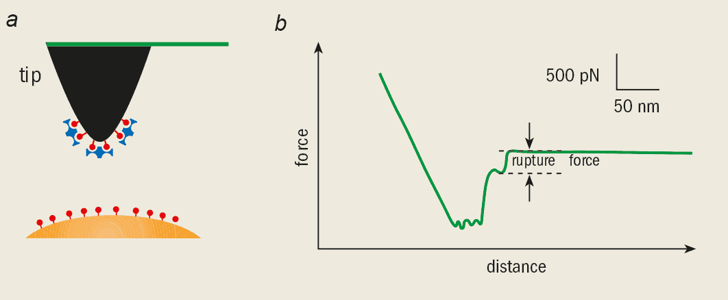
3 Specific interaction (a) The tip of an atomic force microscope (black) is covered in a vitamin-B molecule called biotin (red) and then covered in a protein called streptavidin (blue), which binds strongly to it. The bead-shaped substrate is also coated with biotin. Usually just one bond is formed when the tip and substrate are brought into contact, (b) The final jump in the force-extension curve corresponds to the force needed to break a single (or only a few) molecular bonds.
To measure the strength of this bond, the AFM tip was coated first with biotin and then with streptavidin. A bead-shaped substrate was also coated with biotin so that it would readily bond to the coated tip (figure 3a). Since a biotin molecule can bind to a streptavidin molecule at four different sites, bonds can be formed between the bead and the tip. The trick is to use very small amounts of biotin and streptavidin so that just one bond (or a few at the most) is formed when the AFM tip is brought into contact with the bead.
The adhesion force was measured as the tip was pulled away from the substrate until the bonds eventually broke. The Munich researchers found that the strength of an individual bond was around 200 pN. The rupture force changes when we use slightly different molecules, but simulations give us a detailed understanding of the actual processes that happen as the bonds break.
Several other groups have adopted this experimental scheme to measure the molecular rupture force for many other systems. Hans-Joachim Güntherodt’s group at the University of Basel in Switzerland measured the strength of the bond between various antibodies and their antigens, substances that can interact with specific chemical groups on the antibody. They also determined the force between proteoglycans – molecules that are important for the adhesion between cells.
Recently, Michel Grandbois in our group at Ludwig-Maximilians University in Munich demonstrated that the strength of a single covalent bond, the strongest type of chemical bond, could be measured (see Physics World June 1999). We have found that the rupture force of silicon-carbon bonds and sulphur-gold bonds is just a few nanonewtons. In biological terms, such large forces are not accessible using other sensitive single-molecule manipulation techniques, such as optical tweezers.
Structure and function of proteins
The beauty of an AFM is that we can investigate functioning processes within living biological systems, such as cells and single molecules. In 1994 Monika Fritz and co-workers at the Technical University in Munich observed the biological activity of human platelets (minute particles found in the blood) using an AFM. And at the single-molecule level, Monika Fritz, Paul Hansma of the University of California at Santa Barbara and I demonstrated that the activity of an enzyme that is present in milk and tears could be measured using an AFM. We showed for the first time that the activity of a single molecule could be determined directly using mechanical sensors. This may give important insights into how the shape and motion of molecules change as they carry out a biological function (see, for example, The physics of protein folding).
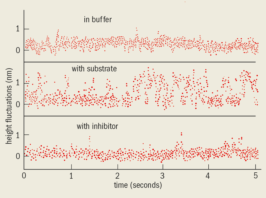
4 Enzyme activity The biological activity of an enzyme called lysozyme is monitored by observing how the height of the tip varies with time, (a) The enzyme is in a buffer solution to prevent it from reacting with the substrate, (b) The height fluctuations are evidence for the reaction between the enzyme and the substrate, (c) An inhibitor is added which binds to the enzyme and destroys the biological activity.
In the experiment the AFM tip was brought into contact with a dense layer of enzyme molecules that were adsorbed on a mica substrate. We kept the horizontal position of the tip stationary and monitored the height of the sample as a function of time by measuring the deflection of the cantilever (figure 4). We saw spike-like fluctuations only when the enzyme reacted with the substrate. No effect was seen when the enzyme was surrounded by a “buffer” solution so that it could not react with the substrate, nor when an inhibitor was added to destroy the activity of the enzyme.
Meanwhile, Magdalena Bezanilla and co-workers in Helen Hansma’s group at Santa Barbara followed how an enzyme called DNAse reacted with DNA that was adsorbed on a mica surface. They found that the enzyme digested the DNA molecules into smaller pieces until they disappeared entirely from the surface.
In 1997 Sandor Kasas and co-workers in the same group used an AFM to follow what happens in one of the first stages of protein synthesis. Living cells manufacture proteins from amino acids, according to the genetic information that is encoded in the DNA. Ribonucleic acid (RNA) acts like a messenger, carrying this information from the DNA to the protein factories within the cells. An enzyme called RNA polymerase reads the genetic information on the DNA strand and then synthesizes the complementary strand of RNA. Kasas and co-workers adsorbed DNA on a mica substrate and incubated it with RNA polymerase. Using an AFM, the researchers directly observed the polymerase move along the DNA strand one step at a time as it read the genetic information.
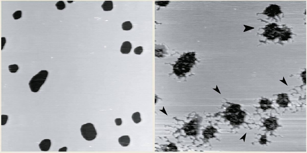
5 Single enzyme molecules (a) A layer of fatty-acid molecules, one of the major constituents of cell membranes, is adsorbed on a mica substrate. The holes have a depth of 5 nm, corresponding to twice the length of the fatty-acid molecules. The image is formed by scanning the tip of the atomic force microscope across the sample, (b) When the enzyme phospholipase A2 is added, it digests canals through the membrane, suggesting that we are observing the activity of a single enzyme molecule.
Meanwhile, Michel Grandbois and Hauke Clausen-Schaumann have studied the effects of phospholipase A2, the active ingredient in rattlesnake venom, with an AFM. When they added phospholipase A2 to a little patch of fatty-acid molecules that were adsorbed on a mica substrate to mimic a cell membrane, they saw the membrane being slowly digested. The activity of a single enzyme molecule was inferred from the creation of little canals in the patch as a single molecule digested its way through the fatty acid (figure 5).
Stability of proteins
Proteins are made from long, 1-D chains of molecules that fold up into certain 3-D structures. The detailed chemical and biological properties of the protein depend on how the molecule folds. Mechanical stability is very important for proteins that form muscle fibres, like myosin and kinesin, and for those that have to withstand forces (like cell-adhesion molecules that stabilize and form the contact between cells in tissues, for example).
6 Unfolding proteins One end of the protein titin is bound to a gold substrate while the other end is held by the tip of the atomic force microscope. The protein fragment resembles beads on a string, where each bead is a tightly coiled portion of the molecule. As the molecule is stretched, the force increases until one of the coils unravels. The process continues until the next coil unfolds and results in the saw-tooth pattern.
Muscle fibres contain two types of filament that slide against each other, causing the muscle to stretch or contract. A protein called titin acts as a buffer between the two filaments and protects them. But Harold Erickson at Duke University in the US has speculated that the mechanical properties of the titin molecule give the muscles their elasticity.
The structure of the protein molecule resembles a string of beads. The muscle’s elasticity may be due to either the random coils (the beads) or the “repeat region” made up of units called domains. The mechanics of the random coil has been determined by experiments using optical tweezers, whereas Mathias Rief and co-workers at Munich have investigated the properties of the domains using an AFM.
Short fragments of the titin molecule, either 4 or 8 molecules long, were first bound to a substrate. Rief then picked up the fragments using the AFM tip and pulled them until the individual domains unfolded (figure 6). The measured force increased as the polymer was stretched until one of the random coils unravelled. At this point the force dropped and the process continued, resulting in a force-extension curve with a saw-tooth pattern.
The titin force-extension curves are best described by the “worm-chain model” – another one of the standard models from polymer theory. In this model the elasticity of the polymer molecule is described by the energy needed to bend the molecule, rather than the energy required to rotate the bonds.
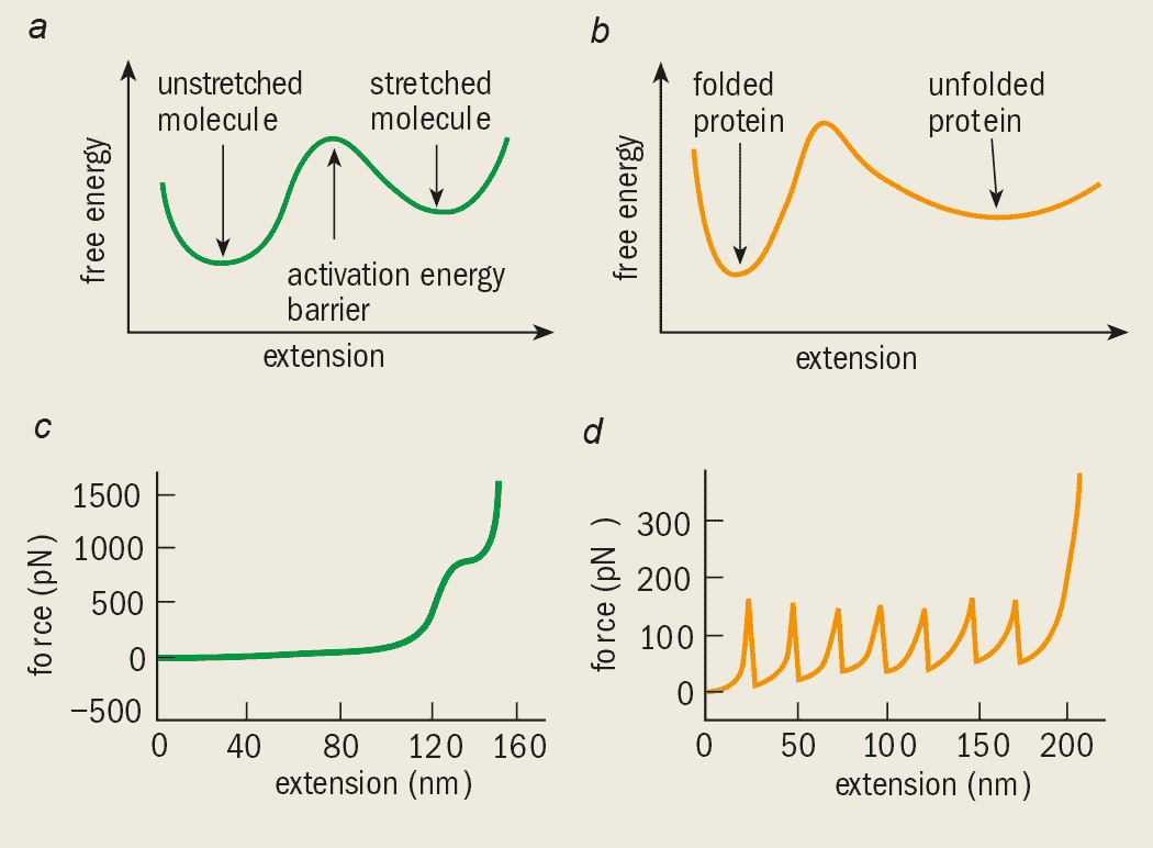
The experiment also determined the stability of a protein domain by measuring the force needed to unfold it. Simulations performed by Klaus Schulten and co-workers of the University of Illinois at Urbana Champaign showed that hydrogen “bridges” in the protein domain are important for its stability. By comparing experimental results with such simulations we gain a much clearer and more detailed understanding of the protein at the molecular level. Although the chemical and biological properties of dextran and titin are very different, the same physics underlies the elastic behaviour and can be understood by comparing the AFM results with a two-state model (figure 7).
7 Physical similarities Simulations of the dextran polymer and titin protein reveal that the elastic properties of the two molecules are governed by the same underlying physics, (a) and (b) show the energy versus stretched distance of the dextran and titin molecules. In both cases a minimum or valley in the energy landscape characterizes the initial (unstretched or folded) and final (stretched or unfolded) states. The “hill” in between the two states is known as the activation energy barrier, which usually keeps the system in one of its two states. By pulling the molecule with an atomic force microscope we can overcome this barrier as shown in the simulated force-extension curves for (c) dextran and (d) titin. The results from the simulations agree well with experimental data, although the pulling speed has an influence on the results.
Future outlook
The atomic force microscope has opened up a whole new class of experiments with single molecules in the physical and engineering sciences. But perhaps biologists have profited the most from the development the AFM, which allows them to study a wide range of living biological processes with unprecedented accuracy for the first time. In addition, AFM experiments give biologists the opportunity to study molecules in forms that are rarely seen in nature. In their natural habitat, molecules spontaneously change shape, and may even completely unfold and refold again. Conventional methods in biology examine an ensemble of many molecules, and therefore “average out” these shape fluctuations. However, by studying single molecules we can examine a given conformation in detail. Moreover, we can manipulate the molecules into states that are normally only present for a short period of time and study them in detail using an AFM.
Since atomic force microscopy is a relatively new technique, we can be sure of many more exciting developments that will undoubtedly shed light on the physics of life.
“The entire scope of human experience can be viewed as a collective effect resulting from elementary particles dutifully following well-understood equations.” Statements like this excite deep-seated passions and tend to bring out strongly polarized views. The Pearly Gates of Cyberspace by Margaret Wertheim is a product of this passionate debate.
This debate is one I usually find quite tiresome. As someone who is comfortable with reductionist views, I readily concede that comments like the one above shed very little useful light on many aspects of our lives, particularly those that we might call “spiritual”. Still, I hope that bridging the mind/body gap will lead to interesting insights into both psychology and physical collective phenomena. I expect the wide range of medical and other researchers who are investigating this frontier will make exciting progress in my lifetime. That’s the fun part.
But no matter what progress is made, it seems reasonable to expect there will remain vast domains of human experience that will be more effectively discussed in broadly “spiritual” terms, despite the existence of a reductionist perspective. What I find tiresome in all this is the way many people see these issues as an opportunity to preach about matters that are far removed from the really exciting problems. This rather empty debate seems to occupy an extraordinary amount of some people’s time, and seems to receive a huge amount of publicity. Statements like: “The failure of modern science to incorporate this immaterial ‘I’ – this ‘self’, this ‘mind’, this ‘spirit’, this ‘soul’ – into its world picture is one of the premier pathologies of modern Western culture…” (quoting Wertheim) are the sort of things I am talking about. Whether or not our culture pays sufficient attention to spiritual matters, to set modern science up as the villain seems quite beside the point.
So, is The Pearly Gates of Cyberspace simply another scolding of the science community on behalf of those whose humanity is being “crushed” by our rationalistic zeal? Not at all! Wertheim manages to take what seems like a tired old subject and introduce an entirely fresh and original perspective.
The traditional mind/body debate is argued between those who cannot imagine that the richness of our personal experience is built on a mechanistic world of atoms and molecules, and those who are enthusiastic about that possibility. Wertheim steps in with an entirely different angle. To start with, she chooses to focus on the concept of “space”. For Wertheim, it is our 3 + 1 dimensional concept of space and time that most clearly characterizes the limitations of modern science. She wonders where there could possibly be room for a soul in such a space, and concludes that, with this kind of view of the world, we are bound to stifle the human spirit.
But physicists are perhaps even too eager to consider all kinds of different spaces. There are, for example, the Hilbert space of atomic states, the Fock space in field theories, and the spaces of gauge transformations (Abelian or non-Abelian). There is the space of “all vacuua of M-theory”, or the space occupied by the phase diagram of helium-3, and so on. What Wertheim seems to have completely ignored is that we are quite happy to use a much more generalized idea of space to describe different states of matter. For the reductionists among us, it is in this space that we might hope to find our spirit romping around, but somehow Wertheim manages to avoid this point. Well, she gingerly acknowledges the “extra dimensions” models for the matter fields but still argues vigorously that even the extra dimensions are not a suitable place for the soul to reside.
But the real thrust of Wertheim’s book is that she feels she has at last discovered the true space for the human soul: cyberspace! I imagine most readers are just as stunned as I was about this. Even the most cranky among us will admit that some interesting (possibly even amazing) advances will be required before we can understand how the human mind can be built out of the microscopic world we know. Many will feel that within that gap lie endless possibilities for even the subtlest spiritual nuances to emerge.
But cyberspace? Cyberspace today is perhaps the most boring mechanistic object in our entire world. Just a bunch of files shifting from one computer to another in a way that (if sometimes delayed) is usually accurate to the point of tedium. The microscopic perspective on this process is perfectly well understood due to the efforts of many scientists and information-technology engineers. Wertheim imagines that she has a “position” in cyberspace that does not obey “F = ma”, and finds this deeply liberating. Why this aspect of cyberspace is so exciting, while the flights of the thoughts in our own minds do not seem to count for much, is completely beyond me. If I am looking for spiritual enrichment I will take my brain (or anyone else’s) over cyberspace any day. Perhaps some day that will change, but then you could write another book.
In the process of developing her argument, Wertheim undertakes to review the abstract notions of spiritual space that have existed throughout history, discusses parts of the history of science, and surveys the great opportunities presented by the Internet and other information technology. Each of these is an interesting topic in its own right, but my aversion to the overall premise of the book made it hard to enjoy any of these discussions. None of them seemed particularly inspired.
The Pearly Gates of Cyberspace gave me only one enjoyable moment: this was when I learned of the plan by the German-born mathematician Theodr Kaluza to shed the “crazy theorist” label attached to his proposal, with Oskar Klein, about a possible fifth dimension. A non-swimmer, Kaluza decided to teach the world about the value of pure theory by carefully studying the “theory of swimming”. When he was satisfied with his understanding, he threw himself into the sea (we are not told how deep it was) and indeed he could swim! Boy, theorists are a crazy bunch.
Aside from this entertaining moment, I found The Pearly Gates of Cyberspace downright painful to read. I doubt any reader would enjoy it.
1 Physics looks at the brain Functional magnetic resonance imaging(fMRI)combines two physics-based techniques: magnetic resonance imaging (MRI) and positron emission tomography (PET). Conventional MRI offers high spatial resolution while PET shows which regions of the brain are active. fMRI can therefore be used to monitor brain activity during various tasks. These images, taken during a recognition task, show that most areas of the brain are active after a relatively short delay of 4-6 seconds (top), whereas the anterior prefrontal regions only become active after a delay of about 8-9 seconds (bottom). (B R Rosen et al. 1998 Proc. Natl Acad. Sci. USA95 773-780)
The aim of most biomedical research is to uncover new knowledge that will lead to better health. At the National Institutes of Health (NIH) in the US we do this by supporting research on the prevention, detection, diagnosis and treatment of disease and disability, from the rarest genetic disorder to the common cold, as well as research on the basic principles of biology.
In this article I would like to discuss my conviction that we can only wage an effective war on disease if the scientific community harnesses the energies of many disciplines, not just biology and medicine. These allied disciplines range from mathematics, engineering and computer science to sociology, anthropology and the behavioural sciences. But the weight of historical evidence and the prospects for the future place physics and chemistry most prominent among these disciplines.
Physics and biology
I will discuss the effects of physics on the medical sciences from three perspectives. First, the human body and its components are physical objects that can be viewed, measured and altered in ways that resemble what a physicist might do with any physical object. Second, I will remind you of an enormously important phase in the history of biology in which physicists transformed the study of living things by helping to discover the principles of heredity. Third, I will describe some contemporary problems in the biomedical sciences that I believe present challenges to physicists, young and old. I will also explain the ways in which the NIH is attempting to ease the path from a formal training in physics to an active, investigative role in biomedical sciences.
I am only the latest in a long line of commentators who have made the really quite obvious point that, for at least several hundred years, physicists – and especially their principles, methods and machines – have been illuminating our views of the human body and of every other living thing.
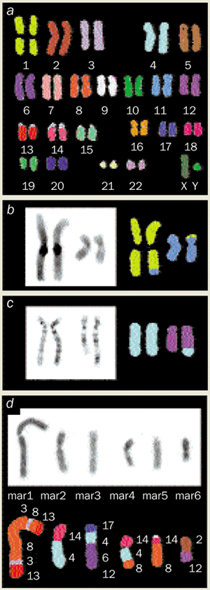
2 Physics looks at cancer Spectral karyotyping is a technique that allows biologists to rapidly identify each of the 23 pairs of normal human chromosomes, and the origins of recombined chromosomes that often appear in cancer cells. The technique relies on a variety of techniques from physics and chemistry, including Fourier spectroscopy, charge-coupled device imaging and optical microscopy, (a) Spectral karyotyping of normal human chromosomes. Note that every chromosome is a different colour, (b) Spectral karyotyping of chromosomes from the father of a child with mental retardation. Notice that small fragments of chromosome 1 (which is yellow) have been translocated to chromosome 11 (which is blue), and vice versa. Also shown are images of chromosomes from patients with (c) ataxia, a muscle disease, and (d) breast cancer. (E Schrock et al. 1996 Science273 494-497)
This notion was brought home to me very early in life when my father – a general practitioner whose office was directly connected to our house – showed me how X-rays and fluorography could reveal the bones and lungs of our pets and his patients, and help make diagnoses of disease. Röntgen and Edison had been pioneers in this respect. The significance of using the discoveries of physics to perceive biological function was further impressed on me at college, when one of my first independent projects required that I try to explain the repeating peaks and valleys of my electrocardiogram as a record of voltage changes in the salty sea of the human body. And at medical school I learned that the doyens of our biochemistry department had become famous by being the first to tag red blood cells with easily detected radioisotopes to learn how long such cells survived in the body.
These few personal memories are just a sampling of the hundreds of physics-based methods that have been applied to view living bodies without the disruption of anatomical dissection or to visualize very small components of living things.
A more systematic rendering of this topic was offered by the distinguished Stanford physicist Robert Hofstadter, in a talk to the National Academy of Sciences in 1983 (see table). It is instructive to note how many of the methods can be classified as techniques that permit us to visualize the inside of the human body at successively higher levels of resolution, or allow us to see smaller and smaller elements of bodily components.
The methods of “macro-imaging” include conventional X-radiology, computerized tomography scanning, ultrasound, positron-emission tomography (PET) and magnetic resonance imaging (MRI). The impact of these procedures on medical practice is unquestioned and continues to grow as new methods and new applications appear. Two recent examples convey the exciting potential for both clinical and investigative work – the combined use of PET and MRI to provide images of the human brain at work (figure 1), and the use of MRI to analyse both structural and functional characteristics of the human heart in diseased states.
“Micro-imaging” began with the use of optical principles to devise the light microscope, but has progressed to much higher levels of resolution with electron microscopy, X-ray crystallography and nuclear magnetic resonance.
Sometimes a collection of methods proves important, as in the combined use of molecular hybridization, fluorochrome chemistry, wave optics, and computer science in “spectral karyotyping”. This procedure allows the rapid identification of each of the 23 pairs of normal human chromosomes and also the origins of recombined chromosomes that often appear in cancer cells (figure 2).
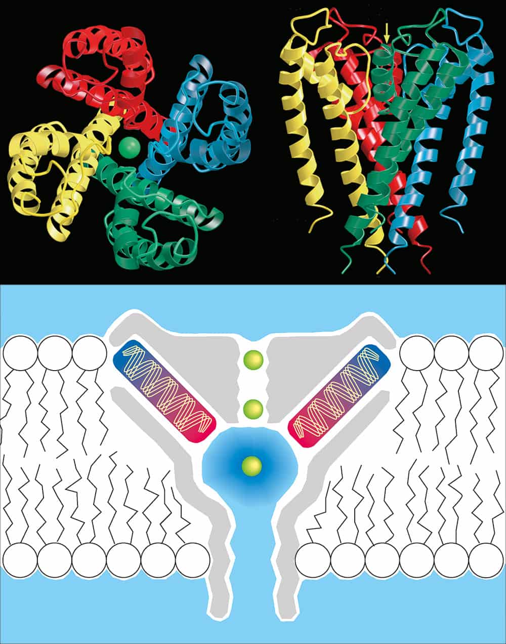
Long-awaited success in using a time-honoured technique, X-ray crystallography, to resolve the structure of proteins embedded in biological membranes has recently transformed the study of cell function and disease. I used an important example of this progress – the analysis by Rod MacKinnon and co-workers at Rockefeller University in New York (see Doyle et al. in further reading) of potassium channel proteins to understand how the channels can be so efficient and yet so selective (figure 3) – when justifying further investments in research to Congress this year.
Despite the centrality of such contributions of physics to modern biology and medicine, I recognize the danger that my emphasis might be interpreted as limited and perhaps even insulting, because (some might say) I have portrayed physicists as merely the developers of tools of measurement that allow biomedical scientists to do the really important work. There are reasons for my sensitivity to this issue: in a 1967 commentary on the role of physics in biology and medicine, for example, Sergei Feitelberg, a physicist from Mount Sinai Hospital in New York, noted that while such “spectacular developments created a clear and unequivocal need for physicists and their help, the role of the physicist was that of a glorified technician engaged in methodology and instrumentation, dignified only by the strangeness of his doings and the mysteriousness of his tools”.
I do not accept that interpretation. In fact, I would argue that we need to show our appreciation of physics-based technology by investing NIH funds more aggressively in its development. We have begun to do just that through a new Bioengineering Consortium and a trans-NIH emphasis on technology development. Still, I would like to address a deeper set of contributions that physics makes to biology – through the efforts of physicists who themselves seek to understand the rules of living systems.
Correlations between physics and medicine
Physics
Medicine
Statics (mechanics)
Orthopaedics
Dynamics (mechanics)
Heart motion
Elasticity and strength of materials
Orthopaedics
Fluid statics
Blood pressure
Fluid dynamics
Blood flow in vascular system
Surface tension
Capillary action
Sound and acoustics
Stethoscope, ultrasound, acoustic microscope
Electricity
All life processes, ion transfer at membranes
Magnetism
Nuclear magnetic resonance imaging
Light and optics
Light microscopy, laser therapy, fibre optics
Heat and thermodynamics
Heat balance
Kinetic theory and statistical mechanics
Brownian motion, osmosis, diffusion of gases
Atomic physics and spectroscopy
“Chemical shift” in NMR imaging, lasers in medicine
Molecular physics
Genetics, antibodies, protein structure, electron microscope
This table was presented by Robert Hofstadter of Stanford University at a conference on biological imaging organized by the National Academy of Sciences in October 1983. Hofstadter had shared the Nobel Prize for Physics in 1961 for his work in nuclear physics. Many new physics-based techniques have become important in biology since then, for example various image capture and analysis techniques developed by astronomers and astrophysicists.
Physicists, heredity and the rise of molecular biology
Exactly 50 years ago, in a speech entitled “A physicist looks at biology”, Max Delbruck, a leading physicist who had made a conversion to biology some years earlier, attempted to describe the transition. In the speech, delivered to the 1000th meeting of the Connecticut Academy of Arts and Sciences, Delbruck said: “A mature physicist, acquainting himself for the first time with the problems of biology, is puzzled by the circumstance that there are no ‘absolute phenomena’….The animal or plant or micro-organism he is working with is but a link in an evolutionary chain of changing forms, none of which has any permanent validity. Even the molecular species and the chemical reactions which he encounters are the fashions of today to be replaced by others as evolution goes on. The organism he is working with is not a particular expression of an ideal organism, but one thread in the infinite web of all living forms, all interrelated and all interdependent. The physicist has been reared in a different atmosphere. The materials and phenomena he works with are the same here and now as they were at all times and as they are on the most distant star.”
Delbruck had been a student of Niels Bohr and then a powerful proselytizer for biology. With the assistance of Bohr’s book Light and Life and, more importantly, Schrödinger’s book What is Life?, he attracted many other physicists to biology. The effects of his missionary zeal were powerful – not just because some very smart people started to do biology, but because they brought to biological problems a quantitative, analytic approach – an approach that created the atmosphere in which principles of molecular biology were discovered by seeking the physical basis of heredity.
The leading physicist Leo Szilard was among the converts, and claimed that what physicists brought to biology was “not any skills acquired in physics, but rather an attitude: the conviction which few biologists had at that time, that mysteries can be solved” (see Fleming in further reading).
Delbruck and his friends were gripped by some fundamental questions: what is the physical form in which hereditary information is stored? How is it reproduced when a cell divides, or when a single virus particle invades a cell and makes hundreds or thousands of copies of itself? How is the information reassorted during sexual reproduction? How does the information change when mutations occur?
3 Physics looks at cell membranes The diffusion of potassium ions across cell membranes underlies many fundamental processes in biology, including electrical signalling in the central nervous system. The ions move through proteins known as K+ channels: however, sodium ions, which are smaller than potassium ions, cannot pass through. Researchers at Rockefeller University in New York have used a variety of techniques, including synchrotron radiation, to establish how these K* channels work. They found that four identical subunits create an inverted teepee. This teepee cradles a selectivity filter that is 12 A long and contains two K* ions (green dots) about 7.5 A apart. For full details of this experiment see D A Doyle et al.1998 Science280 69-77
Answers to many of these questions came from the “phage school” that Delbruck founded. The phage school was a group of former physicists and some biologists who shared his passion for reducing the problem of heredity to simple rules, physical entities and conserved energy by studying the replication and genetic behaviour of bacterial viruses (also called bacteriophage or “phage”) in their bacterial hosts. The studies culminated in findings that form the pillars of modern molecular biology: the identification of deoxyribonucleic acid (DNA) as genetic material, a description of the physical organization of DNA through X-ray crystallography, the deduction of the principles of base pairing and the strategy of replication from the organization of the double helix, and the deciphering of the genetic code as triplets chosen from a set of four nucleotides.
Delbruck and his phage school were important, but there were, in fact, multiple intellectual lineages connected with physics that helped to create the modern world of molecular biology (see Keller in further reading). For instance, Warren Weaver was a mathematical physicist turned science administrator who, in 1932, first used the term “molecular biology”. He chose this phrase because he foresaw “that the moment would arrive when the distinction between chemistry and physics and even mathematics on the one hand and biology on the other would be so illusory and in fact so unfortunate” that he did not want to use the word “biology” to describe the programmes he was supporting at the Rockefeller Foundation.
British scientists with a strong physical bent, such as Astbury, Bragg and others, used X-ray diffraction to study the organization of fibres of many kinds, mainly proteins found in textiles, in an intellectual lineage that led to Wilkins and Franklin and, of course, DNA. The American geneticists T H Morgan and H J Muller used physical agents – namely X-rays – to induce mutations in fruit flies. Muller’s affinity for the principles of physics was especially strong. He was fond of noting the potential similarities of mutation of genes to transmutation of elements, calling the prospect of understanding these events in physical terms “the two keystones of our rainbow bridges to power” (see Carlson in further reading)
Bringing physics, not just physicists, to biology
To the birth of modern molecular genetics, physicists contributed their analytic skills but they were not really doing physics, and many were not even using the computational or imaging tools of physics as many biologists do. Delbruck and his colleague Salvador Luria laboriously counted virus infections by hand and eye, just like any other biologist. But contemporary biology, especially the deciphering of genomes by nucleotide sequencing, is about to change that. Biology is rapidly becoming a science that demands more intense mathematical and physical analysis than biologists have been accustomed to, and such analysis will be required to understand the workings of cells.
This change was clearly foreshadowed in Delbruck’s 1949 lecture in Connecticut. He first described his awe at the complexity of biology: “The closer one looks at [the] performances of matter in living organisms the more impressive the show becomes. The meanest living cell becomes a magic puzzle box full of elaborate and changing molecules, and far outstrips all chemical laboratories of man in the skill of organic synthesis….”
But Delbruck also sounded a warning: “Biology is a very interesting field…[because of] the vastness of its structure and the extraordinary variety of strange facts…but to the physicist it is also a depressing subject, because…the analysis seems to have stalled around in a semi-descriptive manner without noticeably progressing towards a radical physical explanation…we are not yet at the point where we are presented with clear paradoxes and this will not happen until the analysis of the behaviour of living cells has been carried into far greater detail.”
In the past 50 years, and especially in the past 20, molecular and cell biologists have moved much closer to the “radical physical explanation” of cell behaviour that Delbruck sought. Certainly the chemical elements – especially the genes, the ribonucleic acid (RNA), and the proteins – and some of their basic functions are coming into view. What is lacking is a sense of how these functions are integrated to allow cells to manifest their physiological traits.
I would like to mention three of the several arenas of biology in which I believe the skills of physicists and their close cousins can be most productively used.
The first is perhaps the most reductionist. Methods are now available for examining the physical and chemical properties of single macromolecules and single complexes of large molecules. These advances are important because they avoid the need to synchronize a population of molecules to measure function. Several of these methods and their applications are reviewed in a special section on “single molecules” in the 12 March 1999 issue of Science. They include laser traps (“optical tweezers”) to study the energetics of molecular motors used for transport, for contraction and for flagellar motion. Steven Chu of Stanford University, who shared the 1997 Nobel Prize for Physics, has made significant contributions to this problem in collaboration with his colleague the cell biologist Jim Spudich.
Laser traps can also be used to measure the force of an enzyme complex, such as the one that copies DNA sequences into RNA. Fluorescence spectroscopy and scanning tunnel microscopy can visualize the conformation of single large molecules, and methods now in development may soon be able to determine the order of bases in single, long DNA molecules.
Second, the computational experience of physical scientists is needed to help interpret complex data sets and the process of “gene expression”. One of the consequences of projects to sequence the genomes of human beings and many other species is the opportunity to understand the processes by which the genes of an organism are expressed. Such information can help us to understand, for example, why some cells develop into muscle tissue, while others become brain cells. New methods, built on the availability of a piece of DNA from each gene, allow us to measure the extent to which genes are read to form RNA (and subsequently protein) in different tissues and under different environmental conditions.
These micromethods, called “expression arrays”, are coming into wide use to study bacteria (with several hundred to a few thousand genes), yeast (with about 6200 genes), worms (with about 19 100 genes) and vertebrates (which are predicted to contain about 80 000 genes). Some progress has been made through computer-based “cluster analysis” (see Eisen et al. in further reading) to begin to interpret the voluminous data that such experiments generate, but biologists are generally unused to such complex data sets. Recently, I spent an evening at the Carnegie Institution’s observatory at La Serena in Chile, watching astrophysicists gather amazingly similar data sets to search for supernovae and to measure the chemical composition of distant stars. We are all likely to benefit from an interdisciplinary exchange of computational approaches.
The third area of opportunity for physicists in biology is the one that most closely approaches Delbruck’s goal of developing a “radical physical explanation” for cell function. In the past 20 years, mainly through efforts to identify the genes and proteins that control cell growth and responses to hormones, biomedical investigators have constructed many so-called signalling pathways that link molecular interactions at the cell surface to changes in gene expression in the nucleus.
While there is consensus that these linear pathways are over-simplified, the way forward is far from clear. The pathways doubtless have many unrecognized components; the information is certainly flowing between, not just along, the several pathways; and the pathways are probably regulated in complicated ways through feedback mechanisms and other means. A few investigators are beginning to grapple with these issues (see Bhalla and Iyengar, and Weng et al. in further reading) but there is an obvious need to apply experiences with potentially analogous complex machines.
Finale: moving between disciplines
In talking about the effects of one field on others, I have generally ignored the “boundary problem” – how do we distinguish among fields? We do this now, in part, by self-identification, just as we deal with ambiguity about race, ethnicity and religion. Self-identification in science is commonly linked to the source of one’s graduate degree, and departmental names on diplomas can become limits to exploration in adjacent fields. But many of us in biology expect that, as studies of cells and molecules become more obviously in need of several disciplinary approaches, it will become increasingly difficult to label the science and to predict the kinds of degrees the people doing it should have.
At the NIH, we have become concerned about how people should be trained in college and in graduate studies to pursue biological problems over the next 50 years, and we are discussing the need to study this issue with the National Research Council. I also agree with Leon Lederman, who has been leading the movement to establish a more logical order for teaching the sciences in US high schools: that is physics, chemistry and then biology. But these activities will come to fruition only after many years, and it is important to also consider the more immediate need to transport intellects across artificial disciplinary boundaries.
I sense increasing interest in attempting to open borders that have been traditionally hard to cross. In the US, workshops on computational biology and approaches to complex systems have recently been organized by the National Institute of General Medical Sciences and the Department of Energy. New funding opportunities for interdisciplinary work are available through the Bioengineering Consortium and other programmes at the NIH. At present, total NIH funding of physics projects is estimated to be about $287m.
There are many anecdotal accounts of successful interdisciplinary training programmes. Within our intramural research program at the NIH, physicists and physics trainees from the US and abroad do graduate thesis work, take courses in biological topics, and engage in post-doctoral training that promotes interactions with biologists and clinicians. Much of this activity occurs under the direction of some of our most prestigious scientists – such as Ad Bax, Bob Balaban, Bill Eaton and Adrian Parsegian – and includes work on small-molecule and protein NMR, brain and cardiac MRI, and other topics, leading to good job prospects for trainees.
On the occasion of the 100th anniversary of the American Physical Society, I thank physicists for their many contributions to biology and medicine – for providing the tools that allow us to see and probe living things, and for training great minds that have uncovered some of the most fundamental principles of biology. I now encourage physicists to work collaboratively with biologists as we strive to achieve Delbruck’s “radical physical explanation” for biological systems.
The meeting gave the pioneers of “econophysics”, such as Jean-Phillippe Bouchard, János Kertész, Rosario Mantegna and Eugene Stanley, an official forum to present and debate their research on some of the most fascinating dynamical systems. The search for a scientific model that can accurately predict future movements in the financial markets is the investor’s equivalent of the quest for the holy grail. And physicists are now increasingly involved in this pursuit.
In Dublin 187 delegates gathered for two days to listen to 32 presentations and study 55 posters. These numbers illustrate the wide interest in the field among physicists in both academia and the finance industry. Indeed, the availability of financial data has spawned many empirical studies to which physicists bring new techniques and insight. These range from fractals and hydrodynamics to the Ising model of atomic spins in magnetic alloys.
The conference was dominated by two main approaches: the exploration of empirical regularities of financial data, and the numerical modelling of markets. The latter is mainly inspired by game theory – which looks at the way in which decisions influence economic gains and losses – and, in particular, the work of Brian Arthur of the Santa Fe Institute in the US. He devised a model known as the El Faro bar problem in which a large number of customers compete for a small number of seats in a bar. Indeed this type of problem, where scarce resources are allocated and managed, is the crux of economics. Now Damien Challet and Yi-Cheng Zhang from the University of Fribourg in Switzerland have reformulated the model and suggested improvements that will make it easier to implement.
The aim of many physicists working in this field is to develop statistical models that predict the probability that the price of stocks or shares will go up or down. Properties like the distribution of extreme events, such as stock-market crashes, and scaling behaviour have been explored with very large sets of high-frequency data. For instance, Luis Amaral of Boston University in the US reported results on the behaviour of about 40 million equity returns from the New York Stock Exchange. In simple terms, he compared how fluctuations in the prices of stocks and shares compared with a Gaussian distribution. His results confirm that financial assets are definitely riskier than the Gaussian random-walk behaviour would predict. Similar results have been found from studies of the foreign exchange market by Casper de Vries at the Tinbergen Institute in the Netherlands and the Olsen & Associates group in Zurich.
Rosario Mantegna from the University of Palermo in Italy showed that the relation between stock indices in different markets is remarkably stable over time. Such behaviour indicates that this correlation has a strong information content.
Meanwhile, Jean-Philippe Bouchaud, from Science & Finance in Paris, presented a study of interest rates and how they vary with the maturity of a loan. The interest rate that a customer is charged when they borrow money for one year is not the same as that charged to customers who borrow for 10 years. Yet the rates are not completely independent and a plot of the interest rate versus the maturity of the loan forms a so-called yield curve. Understanding the dynamics of this curve represents a real challenge. Currently the models that attempt to do so are rather crude and do not fit the data very well. Some inspiration from string theory could help here. (For more details on Bouchard’s work see Physics World January 1999.)
Eugene Stanley of Boston University explained how statistical physics could contribute to the science of economics. In particular, he extended his earlier work with Mantegna on the scaling behaviour of stock indices (Nature 1996 383 587). In finance, the time interval over which returns are measured constitutes a “fundamental” variable. In other words, the size of the return scales with the length of time over which an investment is made. Several groups have studied this behaviour since Mandelbrot’s work on cotton prices. In the early 1990s Olsen & Associates examined foreign exchange rates and interest rates, while Stanley’s group considered stock indices. All of these studies concur that financial returns cannot be viewed as a random walk.
Doyne Farmer, one of the founders of the Prediction Company – a private firm that conducts research into financial markets – gave the first talk of the conference. After several years working in the development of automatic trading systems and studying financial markets, Farmer has developed a model of the market that was inspired by evolutionary models in ecology. Farmer has formulated a theoretical framework that attempts to encompass both short and long evolutionary timescales. One of the main ideas of Farmer’s model is to define the rules for setting the price of goods according to supply and demand, and then study the evolution of different trading strategies given these rules. His model shows some interesting characteristics, like outbursts – where the volatility of a price rises well above its average value – and long oscillations, similar to those observed in the market.
Wolfgang Breymann of the University of Basel in Switzerland has extended his so-called information-cascade model, which made it possible to study market dynamics on a short timescale (S Ghashghaie et al. 1996 Nature381 767). Now the model can reproduce the volatility of the stock market over longer periods. It assumes that events can happen at different timescales representing the trading behaviour of different market players.
In general, the most successful attempts to model financial markets account for the fact that the various market players have different strategies and attitudes to risk. At a given point in time, therefore, the different players have different opinions on whether to buy or sell. The success of such models has been confirmed by various research, including that of Neil Johnson from Oxford University and Giulia Iori from Essex University, both in the UK. These findings agree with recent developments in mainstream finance, thereby building a bridge between it and the econophysics approach.
Olsen & Associates presented a scale of market shocks, inspired by the Richter scale in geophysics, for quantifying market crises (see right). The idea is to be able to compare and analyse shocks in financial markets and monitor them continuously. Our model calculates the price movement from foreign exchange rates in terms of a scale that is related to the probability of a volatile occurrence. This scale is currently running at our offices and detects turbulence as it occurs. In a study of 1997 data, we were able to demonstrate the relative impact of the Asian crisis on the $/yen and the $/DM foreign exchange rates. We found that the latter was much more stable than the former, as expected. This is a first step towards building a global “early warning system” for financial crises.
Marcel Ausloos and Nicolas Vandewalle from the University of Liège in Belgium have been looking into similar problems. They used their experience in studying rupture phenomena in disordered materials to propose a method that attempts to predict the occurrence of financial crashes.
Some talks at the conference were more oriented towards economic theory. Per Bak from the Niels Bohr Institute in Copenhagen talked about the dynamics of money. He showed that the problem could be treated as a many-body dynamical system where the value of money in equilibrium is not fixed by equations and thus represents a “continuous symmetry”. Yi-Cheng Zhang from the University of Fribourg spoke about a new approach to efficient-market theory. In traditional efficient-market theory, the price of stocks and shares is impossible to predict because it follows a Gaussian random walk. Zhang’s approach, which is based on game theory, allows speculators to exploit inefficiencies of the system and thus make it efficient.
The diversity of the presentations in Dublin is a sign of the vitality of this new research field for physicists. Yet for it to prosper the organizers must seek closer links both to practitioners in the field (which should not be difficult given the number of physicists now working in banks) and to mainstream economists and finance researchers. There is no doubt that both groups would benefit from this interaction.
Atomic interferometers use lasers to place atoms into superpositions of different quantum states. These states acquire different phases as they move in a gravitational field, and this phase difference can be measured by using other lasers to return the atoms to the initial quantum state. The longer the atoms spend in the superposition state, the larger the phase difference and the more accurate the measurement of g. By vertically launching caesium atoms cooled to 1.5 microkelvin in a fountain geometry, Chu and colleagues were able to maximize the length of time spent in the superposition state.
Their results prove that the gravitational force on quantum objects, such as atoms, is the same as that which acts on larger objects. Previous interferometer experiments with neutrons had found that the gravitational force experienced by the neutrons differed by several per cent from that experienced by larger objects. The Stanford team believe they can improve the accuracy of the measurements by one to two orders of magnitude.