

When light hits an object, diffraction causes the beam to spread over an area that depends on the aperture of the lens. The phase filters make this area smaller but also cause a reduction in the beam brightness (see image). Sales wondered what could be the smallest possible focal spot a beam could be subjected to before quantum mechanical effects interfered with the beam. Using standard Fresnel approximations to describe diffraction, and Bessel functions to model the filters, he showed that the theoretical minimum spot size of a circular beam through a filter is 0.94 that of the smallest unfiltered spot size, but approximately 0.5 of the spot size if the beam narrows along one of its axis.
ESA maps out a Martian future
At a cost of ECU 150 m Mars Express is already the cheapest space mission ever proposed for the red planet. However, scientists on the committee are worried that if costs on Mars Express escalate, then other missions – such as PLANCK, a cosmology mission, and FIRST, an infrared telescope – will be threatened. The orbiter will carry seven scientific instrument including a high-resolution camera, a range of spectrometers, and a radar to penetrate below the surface. “Mars Express will confirm Europe’s interest in a major target for space research in the new century, ” said Roger Bonnet, ESA’s director of science.
The orbital instruments are included in the cost of the mission. The committee also approved Beagle 2, a project led by the Open University in the UK, as the only appropriate lander proposal for Mars Express. However, the Beagle 2 team will have to raise £25 m (ECU 35.05 m) for the project, otherwise it will not be allowed on the mission.
Evidence mounts that the expansion of the Universe is accelerating
Astronomers have been puzzling over the expansion rate of the Universe and its mass for decades. If the mass of the Universe is large enough, the expansion will eventually decrease and the Universe will then collapse in on itself. However, if the density of matter in the Universe is less than a certain critical density, it will continue to expand for ever. The ratio of the actual density to this critical density is called Omega.
Many astrophysicists believe that the Universe underwent a period of incredibly rapid expansion shortly after the big bang. One consequence of this “inflationary” model is that the Universe is “flat” with a value of Omega equal to one. However, when all the visible mass in the Universe is added up, it is much less than that needed to give a flat Universe. This is one of the main motivations of the search for invisible or “dark” matter in the Universe.
Supernovae allow astronomers to measure the expansion rate of the Universe and hence determine how much mass there is in the Universe. A distant supernovae observed by Perlmutter and colleagues last year gave a result that contradicted the inflationary model. It suggested that the expansion rate was actually increasing rather than decreasing (Nature 391 51). Further research by other groups confirmed their results.
Some astronomers believe that dust clouds could have altered the luminosity of the supernovae and affected Perlmutter’s calculations. Dust absorbs blue light more readily than light from any other part of spectrum, making objects appear redder than they actually are. However, Perlmutter and Filippenko have now excluded those supernovae that they believe are affected by dust, and they still conclude that the expansion rate is still increasing. Other astronomers are slowly coming round to agree with them. “I’m reaching the point that I’m beginning to believe the two teams, ” says Jeremiah Ostriker of Princeton University.
Some cosmologists believe that the acceleration is caused by quantum effects, which result in a non-zero cosmological constant, Lambda. If the sum of Lambda and Omega equals 1, then the Universe will remain “flat”, as predicted by inflation theory.
Some groups have tried to measure Lambda by studying gravitational lensing. If Lambda is non-zero, then astronomers should see more lensing events than if Lambda were zero. According to Matthias Bartelmann of the Max Planck Institute for Astrophysics in Garching, Germany, computer simulations can predict the number of lensing events you should see for different values of Lambda. His results approximately match observations of the gravitational lensing of radio galaxies carried out by Chris Kochanek and colleagues from the Harvard-Smithsonian Center for Astrophysics in the US (Astrophys J. 495 157). Their results place an upper limit of 0.7 on Lambda and a lower limit of 0.3 on Omega. Both these figures match the supernova data.
Astronomers hope that the European Space Agency’s PLANCK mission and NASA’s Microwave Anisotropy Probe (MAP) will make more accurate measurements of both Lambda and Omega within the next decade.
Clock-watching since the start of time
If you want to learn about atomic clocks or the sophisticated construction of an atomic timescale, then Jo Ellen Barnett’s book is not the right place. However, if you are fascinated about discovering the impact that improved time measurements have had on our daily lives and on our understanding of the world, then this is the book for you. To our knowledge the long history of clocks began about 4000 years ago in Egypt, where sundials were used to divide the daytime between sunrise and sunset into 12 equal parts. Although it was accepted that those hours changed in length over the course of a year, there was no need in those days for equal hours. Timetables for buses or trains did not exist, and organized work in factories had not yet been introduced. This situation lasted throughout the whole of antiquity for some 3000 years. Although people may not have needed accurate time measurements, the concept of time was critically discussed even then. It was generally felt that time belonged to God and could not be sold. Making money with time – for example, by making interest payments or by buying things on credit – was therefore regarded as morally doubtful. Indeed, these activities were banned by Christians, Jews and Moslems until recent times. A new era was heralded about 700 years ago with the appearance of mechanical clocks. The unequal temporary hours, which had run for more than three millennia, were finally replaced by equal hours. However, this was a problem for Christians, who felt that mechanical time had apparently lost its relation to nature, whereas in the old system prayer times were fixed. It took some time before the Church finally allowed mechanical clocks to ring 24 equal hours from its towers. The growing precision of these new clocks – driven forward by Galileo’s discovery that a pendulum swings with a constant time period and by Christiaan Huygens’ construction of the first clock to be based on this principle – finally opened the way to more practical applications. When Columbus sailed to America at the end of the 15th century, he got totally lost because at that time there was no way of measuring longitude on the open seas. Determining the difference of local times between distant places could have solved the problem, but existing clocks were not stable enough to do so. It was only some 270 years later that John Harrison constructed a marine chronometer that was good enough to do this. As a consequence of this, maps of the Earth’s surface could be drawn for the first time, showing the position of distant islands and continents with an accuracy of a few miles. However, by the end of the last century, local times – which differed from town to town – began to be a problem. More and more people were beginning to travel, and their wristwatches, the timetables of railway companies and church-tower clocks in different towns all had to be synchronized in some way. As we all know, the problem was solved by adopting the convention of a prime meridian and introducing time zones that differ by one hour every 15°. However, much local pride had to be overcome before this concept was generally accepted. Indeed, more than one country wanted to host the prime meridian and many people felt that they would lose their proper place under the sun if they had to give up their local time. The end of mechanical clocks for accurate time measurements was marked with the discovery of electromagnetism and quantum theory. Marconi’s invention of wireless telegraphy allowed distant clocks to be synchronized, thus making sophisticated marine chronometers obsolete. Quartz-crystal technology, which was applied for the first time by Warren Marrison in the 1920s, allowed cheap and accurate watches to be mass-produced. Their performance was again surpassed by the development of atomic clocks in the 1950s, in which the frequency of a quartz crystal was stabilized to a particular atomic transition. Since all atoms of the same species are identical and since any aging or frictional effects are eliminated, such clocks differ by just 10-6 s over the course of a year. Even if such accuracy is not needed in everyday life, atomic clocks are now a prerequisite for high-speed data transfer, satellite navigation and deep-space missions. Barnett not only describes the technical problems that had to be solved along this way, but also devotes a large part of her book to the consequences of accurate time measurement. This becomes particularly evident in the second part of the text, which deals with Henri Becquerel’s discovery of radioactivity and with the controversy over the age of the Earth. Whereas conventional clocks measure the present moment only, the fact that radioactive atoms decay at a constant rate allows events in the past to be dated precisely. It is surprising to learn, as we do in this book, that neither the Greeks nor other ancient cultures asked the question of the age of the Earth. Only the Bible, which talked about the creation of the world, raised the problem – and even then it got the answer badly wrong. By counting generations mentioned in the Bible, Jews and Christians believed that the Earth was no more than 4000 to 5000 years old. Even last century, when the study of fossils in different layers of sedimentary rock showed that this estimate was far too small, there was no way of evaluating the absolute age of our planet until Becquerel’s discovery came along. Today, by knowing the half-life of radioactive isotopes, and by measuring the relative proportion of decay products and parent substances in a rock or crystal, we can determine when the material formed. And since some of these isotopes have half-lives of billions of years, radiometric dating can go right back to the creation of our Earth. Studies of meteorites and of rocks gathered on the Earth and the Moon all lead to the conclusion that our planetary system began some 4.5 billion years ago. The length of time that man has been on the Earth can also be determined by similar methods. Compared with the eons that our planet has existed, humans have walked on it for a relatively short period of some million years. A clock telling us the age of the universe may not yet have been found, but Jo Ellen Barnett’s book certainly helps us to appreciate our place on our planet.
Physics in medicine
Throughout this century advances in physics and medicine have gone hand in hand. The most fundamental discoveries in physics have rapidly been exploited by the medical community to devise new techniques for diagnosing and treating a variety of illnesses. And physicists are increasingly listening to the demands of the medical profession when defining the direction of new research.
The best known example of the link between physics and medicine is the use of X-rays to diagnose and treat disease. Less than a year after their discovery by Wilhelm Röntgen in 1895 scientists had found that X-rays could help to treat malignant tumours and cancer. But the impact of X-rays was not fully revealed until the early 1970s, when an imaging technique known as X-ray computed tomography was introduced. This method – now known simply as CT scanning – made it possible to construct three-dimensional images of the human body for the first time.
Recent research efforts have focused on improving the effectiveness of treating cancer with X-rays. As Steve Webb reports (see summary), malignant tumours can be destroyed by focusing several X-ray beams onto the diseased tissue. Progress in generating “shaped” beams has made it possible to match the high radiation dose to the shape of the tumour, but existing techniques cannot cope with the 30% of tumours that have dips or concavities in their surface. To solve this problem techniques are now being developed to vary the intensity of the X-ray beams by moving small pieces of lead or tungsten into the radiation field for precisely controlled periods of time. Such techniques are still at the research stage, and clinical trials are just beginning.
Hyperpolarization reveals all
Basic physics research has also played a crucial role in the development of magnetic resonance imaging (MRI). This technique is based on nuclear magnetic resonance, a quantum phenomenon that was first demonstrated by Edward Purcell and Felix Bloch in the late 1940s. Imaging systems based on this effect were first demonstrated in the 1970s, and MRI has since developed into a powerful clinical tool.
But even the latest equipment cannot image inside the lung. Conventional MRI detects the signals generated by the nuclear spins of protons present in water and fat, but the proton density of the lung is too low to generate clear images. In their article (see summary), Allan Johnson and colleagues explain how this problem can be overcome through the use of “hyperpolarized” gases, an idea that has emerged from experiments in atomic physics carried out over the last 30 years or so. Hyperpolarized samples of helium and xenon can be created with powerful lasers that induce atomic transitions in the gas and hence align some of the nuclear spins. This strengthens the magnetic resonance signal, making it possible to image the air spaces throughout the lung and to diagnose bronchial diseases such as emphysema. The technique is now being extended to monitor blood flow to the lungs and brain.
Nonlinearity and the heart
A less well known application of physics in medicine is the use of nonlinear waves to explain the motions of the heart. The cardiac muscle is essentially a mechanical pump that drives blood throughout the body. Electrical impulses sent from the brain propagate through the muscle, exciting the tissue and stimulating a synchronous contraction of the heart. This electrical excitation of the heart can be described in terms of a nonlinear wave equation that can be combined with biophysical models to simulate wave propagation in the heart.
These numerical studies are helping to understand what happens in a cardiac arrhythmia, a potentially lethal condition in which different parts of the muscle contract at different times. As Arun Holden explains, arrhythmias can be described in terms of “re-entrant” wave propagation, where the same wave of electrical activity repeatedly passes through the same tissue. Numerical models of the heart have revealed how these re-entrant waves can lead to a highly irregular motion of the heart known as fibrillation.
If fibrillation occurs, the traditional approach is to “restart” the heart with a single electric shock. However, the physical basis of this approach is not clear and the strength of the shock can damage the heart tissue. Studies of nonlinear wave propagation indicate that it may be more effective to apply a series of small-amplitude shocks. Another approach might be to modify the properties of the re-entrant wave with drugs.
These are just a few of the ways in which physics has been exploited in medicine. Optical-based techniques are widely used for imaging and analysis, while lasers are increasingly being employed for microsurgery. Ultrasound has also become a common clinical tool, and researchers are now attempting to exploit high-intensity ultrasound for bloodless surgery. Over 100 years since Röntgen discovered the X-ray, the collaboration between physics and the medical community continues to yield new treatments that improve the life of everyone.
Hard facts for hard science
Here are two related problems. First, the number of applicants to UK university courses in the physical sciences has fallen by 26% over the last three years. Second, there is an increasing tide of opinion – not only among opinion-formers, but also among those who set research priorities – that the future of physics in the 21st century lies in its role as a “service industry” for other fields, in particular biomedicine. Is there any reason to worry about this? You might think that we can face the future with confidence, following the government’s recent decision to invest an extra £1.1 billion in scientific research over the next three years (see The 26% fall in the number of applicants will mean that there will be fewer physics departments and fewer physics courses. According to some recent press reports, the provenance of which is unclear, the number of courses in the physical sciences is expected to drop by 5% by next year in the UK, while the number of business degrees will increase by 33%. This drop in numbers and the widespread opinion that physics is not useful is a consequence of an underlying problem that affects us all. Scientists in general – and physicists in particular – must try harder to explain why spending public money on physics is not only justified but essential. We must strengthen the “physics is useful in its own right” argument by pointing out the long-term advantages of physics to culture and society. However, we must also show how physics can benefit short-term wealth creation, which is generally in areas where physics adds value to other disciplines. Most important, we must show more forcibly how pure and applied research are connected, and not shy away from explaining how seemingly esoteric research fields do have practical applications. Making the link from curiosity-driven research to wealth creation should become second nature to us. One danger of stressing the long-term benefits of physics is that some of the claims that physicists make are simply misleading. It is pretty clear that the work of Röntgen and the Braggs led to X-ray crystallography, and from that one can make a plausible intellectual link to molecular biology. However, to claim – as some physicists do – that physical scientists founded molecular biology will infuriate those legions of chemists, biochemists and biologists who laboured for decades to make the subject what it is. Similarly, it is true to say that quantum mechanics grew from work that was then at the frontier of high-energy physics, and that without quantum mechanics, electronics would probably not have gone beyond the cathode-ray tube. However, high-energy physicists who claim the trillion-dollar electronics industry as their own are simply being fatuous. Indeed, the transistor emerged from an industrial, not a university laboratory. The extreme case of this arrogance came from physicists on the Superconducting Super Collider project in Texas, who claimed that scientific progress – and possibly civilization – would halt if its funding were cut. It was, and neither did. What this case showed was that the best way for politicians to cut a physics project is to get other physicists to wield the axe. Particle physicists and astrophysicists have, after all, enjoyed a large fraction of the science funding “pot” ever since the Manhattan atomic-bomb project during the Second World War. This has been to the detriment of other worthwhile disciplines, and particle and astrophysicists have sometimes felt threatened by other physicists jealously clamouring for some of their money. Now all physicists are under threat from the advocates of other sciences. Ironically, progress in the pure physical sciences has recently been so fast that funding for physicists has never been more necessary. There are, after all, many challenges lying round the corner. Particle physicists around the world are gearing up for the Large Hadron Collider, which should come on-line at CERN in Geneva by 2005. This accelerator will put particle physicists in the ideal position to discover the origins of mass, and to understand why the universe is matter and not a mixture of matter and antimatter. Astrophysicists, meanwhile, are rolling up their sleeves and licking their lips at the prospect of ever more finely resolved temperature maps of the universe, as well as a host of new tools to study extreme data. Some of the key questions we face concerning the beginning of time, the structure of the universe, and the nature of matter will be addressed – and perhaps even answered – in the next ten years. There is also increasingly compelling evidence that neutrinos have mass, which, if true, would mean that they completely dominate the universe’s mass (See Weekly News : How can we convince those wise and influential people who question our need to undertake this research? Some of them consider physics to be useful only because it allows better medical imaging, faster electronics or terrific risk-brokerage. Why, they say, should we spend so much money studying stuff that existed for only 10-15 s or – God help us – 10-45 s after the big bang? Our response is that particle physicists and astrophysicists must not simply shrug and say, as they often do, “Well, it’s fun, it’s cultural, it’s what separates us from the apes.” If that sort of frivolity had its place once, it has long since gone. As physicists, it is clear that our top priority must be to try to answer the big questions. That requires huge investment in explaining to people who are not interested in charge-parity violation, quantum gravity or evaporating black holes why these questions are important. To do so effectively, we must also champion the applications of physics and the physical sciences. Some of our colleagues say that the link between the pure “hard” physical sciences and spin-offs is tenuous, difficult to quantify and actually a bit “grubby”. We disagree. Of course, it is vital to get the balance of explanation right: the transistor would not have been invented without the quantum mechanics developed 30 years earlier, but the key contributions came from those industrial scientists who were specifically targeting such a device. However, we must not ignore the fact that there is a good story to tell. And if we can justify our work in terms of tangible spin-offs – what businesses now call “deliverables” – then we get the wondrous quantum gravity stuff for free. The argument we can make goes something like this. The characteristic feature of today’s physics research is that it is at the extreme. In the case of particle physics and astrophysics, this means being at the limits of what is possible in terms of electronics, detectors, materials and measurement. Theorists in these disciplines, meanwhile, exist at the limit of (and sometimes a bit beyond) human comprehension, although even they help to push technological barriers. In other words, by solving the technological problems of particle physics or astrophysics, physicists are solving similar technological problems elsewhere. At the very least this problem-solving process trains people who can also solve problems elsewhere, which is possibly just as valuable. Examples of spin-offs abound. Some are well known and carefully monitored by the funding agencies, such as the number of postgraduates who go into industry or the proportion of the subscriptions to international collaborations – like CERN or the European Space Agency – that benefit high-tech industry. Other spin-offs, such as collaborative research programmes between universities and industry, are deliberately encouraged. However, some spin-offs go completely unrecorded. The Blackett Laboratory, here at Imperial College, has long been a source of new, high-tech firms, such as ICOS, Infrared Industries, Chelsea Instruments, Queensgate Instruments, Kidger Optics and even Psion. But such companies are rarely mentioned in discussions about the “usefulness in science”, even though they employ hundreds of physicists and make profits of millions of pounds. The common thread is that while research is a high-risk endeavour if assessed as a wealth generator, it does pay off handsomely in the end if considered over a broad enough front. Unfortunately, the investor who is asked to pay for this research – usually the taxpayer – does not see this clearly enough. The problem is that it is not currently part of our ethos to explain. Physics should be more than just a part of biomedicine’s back-up team. While physicists obviously ought to be working with the life scientists wherever and whenever they can, neglecting our own discipline on its own merits will be bad for everyone, including life scientists. Grant proposals should therefore emphasize outward-looking as well as inward-looking publications: a publication in Physical Review Letters may bring job satisfaction and the admiration of one’s peers, but a book, press article or media appearance is more likely to maintain government funding. Our subject will die if we persist with an “ivory-tower” culture that sees any publication with coloured illustrations as irrelevant, and any contact with industry as grubby. So what spin-offs can we point to? One in three of us faces cancer. The software of choice in simulating the way that particles used in radiotherapy interact with matter is derived from well known Monte Carlo computational techniques that were developed for high-energy physics. (Indeed, the entire field of computer simulations was one of several benign spin-offs from the Manhattan project.) The huge magnets used in magnetic resonance imaging (MRI) techniques, the gamma-camera technologies used in advanced medical applications, and the detector arrays used to diagnose patients are all based on technologies developed at CERN and elsewhere (see the feature articles in this issue for more on recent developments in medical physics). And who invented the World Wide Web? It was Tim Berners-Lee, while he was working at CERN trying to solve the problems that particle physicists faced in transferring data. Some say that the Web could be CERN’s biggest legacy. After all, the Web’s contribution to global commerce is already much bigger than the CERN budget and is growing faster all the time. In the UK, the Particle Physics and Astronomy Research Council’s mission is also highly conducive not just to spin-off firms but also to specialist post-doctoral analysts. Meanwhile, many former physics students from Imperial earn huge salaries as “rocket scientists” in banks and financial houses. It is important to emphasize that not all pure research can lead to spin-offs and that the connections of a research project in the physical science with wealth creation are not always obvious. However, research funding is a bit like forestry: we plant seedlings because we know that they will grow into saplings and larger trees, even though we cannot say in advance which small fraction of those seedlings will survive. In fact, some seedlings are there merely to protect the eventual winners. The problem is that in much university research – especially in the physical sciences – researchers do not even look for connections and applications. How can “pure” scientists convince those who pay for their research that it is useful? The answer is that they must make the effort. Too few physicists feel obliged to tell the people who pay their salaries why they do what they do. This must change. Physicists ought to be able to say exactly why their research proposal is “of use”, and they ought to be able to do this on two sides of A4 paper and in a language that anybody who is interested can understand. (Black-hole evaporation specialists and quantum-gravity insiders can have three sheets.) They should know how to feed this to a wider audience. We cannot turn our backs on this issue because it is arrogant to expect society’s generosity without explanation. While we welcome the UK government’s extra money for science, we feel that the lucky recipients must redouble their efforts to explain to those from whom we get this money why it is money well spent. It means talking about spin-offs. It means providing explanations. Doing nothing is not an option. If we do not accept the need to inform our masters of our merits then we can be sure that nobody else will.The danger of oversell
Hard science for the millennium
Making the link with applications
Showing off the spin-offs
What can you do?
Magnetic resonance sniffs out bad wine
Recent developments in the use of high magnetic fields and pulsed NMR techniques have made it possible to probe the structure of organic compounds as complex as proteins. Imaging machines based on the NMR principle have also been developed, and now provide a powerful and non-invasive tool for diagnosing a variety of medical conditions. However, less well known are the applications of NMR for analysing food and drink. At the Joint Research Centre at Ispra, we are using a technique to detect whether a wine has been adulterated with foreign substances. This method is based on an NMR measurement of the deuterium content of wine.
Nuclear magnetic resonance is observed for nuclei with non-zero nuclear spin, which includes both the hydrogen nucleus (a proton) and the deuterium nucleus (a proton and a neutron). However, the physical properties of these two isotopes dictate that the NMR signal produced by deuterium nuclei is over 100 times weaker than that produced by the same number of hydrogen nuclei. The natural abundance of deuterium is also extremely low, with typical samples of hydrogen containing only about 0.015% of deuterium. This means that the NMR signal due to deuterium in a natural sample containing hydrogen is about a million times weaker than the signal due to hydrogen.
Despite this drawback, deuterium has very interesting properties for quantitative NMR. Deuterium has a quadrupole magnetic moment rather than a dipole moment, which means that it is unaffected by the nuclear Overhauser effect. This effect – in which radiofrequency radiation applied to the nucleus changes the strength of the resonance – is often exploited to enhance the NMR signal, but it also degrades the precision of quantitative techniques. Indeed, deuterium spectra generally show distinct peaks that are suitable for quantitative purposes.
An important advantage of NMR is that the deuterium content can be determined for each of the sites in a hydrogen-containing molecule that are not magnetically equivalent. For ethanol, for example, it is possible to determine separately the deuterium content of the methyl group (CH2D) and the methylene alcohol group (CHD(OH)) in the deuterium NMR spectrum. The low natural abundance of deuterium means that it is only necessary to consider molecules containing a single deuterium atom.
How does the deuterium content of wine indicate whether it has been adulterated? This is possible because the deuterium content of water in the hydrosphere and biosphere is not a constant. As an extreme example, ice at the South Pole has very low deuterium content, with a deuterium-to-hydrogen ratio of about 90 parts per million (ppm), while ocean water has a value of about 156 ppm. This natural variation is due to thermodynamic and kinetic effects that take place during the water cycle, when water evaporates from the ocean and precipitates over land. The transpiration of water from plants also favours lighter isotopes, leading to a greater abundance of deuterium in the water contained in plants.
The deuterium content of the water in any plant, including the vines used in wine production, therefore depends on several factors that can be related to the geoclimatic conditions during plant growth. Moreover, the water in the plant is used in the photosynthesis of different chemicals, in particular the production of glucose. This transfers the isotopic content of the water to the glucose and other sugars present in the plant, which means that both the metabolism and physiology of the plant influence the final deuterium content of the sugars. The deuterium content of the sugars therefore provides a good indication of their botanical origin.
Although sugars are particularly difficult to study with deuterium NMR, it is possible to detect the deuterium content of the methyl group in ethanol, which is produced during the yeast fermentation of wine. Ethanol is responsible for most of the alcoholic content of wine, and it retains a deuterium-to-hydrogen ratio representative of the sugars from which it is produced. Quantitative NMR can therefore be used to determine whether the ethanol present in wine originates from the sugars naturally present in the grapes or whether other sugars have been added to boost the alcoholic content. This practice – known in the trade as “chaptalization” – is allowed in the European Union, but only within specified limits.
A simple way of using NMR to detect sugar in wine is to compare the deuterium content of the wine being tested with a genuine wine from the same geographical origin. This requires an accurate determination of the NMR signal from the genuine wine, which is being done for all European wine-producing countries by our laboratory and other official laboratories of the European Union. The NMR data of more than 10 000 samples, together with an exhaustive description of the wines, have been collated since 1991 and now provide a powerful tool against fraudulent practice.
Other isotopic indicators, such as the content of oxygen-18 in wine or carbon-13 in ethanol, can be used to help detect other types of fraud, such as watering down the wine or false declarations of geographic origin. These parameters are usually measured by mass spectrometry, but this does not provide the site-specific information given by deuterium NMR.
Isotopic techniques, particularly the NMR analysis of deuterium, can also be used to control the authenticity of fruit juices by first converting the sugars into ethanol using controlled fermentation. Deuterium NMR can also be used to characterize the origin of natural flavours such as vanillin or raspberry. In future the combination of nuclear magnetic resonance and mass spectrometry will almost certainly lead to many other applications in detecting frauds in food.
Nonlinearity in the heart
Your heart beats about once a second throughout your life. These regular contractions pump blood to all parts of the body and are driven by electrical impulses from a natural pacemaker inside the heart. This pacemaker, known as the sino-atrial node, responds to signals from the brain that change the heart rate according to the body’s needs, making it slower when resting and faster during exercise.
The heart essentially functions as a electromechanical pump. Each beat consists of two main actions: a synchronous contraction of the two upper chambers of the heart (the atria) drives blood into the lower chambers (the ventricles); and a synchronous contraction of the ventricles then ejects the blood into the circulatory system. As with all mechanical pumps, this two-stage contraction increases the pressure and ensures that blood can reach all of the capillaries in the body. Blood at a lower pressure is pumped from the right ventricle to the lungs to allow oxygen and carbon dioxide to enter and exit the blood stream.
The rhythmic contractions of the heart are triggered by waves of electrical activity that spread from the sino-atrial node throughout the heart muscle. This rhythm can be so regular that Galileo used his pulse to time the swings of a pendulum in the cathedral at Pisa. However, even the resting heart rate is not strictly periodic. There are small fluctuations in the time intervals between beats that are fractal in nature, and a loss in this variability is a sign of cardiac ill health. This variability is not essential for the heart to function as a pump: if the natural pacemaker of the heart fails, as is common in old age, its function can be replaced by an implanted electronic pacemaker.
However, a cardiac arrhythmia, in which the rhythm of electrical waves that drive the heart is broken, can be lethal. A loss in the synchronized rhythm of the heart causes different parts of the atrial or ventricular muscle to contract at different times, undermining the pumping action of the heart. An arrhythmia therefore leads to the mechanical failure of the heart.
Recent research has shown that cardiac arrhythmias can be explained in terms of nonlinear wave dynamics. This has made it possible to simulate what happens to the heart during an arrhythmia, and could help in the development of new strategies to treat the condition.
Loss of rhythm
Cardiac arrhythmias are detected from the electrical signals generated by the heart – either by recording an electrocardiogram from outside the body, or by measuring the electrical activity inside the surface of the heart using catheter electrodes.
One cause of arrhythmias is damaged tissue in the heart muscle, which can act as an abnormal pacemaker and cause the contractions of the heart to be driven by two different pacemakers operating at different rates. Another cause is a change in the pattern of electrical wave propagation. Electrical activity generated by the sino-atrial pacemaker usually spreads rapidly through the atria, generating an electrical “excitation” in the muscle that triggers the synchronous atrial contraction. This excitation is conducted from the atria to the ventricles through the atrio-ventricular node, but is transmitted so slowly that this node can be considered as a delay line between the excitations in the two parts of the heart. After passing through the atrio-ventricular node, the excitation is rapidly conducted to the ventricular muscle, where it triggers the other synchronous contraction.
An arrhythmia can occur if some of the atrial excitations do not propagate to the ventricles. This means that the atrial contractions outnumber those in the ventricles, resulting in an irregular and uneven pulse.
The electrical excitations that induce the contractions of the heart muscle are conducted, or propagated, through the cardiac tissue by the ohmic coupling between neighbouring cells in the cardiac muscle. Excitation is made possible because the fluids inside and outside the cells are weakly ionic: fluid inside the cells is rich in potassium ions, while fluid between the cells is rich in sodium ions.
The different compositions of the fluids inside and outside the cell are maintained by energy-dependent transport processes across the cell membrane – a biomolecular layer with different permeabilities for different ionic species. Ions diffusing across the membrane generate a membrane potential, which is usually close to the equilibrium potential for potassium ions: about -90 mV. Experiments on isolated pieces of cardiac tissue, or even on isolated cells, have made it possible to control the membrane potential and measure the corresponding ionic currents across the membrane.

The transport processes of ions across the cell membrane depend on the electrical energy supplied to the cell. This voltage-dependent flow can be quantitatively described by the “excitation equation” for a single cell. This equation consists of a complex system of higher-order ordinary differential equations, with about 20 dynamic variables and timescales ranging from a fraction of a millisecond to several hundred milliseconds. The excitation equation depends on the properties of the cell, in particular its permeability to different ions and its chemical and biological properties. This means that different parts of the heart are described by quantitatively different excitation equations.
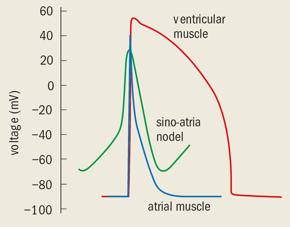
Every cell in the cardiac muscle can become electrically excited when the electrical perturbation exceeds a certain threshold (figure 1). Such a perturbation alters the permeability of the membrane to sodium ions, allowing these ions to flow into the cell until the membrane potential approaches the equilibrium potential for sodium: about 70 mV. At this point, potassium ions start to flow out of the cell, reducing the membrane potential to about -90 mV. The cell recovers to its resting state.
This large-amplitude excursion in the membrane potential, known as the action potential, takes place every time a signal is sent out from the sino-atrial node. It can last for several hundreds of milliseconds, during which time the cell cannot be re-excited and is said to be in a refractory state. (In contrast, pacemaker cells have an unstable resting state and maintain their own rhythm. They respond to a perturbation by changing the phase of their oscillation.)
The ohmic coupling between cells means that an excitation can rapidly spread throughout the cardiac tissue. The ionic currents across the membrane depend on voltage, which yields a nonlinear relationship between the membrane voltage and current. Changes in the potential of one cell therefore generate a local current that can perturb the potential of neighbouring cells.
This knock-on effect can be represented as a lattice of coupled excitation equations, and in the continuum limit the “excitable medium” of the heart can be represented by a partial differential equation analogous to the nonlinear cable equation proposed for nerve impulse propagation by the physiologists Alan Hodgkin and Andrew Huxley in 1952. This is similar to the reaction-diffusion equations used to describe the coupling between chemical reactions and diffusion. For the heart, the diffusion term describes the spread of voltage with distance and the reaction term describes the mechanisms that generate the membrane ionic currents.
Re-entrant excitation and spiral waves
Such partial differential equations predict the formation of travelling waves in the excitable medium. Unlike solitons, these travelling waves are asymmetric, with the rate of rise greater than the rate of fall. The wave velocity depends on the rate of wave formation and on the curvature of the medium in either two or three dimensions. Two of these travelling waves will completely annihilate each other on collision.
The propagation of excitations in heart tissue can be described in terms of travelling waves with a velocity of about 0.5 m s-1 and an amplitude given by the action potential. Since the action potential lasts for several hundreds of milliseconds, a single wave extends over a distance of about 10 cm. This means that a normal human heart is hardly large enough to contain a single wave.
However, in medical conditions associated with a reduced blood supply to the heart muscle, or when the rate at which the waves form is particularly high, the duration of the action potential and the “wavelength” of the travelling wave are reduced. This can lead to re-entrant propagation, in which the same wave of activity repeatedly passes through the same tissue. Such repeated excitation of the atria or ventricles causes them to quiver and writhe, preventing the synchronous contractions that pump blood around the body.
Such re-entrant excitation was first observed in a ring of cardiac tissue before the First World War. However, it was not until 1946 that Norbert Wiener analysed the phenomenon.
A simple way to understand re-entrant excitation is to imagine a single wave propagating around a circular obstacle. The wave repeatedly travels along the same path at a frequency given by the wave velocity divided by the circumference of the obstacle. If the radius is gradually decreased, the frequency increases until the wavefront of a new wave catches up with the tail of the previous wave. At this point the rate at which the wave travels around the obstacle cannot increase any further because the new wave cannot re-excite regions that are still recovering from the previous wave.
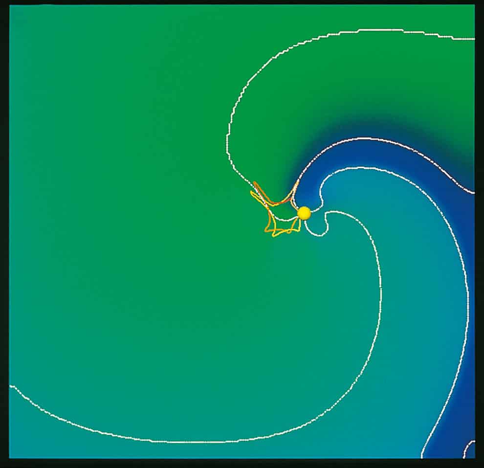
If the radius is made even smaller, the wave is forced to adopt a spiral shape that continues to rotate around a central core (figure 2). The spiral cannot enter the central core because this region is in the refractory state and cannot be re-excited.
Spiral-wave solutions to reaction-diffusion equations are generated in two dimensions by a variety of special initial conditions. They do not require an obstacle to be present, and they can be produced and maintained in media that can be considered to be entirely homogeneous. In a homogeneous medium the spiral can either rotate around a circular core, or the tip of the spiral can “meander” around the central core in a motion that can be biperiodic, quasi-periodic and perhaps even chaotic (figure 3). In an inhomogeneous medium the tip of the spiral can also drift.
Spiral waves have been observed in chemical and physical experiments, and in numerical solutions of reaction-diffusion equations, and they are beginning to be understood mathematically. Re-entrant excitation in the heart can also be idealized as spiral waves that form in the heart wall in biophysical models that assume that the walls of the atria and ventricles are thinner than the wavelength of the re-entrant wave, and so are effectively two-dimensional. Such spiral waves have a spatial extent determined by the duration of the action potential, and a rotational frequency and motion that depend on the excitation model (the equivalent of the reaction-diffusion equation) and its parameters.
Although peculiar initial conditions are required to initiate a spiral wave, once formed, a stable spiral wave will invade and take over the entire medium. This is because the frequency at which spiral-wave sources emit waves is higher than for any other nonlinear travelling-wave source. The most frequent source of waves will come to dominate the medium because they will annihilate waves that are produced less often. This means that a re-entrant arrhythmia, once initiated in the heart muscle, can prove to be lethal.
Waves and arrhythmias
Since the early 1990s a number of numerical studies based on different biophysical models of the atrial and ventricular tissue have shown that spiral waves rotate about 10 times every second. This corresponds with the main frequency component of two forms of re-entrant arrhythmia: atrial flutter and ventricular tachycardia.
Atrial flutter, so named because it feels like the heart is fluttering, is incapacitating but not life threatening. It corresponds to a stable spiral wave rotating in the atrial muscle, and it allows the ventricles to maintain adequate blood circulation. Indeed, atrial flutter might clear itself. It is possible, for example, that the spiral wave drifts upwards until the tip meets the boundaries of the great veins. These regions cannot be excited, thus preventing any further rotation of the spiral wave. If this does not happen, the wave can be eliminated with drugs or, if this fails, by using electrical shocks to regain the regular motion of the heart.
Ventricular tachycardia is associated with a re-entrant wave in the ventricles and is lethal. This is because repeated re-excitation of the ventricles by the same wave propagating around the ventricular wall results in the loss of the pumping action of the heart. Blood pressure cannot be maintained, capillary beds collapse and death follows within minutes.
These two types of arrhythmia can lead to atrial or ventricular fibrillation, in which irregular contractions take over from the rhythmic motion of the heart. Indeed, fibrillation is so severe that it causes the heart surface to look like the surface of a bag of writhing worms. Fibrillation is triggered by electrical activity in the heart, but the mechanism for the breakdown into irregularity is not yet clear.

Some biophysical models suggest that fibrillation results from the fact that two-dimensional spiral waves are not stable. As these waves break down into smaller segments, the end of each broken wave can act as the tip of a new spiral source that can itself break down. In other models the two-dimensional spiral waves are stable but their extension into three dimensions, known as scroll waves, is unstable. Probably the most important cause of fibrillation is the anatomy of the heart. The orientation of the muscle fibre, and hence the velocity of the local scroll wave, changes through 120° from one side of the heart wall to the other. This change in orientation will readily cause a scroll wave travelling through the heart wall to break down (figure 4).
These ideas are largely based on computer simulations of electrical activity, both within idealized models that assume a homogeneous and isotropic medium, and in anatomically accurate models of the heart (see Winfree in further reading). This is because there has been no direct way to observe the spatio-temporal pattern of activity on the heart’s surface, never mind within its walls. In the last few years, however, José Jalife and colleagues at the Health Science Center of State University of New York (SUNY) have used dyes sensitive to changes in voltage to monitor patterns of activity on both surfaces of isolated pieces of ventricular muscle, and they have achieved a spatial resolution of less than 1 mm and a temporal resolution of a few milliseconds. These experiments have observed re-entrant wave propagation and broadly confirm the beliefs fostered by computer simulation.
A collaboration between scientists at SUNY and Leeds University in the UK has used experiments and numerical simulations to show that the pattern of activity in the ventricle at least cannot be described by simple two-dimensional spiral waves. The results show that the activity on the epicardial surface (outside the heart) can be different from the pattern on the inner surface, indicating that the waves are essentially three-dimensional, and in most cases the patterns can be explained by small numbers of scroll waves propagating within the ventricular wall (figure 5). The presence of scroll waves in the human heart has also been inferred from clinical recordings of heart activity.
To sum up, simulations and observations now agree that ventricular tachycardia and fibrillation are caused by self-maintained re-entrant waves of electrical activity in the heart. If death is to be postponed, the only course of action is to defibrillate the ventricle immediately.
Single-shock defibrillation
During fibrillation, different parts of the heart are in different states: some are excited, some are returning to the rest state and others are ready to be excited. The re-entrant nature of the propagation maintains this irregularity: as the re-entrant excitation spreads into excitable areas, recovery processes produce further areas of excitable tissue for the excitation to spread into. To terminate this continuous process, all of the excitable tissue of the heart must be driven into the same state.

One of the most common methods of defibrillating the heart is to deliver a single large electrical shock to the heart. This dramatic piece of medical practice often works, but its physical basis is not clear. The voltage provided by the shock needs to have the same effect on different parts of the heart, but instead it decays exponentially with distance through the heart. Simple arguments on the scale of the heart suggest that the shock should excite the tissue near the negative electrode and favour recovery near the positive electrode, and so the excitation state of the cells will vary from one side of the heart to the other. Furthermore, the current flowing into the cell must be equal to the current flowing out, which suggests that the defibrillating shock has a different effect on different parts of the same cell.
There are two possible explanations for this, but we have no way of knowing which is correct. One explanation is that cardiac tissue is not a homogeneous medium but an inhomogeneous network of coupled cells. This would lead to a variation in activity that could be averaged out by the resistive coupling between the cells. Another explanation is based on the effects of the defibrillation shock on the position of the re-entrant wavefront. The shock essentially pushes the wavefronts as far forward along their direction of propagation as possible, until they are blocked by refractory tissue. The wavefronts then collapse backwards, while the wavebacks continue to move forwards. The two components of the travelling waves would then cancel each other out until all of the tissue is in a resting state.
The quantitative difference between these two pictures depends on role of the cell-to-cell coupling resistance compared with the resistance to current flow outside the cell. Neither of these parameters is known to sufficient precision even for normal tissue, and they are believed to change the conditions that lead to arrhythmias.
Despite these unknowns, defibrillation by a single large shock is widely used. It can also be incorporated into implanted defibrillators, which monitor the electrical activity of the heart and apply an electrical shock if they detect the high-frequency component of fibrillation.
Defibrillation at low amplitudes
The problem with a large shock is that it is damaging and painful, so there is considerable interest in reducing the intensity of the defibrillating shock. This could be achieved by altering the waveform or timing of the shock.

One possible approach, which exploits the mathematical physics of spiral waves, is to apply a series of small-amplitude perturbations at particular times. These times are determined by the time when a spiral wave arrives at a particular recording site. A stable spiral wave will respond to each perturbation by undergoing a small displacement by changing its phase (figure 6). Repeated perturbations at the same phase could direct this drift in the spiral wave source towards the boundaries of the cardiac tissue, at which point it can be extinguished. Numerical simulations by the Leeds group show that the spiral wave source can be moved by 0.5 cm s-1 using perturbations that provide about 10% of the electrical energy given by a single-shock defibrillation, and so could defibrillate a heart within a few tens of seconds. However, problems could arise from localized inhomogeneities that can interact with the spiral waves or prevent them from drifting altogether.
An alternative approach to defibrillation is to extend the linear components of the meandering motion of a spiral wave by using drugs to change the parameters of the excitation equation. Spiral waves would then be more likely to reach the boundary of the cardiac tissue, causing them to be extinguished. This approach is still at an exploratory stage, and numerical studies based on biophysical models of cardiac tissue are now investigating the factors that produce meander.
Waves in practice
The theory of wave propagation in cardiac tissue has developed from little more than a coupled system of differential equations describing the nonlinear responses of single cells. Yet these partial differential equations are pushing our understanding of cardiac arrhythmias beyond what can be revealed with experimental techniques alone. The hope is that this theory can now be exploited to develop better methods for treating cardiac arrhythmias.
What revolution?
Physicists tend to shy away from philosophy (and religion, although there are some exceptions). Any dealings with philosophy – other than those concerning quantum theory – tend to be superficial allusions to Karl Popper’s notion of falsification or to the “paradigm shifts” championed by Thomas Kuhn in his book The Structure of Scientific Revolutions . Put simply, Popper said that theories can never be proved right, only wrong, and that any new theory worthy of the name should make new predictions that can be tested by experiment. Kuhn described science as a cyclic process in which periods of normal science are interspersed with revolutions in which the paradigm of the time is replaced by a new one. An example is the shift from the classical to the quantum paradigm.
Physicists are, by and large, happy with both these notions in the simplistic way expressed here. Falsification certainly happens and paradigms do shift from time to time. However, Steven Weinberg, the Nobel prize winning theoretical physicist, has looked more closely at Kuhn’s work and, in a stylish essay entitled The Revolution That Didn’t Happen, he concludes that Kuhn was wrong about many points.
Weinberg’s assault on Kuhn has been inspired, at least in part, by the recent controversy between various scientists, mostly physicists, and certain sociologists of science on the meaning of “truth” and “reality” in science. It is not so much the basic notion of a paradigm shift that Weinberg objects to, but two of the boundary conditions that Kuhn has attached to it and the consequences that follow from these conditions. Weinberg explains at length how the first condition – that different paradigms are “incommensurable” – is wrong. What concerns him more, however, is Kuhn’s claim that “we may have to relinquish the notion… that changes of paradigm carry scientists and those who learn from them closer and closer to the truth”. As Weinberg writes: “It is just these conclusions that have made Kuhn a hero to the philosophers, historians, sociologists, and cultural critics who question the objective character of scientific knowledge, and who prefer to describe scientific theories as social constructions, not so different from democracy or baseball.”
Weinberg views theories as having a “hard” (as in durable, not difficult) part, such as Maxwell’s equations, and a “soft” part within which the theory is interpreted, for example the ether in the days of Maxwell. Science essentially involves building on the hard part of the theory while revising or changing the soft part as necessary. “The changes in the soft part of scientific theories also produce changes in our understanding of the conditions under which the hard part is a good approximation, ” writes Weinberg. “But after our theories reach their mature forms, their hard parts represent permanent accomplishments. If you have bought one of those T-shirts with Maxwell’s equations on the front, you may have to worry about it going out of style, but not about its becoming false. I can’t see any sense in which the increase in scope and accuracy of the hard parts of our theories is not a cumulative approach to truth.”
Weinberg concludes: “The birth of Newtonian physics was a mega-paradigm shift, but nothing that has happened in our understanding since then – not the transition from Newtonian to Einsteinian mechanics, or from classical to quantum physics – fits Kuhn’s description of a paradigm shift.” How philosophers and others respond to Weinberg remains to be seen – his last appearance in this debate provoked a large reaction – but his attempt to overturn one of the most influential theories in the philosophy of science could result in the paradigm shift to end all paradigm shifts.
Stars hint at Shakespeare’s identity
The foundation of Altschuler’s argument is that although the plays refer to many astronomical events that occurred before de Vere’s death in 1604 – such as the supernova in 1572 and the Earth’s magnetic field, first proposed in 1600 by William Gilbert – there are no references to any such events after his death. William Shakspere, on the other hand, lived until 1616 – but none of the five plays dated between 1609 and 1613 mention major astronomical events that occurred in 1604, 1609 and 1610. The dating of plays after de Vere’s death does not rule out the possibility that he wrote them: the standard dating for Shakespeare’s plays is based on when they were first performed, not when they were written or published. Indeed, many were not published until 1623 – long after both de Vere and William Shakspere had died.
Altschuler also argues that several of the plays – such as Hamlet and Henry VI Part I – refer to a comet that would have been visible from Britain of 1577, but none refer to Halley’s comet, which would have been visible in 1607. Further evidence comes in the form of references to the strange behaviour of Mars in Henry VI Part I, but the absence of any mention of Kepler’s laws of planetary motion, which were published in 1609, or the invention of the telescope and Galileo’s observations of our nearby planets.
Copyright © 2026 by IOP Publishing Ltd and individual contributors