Advances in in silico trials of medical products: evidence, methods and tools

Want to learn more on this subject?

In silico trials (ISTs) for medical drugs and devices have gained increased popularity as cost-effective alternatives to their clinical counterparts. ISTs promise dramatic reductions in the resources needed for assessing novel technologies and for generating evidence in support of regulatory evaluation for safety and effectiveness. Some have suggested significant cost reductions comparing an all in silico approach versus an equivalent clinical trial with humans. Others have argued for, and reported on, incremental implementation of the in silico methodologies that complement or refine the design of clinical trials based on predictions from the in silico trial outcomes.

This webinar has been launched in collaboration with a special issue in Progress in Biomedical Engineering, a new high-impact Reviews journal from IOP Publishing. The special issue is still welcoming article proposals from the community, and participants are encouraged to discuss contributions with the guest editors.

Want to learn more on this subject?

Alejandro Frangi, University of Leeds, UK and KU Leuven, Belgium. Alex is Diamond Jubilee chair in computational medicine and Royal Academy of Engineering chair in emerging technologies at the University of Leeds, Leeds, UK, with joint appointments at the School of Computing and the School of Medicine. He directs the CISTIB Center for Computational Imaging and Simulation Technologies in Biomedicine. He is Turing fellow of the Alan Turing Institute. Alex is the scientific director of the Leeds Centre for HealthTech Innovation and director of Research and Innovation of the Leeds Institute for Data Analytics.

Aldo Badano, US FDA, USA. Aldo holds a senior biomedical researcher service appointment at FDA and currently serves as director of the Division of Imaging, Diagnostics, and Software Reliability, OSEL/CDRH. His interests are in the characterization and modeling of medical imaging acquisition and visualization systems. Aldo is a fellow of SPIE, AAPM, and AIMBE.


Panelists

Marc Horner, ANSYS, USA
Marc Horner is a senior principal engineer, leading technical initiatives for the healthcare industry at Ansys. Marc joined Ansys after earning his PhD in chemical engineering from Northwestern University in 2001.

Abhinav Jha, Washington University in St Louis, USA
Abhinav K Jha is an assistant professor of biomedical engineering, radiology, and electrical and systems engineering at Washington University. His research interests are in developing task-specific computational medical imaging solutions.

Rebecca Shipley, UCL, UK
Becky Shipley is professor of healthcare engineering at UCL, and director of the UCL Institute of Healthcare Engineering, which brings together engineers, medical and clinical scientists to develop medical and digital technologies. Her research interests lie in mathematical and computational modelling in medicine and biology with an emphasis on multidisciplinary approaches which integrate data from biological experiments, medical imaging and patients.

Ed Margerrison, US FDA, USA
Ed is the director for the Office of Science and Engineering Laboratories at the Center for Devices and Radiological Health, US FDA. The Office is responsible for providing technical expertise and analyses in support of the regulatory processes within CDRH. In addition, the c300 scientists and engineers engage in representing the Agency on International Standards organizations, provide scientific guidance for policy, and “futureproof” the Center for technologies making their way into novel medical devices.

Puja Myles, MHRA, UK
Puja is director of Clinical Practice Research Datalink (CPRD), a real-world data research service provided by the Medicines and Healthcare products Regulatory Agency (MHRA). Since 2018 she has been working on the development of high-fidelity synthetic data, used to support the regulation of machine learning algorithms in healthcare.

Ehsan Abadi, Duke University, USA
Ehsan Abadi is an imaging scientist at Duke University. His research focus is in quantitative imaging and optimization, CT imaging, lung diseases, computational human modelling, and medical imaging simulation.

Ilse Jonkers, KU Leuven, Belgium
Ilse Jonkers leads research on the quantification of joint loading, using multi-scale modelling-based analysis to understand the effect of pathological movement on cartilage degeneration. She is also the director of iSi Health, the KU Leuven Institute of Physics-based modelling for in silico health.

Blanca Rodriguez, Oxford, UK
Blanca Rodriguez is professor of computational medicine, Wellcome Trust senior research fellow and head of the Computational Biology and Health Informatics Theme at the University of Oxford. Her team develops human-based interdisciplinary methodologies to accelerate medical therapy development, through industry collaborations.

About this journal

Progress in Biomedical Engineering is a new interdisciplinary journal publishing high-quality authoritative reviews and opinion pieces in the most significant and exciting areas of biomedical engineering research.

Editor-in-chief: Metin Sitti, Max Planck Institute for Intelligent Systems, Stuttgart, Germany

 

Attosecond electron pulses are claimed as shortest ever

The shortest electron pulses ever made in the lab have been claimed by researchers in Germany. By firing ultrashort laser pulses at a tungsten nanotip, the team created electron pulses just 53 attoseconds (53×10–18 s) long and then characterized the pulses using a new technique.

When a short, intense laser pulse is fired at a material it can cause the emission of an extremely short pulse of electrons. These electrons are then driven back into the material before re-emerging – a process that can provide important information about the material’s properties. The challenge in doing such experiments is knowing how long the electron pulses are and characterizing the electrons that are emitted.

Now, Eleftherios Goulielmakis and colleagues at the University of Rostock have developed a new technique to create and study ultrashort electron pulses. In their experiment, an intense laser pulse is fired at a tungsten nanotip that is just 70 nm in diameter at its apex. This causes the emission of a pulse of electrons from the tip. Crucially, the laser pulse is so short that it comprises less than one cycle of the light (about 2 fs). This ensures that the emitted electron pulse is not buffeted around by an oscillating electric field, but instead the electric field gives the electrons a precisely controlled kick in the right direction.

Time-varying shift

After being ejected, the electrons are immediately drawn back to the nanotip, and then backscatter off its surface. This imprints the electron pulse with information about the structure and dynamics of the nanotip. In the next the step of the experiment, the re-emerging electrons are hit with a second laser pulse that is much weaker than the first. This introduces a time-varying shift, or “chirp” in the energy of the pulse, which allows the team to better characterize the pulse using an electron spectrometer.

At the highest peak intensity used for the driving laser pulse, about 1000 electrons were released from the nanotip. The team also found that the electron pulses endured for just 53 as, making them the shortest electron pulses to be made and characterized in the lab. The experiment also marks the first time that the time-varying profile of an attosecond electron pulse has been determined accurately.

The new technique could have exciting implications for science – particularly in electron microscopy. With the ability to probe matter at both picometre spatial scales and attosecond timescales, the technique could open a new window into the nanoworld.

Beyond its research applications, Goulielmakis and colleagues hope their discoveries could pave the way for ultrafast microcircuits in which electron pulses travel directly through a vacuum rather than in conducting wires. This could lead to new types of electronic devices that operate thousands of times faster than conventional electronics.

The research is described in Nature.

The chatbot revolution: how physicists are using large language models in academia

In this episode of the Physics World Weekly podcast, we explore the use of large language models (chatbots) in physics. Our guest is the theoretical physicist Matt Hodgson, who uses chatbots both as a teaching tool and as an aid in writing computer code.

Based at the UK’s University of York, Hodgson points out that the physics community has been late to the game when it comes to chatbots. While he believes that the artificial-intelligence systems are having a mostly positive effect on academia, he says that we should be aware of potential downsides of chatbot use – particularly in the early stages of undergraduate education.

New technique produces colour X-ray images quickly and efficiently

A new technique produces X-ray images in colour quickly and efficiently using a specially-structured device called a Fresnel zone plate (FZP). The technique could have applications in nuclear medicine and radiology, as well in non-destructive industrial testing and materials analysis.

X-rays are frequently used to determine the chemical composition of materials thanks to the characteristic “fingerprint” of fluorescence that different substances emit when exposed to X-ray light. At present, however, this imaging technique requires focusing the X-rays and scanning the whole sample. Given the difficulty of focusing an X-ray beam down to small areas, especially with typical laboratory X-ray sources, this is a challenging task, making images time-consuming and expensive to produce.

Single exposure and no need for focusing and scanning

The new method, developed by Jakob Soltau and colleagues at the Institute for X-ray Physics at the University of Göttingen, Germany, allows an image from a large sample area to be obtained with just a single exposure, while doing away with the need for focusing and scanning.

Their approach uses an X-ray colour camera and a gold-plated FZP placed between the object being imaged and the detector. FZPs have a structure of opaque and transparent zones that are often used to focus X-rays, but in this experiment, the researchers were interested in something else: the shadow the FZP casts on the detector when the sample is illuminated.

By measuring the intensity pattern that reaches the detector after passing through the FZP, the researchers gleaned information on the distribution of atoms in the sample that fluoresce at two different wavelengths. They then decoded this distribution using a computer algorithm.

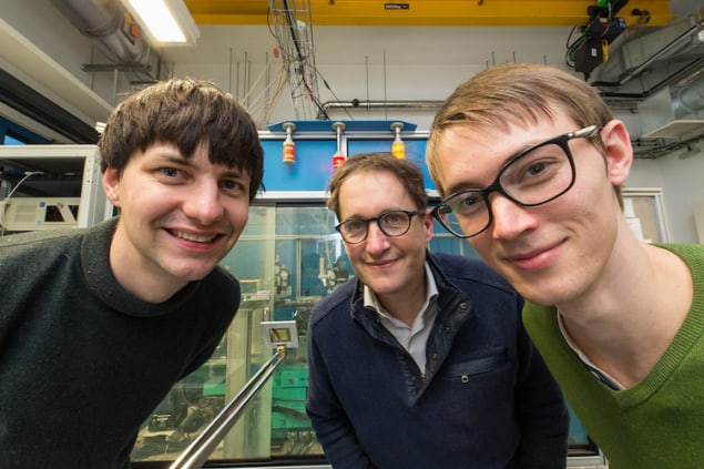
Jakob Soltau, Tim Salditt and Paul Meyer in the laboratory where they carried out this research

“We know the set of algorithms that can favourably be used for this very well from phase-retrieval in coherent X-ray imaging,” Soltau explains. “We apply this to X-ray fluorescence imaging using the X-ray colour camera in our experiment to distinguish between the different energies of the detected X-ray photons.”

Thanks to this full-field approach, the researchers say that just one image acquisition is enough to determine the chemical composition of a sample. While the acquisition time is currently on the order of several hours, they hope to reduce this in the future.

Potential for imaging biological tissues

The team says the new technique has many potential applications. These include nuclear medicine and radiology; non-destructive industrial testing; materials analysis; determining the compositions of chemicals in paintings and cultural artefacts to verify their authenticity; analysis of soil samples or plants; and testing the quality and purity of semiconductor components and computer chips. In principle, the technique could also be used to image incoherent radiation sources such as inelastic X-ray (Compton) and neutron scattering or gamma radiation, which would be useful for nuclear medicine applications.

“As a research group, we are very interested in the three-dimensional imaging of biological tissues,” Soltau tells Physics World. “Combining tomographic imaging, for example, with a detector recording the transmitted X-ray beam to obtain a map of the electron density (a technique known as phase contrast propagation imaging) with our novel full-field fluorescence imaging approach would allow us to image structures and (local) chemical compositions of the sample in one scan.”

In this first demonstration of the new technique, which is detailed in Optica, the Göttingen team achieved a spatial resolution of about 35 microns and a field of view of around 1 mm2. While the number of resolution elements imaged in parallel remains relatively low, this could be increased by using a FZP with smaller zone widths or by increasing the sample area being illuminated towards larger fields of view. Another challenge will be to reduce acquisition times without increasing unwanted background noise from elastically scattered radiation.

The researchers would now like to try their technique with synchrotron radiation, which is much more intense than the X-ray light available in most laboratories. A further advantage is that synchrotron radiation consists of high-energy beams of charged particles generated using electric and magnetic fields, giving it a narrow bandwidth that should allow for higher spatial resolution and shorter acquisition times. The team has booked time on DESY’s PETRA III synchrotron beamline in June for this purpose.

Wearable ultrasound sensor provides continuous cardiac imaging

Cardiac ultrasound imager

A wearable cardiac ultrasound imaging system that can even function during a workout has been developed by researchers from the University of California San Diego. The team hopes that the postage-stamp-sized sensor, which can assess both the heart’s function and structure, will make long-term cardiac scanning accessible to a large population.

According to the British Heart Foundation, in the UK alone, 7.6 million live with a heart or circulatory disease and some 460 people die each day from the same. While cardiac diseases are the leading cause of death among senior citizens, they are also becoming increasingly common among the young.

Hongjie Hu

“The heart undergoes all kinds of different pathologies,” explains co-first author and materials scientist Hongjie Hu. “Whether it be that a strong but normal contraction of heart chambers leads to the fluctuations of volumes, or that a cardiac morphological problem has occurred as an emergency, real-time image monitoring on the heart tells the whole picture in vivid detail.”

The problem with cardiac imaging is that echocardiograms typically require highly trained technicians and bulky scanning machinery, whereas CT and PET scans can be uncomfortable for some and come with the added factor of exposing patients to radiation.

Furthermore, many issues with heart function are intermittent, or only become apparent when the body is in motion, the researchers note. Large, fixed pieces of equipment are ill-suited for long-term monitoring, and certainly cannot image moving patients.

In contrast, says project lead and nanoengineer Sheng Xu, the new sensor can be worn for 24 hours in one go, enabling “anybody to use ultrasound imaging on the go”.

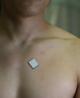
Central to Xu and colleague’s system is a wearable, stretchy and adherent patch – 1.9 × 2.2 cm in size, just under a millimetre in thickness and as soft as skin – that emits and receives ultrasound waves. The patch images the structure of the heart in real time and with high spatial and temporal resolution.

Furthermore, artificial intelligence algorithms built into the device allow it to determine how much blood the heart is actually pumping – a vital measurement, as failure to pump enough blood is often at the root of many cardiovascular diseases.

Wearable ultrasound sensor

The design of the patch makes it ideal for use on bodies in motion. As co-author and nanoengineer Xiaoxiang Gao notes, it can be attached to the chest with minimal constraint to the subject’s movement, even providing a continuous reading of cardiac activities before, during and after exercise. At present, the patch needs to be connected to a computer via cables, but the researchers say they have developed a wireless circuit to remove this limitation.

“The increasing risk of heart diseases calls for more advanced and inclusive monitoring procedures,” says Xu. “By providing patients and doctors with more thorough details, continuous and real-time cardiac image monitoring is poised to fundamentally optimize and reshape the paradigm of cardiac diagnoses.”

Cardiovascular imaging expert Alastair Moss of the University of Leicester, who was not involved in the present study, says that the system has the ability to “transform” the way doctors monitor and treat heart disease among high-risk patients. “High-quality imaging can help save lives for people with heart disease,” he says. “It is astonishing to think that we will be able to take echocardiography outside of traditional healthcare settings and place it directly in the hands of patients.”

Steffen Petersen – a cardiologist at Queen Mary University of London, who was also not involved in this study – agrees, lauding in particular the patch’s ability to provide continuous data feeds during daily activities. He adds: “The potential for such technology in cardiology is huge and assessing cardiac structure and function in such a way will lead to further inventions and clinical indications.”

With their initial study complete, Xu and colleagues are looking to improve the sensor design, miniaturize its power system, and generalize the deep-learning model so it can be used by a larger population of patients. They will also be moving to commercialize the technology – via their start-up Softsonics – over the coming years, with the expectation that a single unit will likely cost around $80 (£66).

The study is described in Nature.

Innovation underpins novel biophysics research

The 67th Annual Meeting of the Biophysical Society aims to provide a dynamic forum for researchers who are working at the interface between the life, physical and computational sciences. Running from 18 to 22 February in San Diego, US, the meeting offers a valuable opportunity for attendees to share their latest research findings while also learning about the emerging techniques and applications being developed by scientists from all over the world.

“The last few years represent an epochal moment for science in human history,” comment programme chairs Baron Chanda and Janice Robertson, both from Washington University in St Louis. “Basic science discoveries were translated into treatments for human diseases at an astonishing pace, and advances in biophysics at various levels has been central to these developments.”

The conference, which is expected to attract upwards of 5000 delegates, combines technical symposia and interactive daily poster presentations with programmes focusing on career development, education and science policy. “This meeting will showcase incredible breakthroughs in new drug developments and protein structure prediction,” continue Chanda and Robertson. “Other symposia will highlight the emerging complexity of cellular membranes, RNA and genomic organization.”

Scientific workshops during the event will feature developments in computational drug discovery, and spectroscopic and microscopy approaches for high-throughput research. The Saturday Subgroup symposia enable attendees to meet within their scientific communities, while social activities throughout the event allow more informal networking with friends and colleagues.

Meanwhile, the technical exhibit offers an opportunity for delegates to explore the latest innovations in equipment and techniques. Some of the vendors are also hosting presentations to explain how their solutions can be used for biophysical studies – read on to find out more.

Precision instruments enable biophysical research

For 25 years Mad City Labs has provided precision instrumentation for biophysicists, including nanopositioning systems, micropositioners, atomic force microscopes (AFMs) and RM21 single-molecule microscopes. For a start, the company’s piezo nanopositioners feature PicoQ sensors, which combine ultralow noise with high stability to yield sub-nanometre precision. Their stability makes them ideal for biophysics and microscopy applications, and they also provide the ideal building blocks for nanoscopy when paired with the company’s micropositioners.

Mad City Labs

AFMs from Mad City Labs achieve atomic-step resolution while also being affordable and fully customizable with automated software and calibration, and for full flexibility all of the company’s nanopositioners and micropositioners are compatible with its AFM instruments. Meanwhile, the RM21 single-molecule microscope offers high stability, precision alignment and direct access to the optical pathway, plus they can be used with nanopositioners to enable advanced nanoscopy methods.

Also available is the MicroMirror total internal reflection fluorescence (TIRF) microscope, a multi-colour instrument that combines an excellent signal-to-noise ratio with efficient data collection. An array of options are available to enable the microscope to be used for different single-molecule techniques. A further addition to the company’s product range is the Mad-Deck XYZ stage, an automated stage platform that is ideal for Interferometric scattering microscopy.

During the Biophysical Society meeting Mad City Labs will host a presentation to showcase research that has been enabled by the company’s microscopy solutions. “Open & Flexible Microscopy Systems for Combined Single Molecule Methods” will run on Sunday 19th February, 1:30 – 3:00 pm, and will feature three talks by leading academics:

  • “Quantification of Lipid Diffusion Dynamics on Live Cell Membranes through Interferometric Scattering Microscopy” by Francesco Reina of the Leibniz Institute for Photonic Technologies and the Friedrich Schiller University Jena, Germany
  • “A Single-Molecule View on Dynamic Chromatin Access of Epigenetic Regulatory Factors” by Beat Fierz at the École Polytechnique Fédérale de Lausanne, Switzerland
  • “Magnetic Tweezers Investigations of the Type 1A Topoisomerase of Mycobacterium Smegmatis” by Maria Mills at the University of Missouri, US

More details and full abstracts are available online.

  • Find out more by visiting Mad City Labs at booth 508

Confocal microscope simplifies time-resolved imaging

Quantitative time-resolved fluorescence techniques such as fluorescence-lifetime imaging microscopy (FLIM) are increasingly being used in cell biology to monitor processes such as phase separation, conformational changes, and the interactions between proteins. So far, however, expert knowledge has been needed to obtain accurate and reproducible results from these tools, which has slowed down their widespread adoption.

The Luminosa confocal microscope

PicoQuant’s new Luminosa confocal microscope overcomes that challenge by combining state-of-the-art hardware with cutting-edge software to deliver high-quality data while also simplifying daily operation. The software includes context-based workflows to improve the reproducibility of experiments, while features such as sample-free auto-alignment and calibration of the excitation laser power make experiments more efficient. At the same time, every optomechanical component is fully accessible to enable the development of new methods.

Application specialists Marcelle König and Evangelos Sisamakis will present two use cases during the meeting. First, they will show how the instrument can make it easier for researchers to use single-molecule fluorescence resonance energy transfer (smFRET) in their experimental studies. For example, the FRET efficiency and stoichiometry can be calculated during the experiment, corrected according to standard protocols, and displayed in real time.

Second, they will describe how Luminosa can be used to streamline FLIM experiments. The instrument’s rapidFLIM module can record images at several frames per second with high photon-count rates, which the software handles with a novel dynamic binning format to enable high-speed automated analysis of FLIM images. Join their talk on Sunday 19 February, 12:30 – 2:00 pm, in Room 10.

  • For a live demonstration of the Luminosa confocal microscope, and to find out about other innovations from PicoQuant, visit booth 309.


Compact mass spectrometer could search for life on distant moons

A compact prototype instrument that could one day search for signs of life in the solar system has been unveiled by a team of researchers in the US, France, and Germany. Led by Ricardo Arevalo at the University of Maryland, the team’s Orbitrap LDMS instrument can non-invasively identify complex molecules, despite taking up just a fraction of the size and weight of commercially available counterparts.

The prospect of finding evidence of life existing elsewhere within the solar system is a major driving factor for future space missions. In the coming decades, scientists hope to search for evidence of biological processes in extraterrestrial environments including the vast subsurface oceans of Europa and Enceladus – moons of Jupiter and Saturn respectively.

Key to the success of these missions will be the ability to detect a variety of biomarkers: including proteins, isotopic ratios and complex structures that indicate microbial activity. Crucially, missions must be able to distinguish larger, more complex organic molecules such as proteins from smaller, less useful signatures like amino acids – which can still be assembled by non-biological reactions.

Micron scale analysis

The instrument is called Orbitrap LDMS and employs a technique called laser desorption mass spectrometry (LDMS). This uses a pulsed ultraviolet laser to ionize small fragments of solid samples. An important benefit of LDMS is that the laser light can be tightly focused on the surface of a sample, allowing different components of the sample such as grains, dust particles and other tiny structures to be analysed at the micron scale. LDMS also minimizes the contact between the instrument and the sample, thereby reducing the chances of sample contamination.

After the ions are created, they are directed to an Orbitrap analyser, which is a mass spectrometer that was invented in the 1990s by team member Alexander Makarov, who is now at Thermo Fisher Scientific in Germany. This instrument traps the ions in orbits around a spindle-like electrode. The motions of the ions are tracked, and this information is used to determine the masses of the ions. This mass data can then be used to determine the nature of the molecules.

Optimized for space

Both LDMS and Orbitrap are established technologies, but they had to be combined in a way that is appropriate for use on space missions – minimizing mass, volume and power requirements.

Now, after eight years of development, Arevalo’s team has unveiled a prototype for a miniaturized Orbitrap LDMS. Measuring just a few centimetres in size, the prototype was able to detect target biosignature molecules at appropriate densities for studying the subsurface oceans of Europa and Enceladus. As the researchers hoped, the quality of this analysis matched the performance of existing commercial systems.

Arevalo and colleagues hope that Orbitrap LDMS will be included aboard future interplanetary space missions. These include NASA’s proposed Enceladus Orbilander mission, which could reach the surface of Enceladus in the early 2050s. In the shorter term, it could also be employed by missions like the NASA Artemis programme, to deepen our knowledge of the chemical make-up of the lunar surface.

The research is described in Nature Astronomy

Visible-light lasers shrink to chip scale

Illustration of the integrated laser platform showing different colours of visible light emerging from a single chip

Researchers in the US have created the first high-performance, tuneable and narrow-linewidth visible-light lasers that are small enough to fit on a photonic chip. Developed by a team at the Columbia University School of Engineering and Applied Science, the new lasers operate at wavelengths shorter than the red part of the electromagnetic spectrum and could be employed in technologies such as quantum optics, bioimaging and laser displays.

“Until now, lasers with performance similar to ones we have developed were benchtop-sized and expensive, which made them unsuitable for high impact technologies such as portable atomic clocks and AR/VR [augmented reality and virtual reality] devices,” explains Mateus Corato Zanarella, a member of Michal Lipson’s nanophotonics group at Columbia. “In our work we show how we can use integrated photonics to drastically shrink the size of complex laser systems.”

Integrated photonics has already revolutionized the way we control light for applications such as data communications, imaging, sensing and biomedical devices, he adds. By routing and shaping light using micro- and nanoscale components, it is now possible to shrink full optical systems down to objects that can fit on a fingertip. Despite great advances, however, high-performance chip-scale lasers have been lacking – meaning that a key component for complete miniaturization remains out of reach.

Tuneable and narrow linewidth light of wavelengths shorter than red

Columbia’s new on-chip laser platform is the first to demonstrate tuneable and narrow linewidth light at wavelengths shorter than red, with the smallest footprint and shortest wavelength (404 nm) of an integrated laser platform. It is composed of commercial Fabry-Perot laser diodes as the light sources and a photonic integrated chip (PIC) with micron-sized silicon nitride resonators. The latter component is designed to modify the laser emission to be single-frequency, easily tuneable and narrow in linewidth through a physical process known as self-injection locking. Without this PIC, the device would emit at several wavelengths and would not be easily tuneable.

“Each laser diode originally emits impure light of different shades of a colour and we design our PIC to ‘purify’ that emission,” Zanarella tells Physics World. “When we combine the diode and the chip, the selective and controllable optical feedback provided by the PIC forces the laser to emit a single colour of high purity instead of multiple shades.”

High-end applications

The researchers say they can generate and control pure light at colours from near-ultraviolet to near-infrared in a precise and fast fashion – up to 267 petahertz/second. Such light could be employed in high-end applications such as portable atomic clocks that were previously not possible because of the of the size of the required laser sources. Other potential applications include quantum information, biosensing, underwater laser ranging (LiDAR) and Li-Fi (visible light communications).

“What’s exciting about this work is that we’ve used the power of integrated photonics to break the existing paradigm that high-performance visible lasers need to be benchtop and cost tens of thousands of dollars,” Zanarella says. “Until now, it’s been impossible to shrink and mass-deploy technologies that require tuneable and narrow-linewidth visible lasers. A notable example is quantum optics, which demands high-performance lasers of several colours in a single system. We expect that our findings will enable fully integrated visible light systems for existing and new technologies.”

The Columbia researchers now intend to turn their chip-scale laser into standalone units that can be easily deployed in practical applications. They have also filed a patent for their technology, which they describe in Nature Photonics.

From tech fear to tech leader: a step-by-step guide

We know from countless reports and surveys that women are under-represented in science, technology, engineering and mathematics (STEM). The reasons for this are manifold, and most attempts to rectify it concentrate on education in schools, particularly of younger children. Computer scientist and entrepreneur Anne-Marie Imafidon has likewise focused her outreach and leadership efforts on girls and non-binary young people through her organization Stemettes. Since 2012 more than 50,000 of them have attended technology workshops, hackathons and other free events run by Imafidon and her team.

The success of Stemettes gave Imafidon a platform to speak more widely about diversity in STEM, particularly on how to get more women into positions of power in the technology sector. It is this that she focuses on in her new book She’s in CTRL: How Women Can Take Back Tech. In it, Imafidon first addresses why STEM workplaces need to be more representative of wider society than they currently are.

The examples are numerous. Most crash-test dummies are based on an average male body, leading to unforeseen types of injury among women in car accidents. Facial-recognition technology used in large warehouses doesn’t work well with darker skin, causing Black employees to be penalized unfairly. Several health apps, meanwhile, make assumptions about menstruation that cause them to be unusable for people who have menstrual cycles that are irregular or longer than the average range (for example, sufferers of polycystic ovary syndrome), or who are menopausal.

Some of this will be familiar from other excellent titles such as Invisible Women by Caroline Criado-Perez or Inferior by Angela Saini. Imafidon’s book is different in that she explicitly argues that if a woman or person of colour had been on the tech team behind these products, such problems wouldn’t have occurred in the first place. The companies would have saved themselves money and reputational damage, and the world would have become a better place for everyone.

Getting comfortable with tech

Imafidon assumes two things about her reader: that they are female (which is fairly likely, given the subtitle), and that they are non-technical, perhaps even fearful of tech. While I doubt many readers of Physics World are scared by technology, don’t skip past this book just yet – it contains information and insights that even the most tech-savvy among us could benefit from. As Imafidon says, “It’s important to make sure that tech is not built by the few and feared by the many.”

Though I’m a decade older than Imafidon, I recognize her descriptions of having computers both at school and at home in the 1990s, when that wasn’t universal or even common. And I can see that gave me (as it did her) a familiarity and comfort with certain types of technology that isn’t ubiquitous for people in their 30s and older.

Unlike Imafidon, however, I did not pass A-level computing aged 11, and I don’t have a long roster of honorary doctorates on my CV. She is an impressive woman who has done exactly what she is encouraging her readers to do. Yet even this admirable role model experienced micro- and macro-aggressions in response to her early interest in technology, for flying against what is expected of a woman – especially a Black woman like her.

As with the early home computers, I can relate to Imafidon’s memory of schools discouraging girls from STEM. It’s in the way teaching materials are written (using male pronouns, or traditionally male interests in example text); the pupils who are called on in class, encouraged to do extra projects or simply get more of their teachers’ time; the careers and higher-education advice that is given to pupils. And the women who do make it past all that discouragement to get a STEM qualification are less likely to be hired in technical roles – or promoted to a management position in a STEM-based company – than equally qualified men.

A lifetime of discouraging comments or being ignored in technical conversations means many women have low confidence in their technical abilities, or their right to be in a technical space. This is who Imafidon is addressing. Even women working in and enthusiastic about STEM can be deterred from trying new technical solutions, or widening their technical knowledge. And Imafidon’s advice is useful for us too.

Imafidon explains how women who aren’t currently in a technical role can start engaging with technology, as well as how women already in STEM can make sure they’re holding the door open behind them for other under-represented people. She does this via practical exercises at the end of each chapter, turning her book from a series of interesting essays into a useful guide. She introduces the first of these “getting started” sections with an acknowledgement that some suggestions will be easier to follow than others. Every exercise has diversity and inclusion baked into it. They are all zero or low-cost.

However, I found some of Imafidon’s assumptions about her readership frustrating. In fact, the book almost lost me during its early chapters, as they lean hard into persuading the reader to care about, and want to engage with, technology. I’m not sure who would look at the title of this work and start reading without already being on board with the concept. Imafidon also repeats a little too often the idea that anyone can – and should – be an entrepreneur. While I agree that entrepreneurship is currently too male and white, it’s not the only option in the technology sector, and not every reader will have the opportunity, or the desire, to take that route.

Yet I’m glad I persevered. I enjoyed learning about the many female and non-binary tech leaders that Imafidon profiles, such as Tobi Oredein, who found herself excluded from her chosen specialism of lifestyle journalism. In response, she launched her own lifestyle website for Black British women, Black Ballad, but then ran into the problem that quality journalism relies on good data, and there just wasn’t data available on the subjects she wanted to cover. So she applied for grants to collect the data and made those data available online to other journalists and academics. Data collection and management is now as large a part of her role as journalism.

Physics World contributing columnist Jess Wade also gets a mention for her project creating a Wikipedia entry every day for a woman or other under-represented person in STEM. Of Wikipedia’s many thousands of profiles of people, around 80% are of men. Sadly, it took a woman to challenge this. It is likely related that 90% of Wikipedia’s voluntary editors are men. Editing Wikipedia requires free time – which, on average, women have less of than men – and a little technical knowhow.

In She’s in CTRL, the examples of women in STEM from history (or “herstory” as Imafidon styles it) are familiar but told from a slightly different angle. For example, Florence Nightingale is widely lauded for her nursing but she also made innovations in statistics and data visualization. Indeed, Nightingale was elected as the first female member of the Royal Statistical Society in 1859.

One last warning concerns the pun in the title. Imafidon repeats it frequently throughout the book, which is perhaps not unexpected given that her podcast for the Evening Standard has the equally puntastic title Women Tech Charge. Still, if you’re comfortable with a little wordplay and a hefty dose of self-improvement, there is bound to be some useful advice for you in She’s in CTRL.

  • 2022 Penguin Random House 368pp £16.99hb

Interactions between ultracold molecules controlled by physicists

A way of colliding ultracold molecules while controlling the rate at which they react has been developed by physicists at the Massachusetts Institute of Technology (MIT) in the US. Researchers at Germany’s Max Planck Institute for Quantum Optics have made a similar discovery using an different experimental technique. Their research opens new pathways for enhanced control of chemical reactions.

Chemical reactions are immensely complex, with huge numbers of atoms and molecules colliding with each other while being driven by kinetic forces. This complexity makes it very difficult to focus on reactions at the atomic and molecular level.

To get around this complexity problem, researchers can cool atoms and molecules to microkelvin temperatures to limit the possible quantum states the reactants can be in. Reactions involving these ultracold atoms and molecules can then be partially controlled using lasers or magnetic fields, providing important information about chemical processes.

One challenge in studying ultracold molecules is that they have rotational and vibrational quantum states. This makes molecules much more difficult to control than atoms, and this has prevented ultracold experiments from moving beyond simple atom–atom and atom–molecule reactions.

Feshbach resonances

Now, a team at MIT led by the Nobel laureate Wolfgang Ketterle has developed a new way of controlling of ultracold molecules. The technique uses Feshbach resonances, which occur when two colliding atoms or molecules briefly form a bound state. Feshbach resonances are widely used in the study of ultracold gases because they can be used to fine tune interactions between atoms.

Applying Feshbach resonances to ultracold atoms was pioneered by Ketterle in 1998, when he made the first ever observation the phenomenon in ultracold sodium atoms. Since then, researchers have been searching for similar resonances in collisions involving both atoms and molecules. Last year, Ketterle and colleagues used Feshbach resonances to create reactions involving sodium atoms and sodium-lithium molecules. They found that quantum interference effects related to multiple bounces between colliding particles can be constructive or destructive. This either enhances or suppresses the reactions by factors of about 100.

Now the MIT researchers have found a Feshbach resonance in collisions between pairs of ultracold sodium-lithium molecules. It occurs within a very narrow range of the applied magnetic field. When the researchers looked over a magnetic-field range of more than 1000 G, they found an increased reaction rate between molecules in a narrow 25 mG window. The team concluded that the Feshbach resonance encouraged the molecules to move into a relatively long-lived intermediate complex that in turn increased the number of molecular reactions up to 100 times.

Big surprise

Further analysis of the new data yielded a surprising discovery. Precisely at the resonance, two states of the molecule have exactly the same energy and therefore can both take part in the collision. Even though the result was unexpected, Ketterle points out that sodium–lithium is the lightest ultracold molecule being studied. As a result, it has the smallest density of states and that therefore it is highly likely that the molecule has an isolated state that is long-lived.

To understand their observations, the team developed a model that describes the resonance caused by the magnetic field and the decay of the intermediate complex into an open channel causing the molecule to disappear.

Their model is analogous to light resonating within a Fabry-Perot cavity – a device comprising two thin mirrors that will transmit light at a specific resonant wavelength. The lifetime of the intermediate complex is analogous to round-trip time that a photon spends inside a resonant cavity.

While this model explains the results, some open questions remain. For example it would be useful to know if these narrow resonances are unique to molecules with small atoms – molecules that have a lower density of states. It would also be of interest to explore whether other magnetic-field values create long-lived complexes. Undoubtedly these questions will spark a wave to excitement in the field of ultracold chemistry and could lead to new applications and physical insights.

In control

Ketterle believes that the research will prove to be important for quantum science, physical chemistry and chemistry. But he acknowledges that more work needs to be done and that without a full understanding of the resonance it is difficult to make predictions for other molecules. However, he says that his team’s observation has made it more likely that resonances and long-lived collision complexes exist in other molecules.

“The field is currently progressing towards control at the quantum level over more and more complex systems.  Our work is a step to achieve quantum control over molecular collisions and reactions and to map out more broadly the collisional properties of these molecules with the goal of finding a deeper understanding”, he tells Physics World.

Bo Zhao from the University of Science and Technology of China lauds the team’s  discovery of a magnetically tunable Feshbach resonance between ultracold ground-state diatomic molecules, adding that the work is an important advance in ultracold molecules and ultracold chemistry. He states that Feshbach resonances between molecules could lead to many new research possibilities, including the study of strongly interacting molecular gases.

The research is described in Nature. In the same issue of the journal, Xin-Yu Luo and colleagues at Germany’s Max Planck Institute for Quantum Optics describe a similar scheme for controlling the reaction rate of ultracold sodium–potassium molecules. In this research, the team used oscillating microwave radiation to create the resonance.

Copyright © 2025 by IOP Publishing Ltd and individual contributors