A lightweight, easily transportable antenna that can communicate reliably with either satellites or terrestrial devices depending on its spatial configuration could prove useful for coordinating disaster relief efforts. The helix-based device, which resembles a child’s finger-trap toy, switches between its two operating modes as it is extended and contracted and could also be deployed in space or in areas that currently lack good communications infrastructure.
Developed by researchers at Stanford University in the US and the American University of Beirut (AUB) in Lebanon, the new antenna has a mass of just 39 g and consists of counter-rotating helical strips that are connected by rotational joints and made from polymer fibre composites containing a conductive mesh. In its shortened configuration, it resembles a ring just over 2.5 cm thick and 12 cm across and can be used for targeted satellite communications. When extended, it forms a thin cylinder about 30 cm tall that sends signals in all directions, like a WiFi router. This adaptability is crucial for post-disaster search and rescue operations, explains Joseph Costantine, an electrical and computer engineer at AUB who co-led the antenna’s development together with Stanford aerospace engineer Maria Sakovsky.
When deployed, the antenna is mounted on a custom ground plane that reflects radio waves while allowing the antenna base to slide and change shape. It is stable in each of its configurations, and can be switched between them simply by pulling or pushing on it, with no need for electrical power. It can also be connected to a transceiver (for sending and receiving signals), a laptop and other electronics to make a complete package with a mass of about 1 kg. This is much less than the metallic dishes typically employed in disaster-struck areas, which weigh in at around 20 kg and require a lot of power to operate.
Lightweight and compact
The antenna’s small size and low mass mean that it might also be used in space, where fuel and cargo limitations mean that everything needs to be as light and compact as possible. It could, for example, replace multiple antennas on a satellite with a single one, Sakovsky says.
Looking forward, the researchers aim to broaden their focus to include multi-stable structures with hundreds of operating states, not just two, leveraging their properties to pioneer the development of morphing, intelligent surfaces. “These will be able to dynamically steer electromagnetic beams to alter wave polarization, for instance,” Costantine tells Physics World. “Our goal is to extend the reach of signals emitted by such antennas, enabling faster data rates and more efficient communication schemes with minimal power requirements and enhanced adaptability.”
Several (online) adaptive radiotherapy techniques have been developed to maximize healthy tissue sparing in the presence of interfractional or intrafractional motion during SBRT. Adaptive radiotherapy results in increased treatment complexity and may be susceptible to motion-delivery interplay. To guarantee the quality and safety of these treatments, motion phantoms with integrated time-resolved dosimeters are required.
Currently, available phantoms and dosimeters are often not suitable to validate adaptive treatments on an MR-linac due to a lack of MR-compatibility or a motion component. An alternative dosimeter is an MR-compatible and time-resolved plastic scintillation dosimeter (PSD). The PSD’s scintillator emits an optical photon flux proportional to the received energy when it is excited by ionizing radiation.
However, a single PSD would provide insufficient volume coverage to validate an adaptive workflow. To improve this coverage, we developed the novel MRI⁴ᴰ scintillator cassette together with IBA QUASAR (London, ON) and Medscint (Quebec City QC, Canada). This device combines radiochromic film with four PSDs. Furthermore, it seamlessly integrates with the IBA QUASAR MRI⁴ᴰ Motion Phantom, providing simultaneous spatial, temporal, and motion-included dosimetry.
In this webinar, we will demonstrate the suitability of the HYPERSCINT RP-200 scintillation dosimetry research platform in a 1.5 T MR-linac. Then we will also show the performance of the new commercially available MRI⁴ᴰ scintillator cassette.
Prescilla Uijtewaal is a final-year PhD candidate at the University Medical Center Utrecht (UMCU) in the Netherlands under the supervision of Dr Martin Fast. She obtained her master’s degree in biomedical engineering at the Technical University Delft. In her current work, Prescilla investigates the feasibility and dosimetric benefits of MR-guided MLC tracking on the MR-linac. Additionally, she focuses on the validation of online adaptive radiotherapy workflows by developing and testing a PSD-based QA device. Her work is published in renowned, peer-reviewed, international journals. She also presented on MR-guided MLC tracking and dosimetry-focussed work at recent ESTRO and AAPM meetings.
Computer simulations and laboratory experiments done in Switzerland have advanced the design of a new type of positron source that could be used in next-generation lepton colliders such as the proposed Future Circular Collider (FCC) at CERN. Developed by Nicolas Vallis and colleagues at the Paul Scherrer Institute (PSI), the design uses high-temperature-superconductor magnets to collect positrons and focus them into a tight beam. The team says that its source could be fully operational by 2026.
Positron sources for accelerators rely on an effect called pair production, whereby a high-energy photon interacts with an atomic nuclei to create a positron and an electron. This is usually done by firing a high-energy electron beam into a dense solid target. Electrons that are deflected by atoms in the target will radiate photons, which then interact with other target atoms to create the electron/positron pairs.
Although this approach creates lots of positrons, they fly off in many directions. If the positrons are destined for use in a particle accelerator, they must be gathered and focussed into a beam. This process is very inefficient, with most positrons being lost.
Magnetic and mechanical challenges
Today, the gathering and focusing is done using electromagnets called solenoids. “However, the strength of conventional magnets, even in the multi-Tesla range, only allows a small proportion of the generated positrons to be captured,” Vallis explains. “Moreover, their mechanical implementation is in conflict with the target, keeping it away from its optimal location inside the magnetic field.”
Building better positron sources is a goal of physicists and engineers working on designs for future lepton colliders. These include the International Linear Collider and a version of the FCC called FCC-ee, which would collide positrons with electrons. The PSI Positron Production, or P-cubed experiment is one such design effort.
“One of the challenges we face is to produce, capture and transport positrons in sufficiently high quantities to achieve the desired luminosity,” Vallis describes. “P-cubed addresses this problem and proposes a new positron source and capture system with the potential to enhance the current positron yield by an order of magnitude.”
Latest advances
The team’s approach is based on the latest advances in solenoids made from high-temperature superconductors (HTSs). These can generate much higher magnetic fields than solenoids that use conventional conductors.
In their latest research, Vallis and colleagues describe how their prototype positron source will be implemented at PSI’s SwissFEL X-ray free-electron laser. Pulses from SwissFEL will accelerate bunches of electrons towards a solid target that will be surrounded by the new HTS solenoid. The solenoid’s magnetic field will then focus positrons into two successive RF cavity accelerators to create a positron beam
In addition to the solenoid’s strong magnetic field, Vallis says “its mechanical design allows for a full immersion of the target in the magnetic field, enabling optimal conditions for positron capture”.
Further improvements
With this set-up, the researchers could also examine how other components could help to improve positron yields. These include large aperture accelerating cavities, and novel arrangements of detection instruments. The P-cubed experiment is currently being installed at SwissFEL and it should begin operation in early 2026.
“If the experimental findings live up to our expectations, P-cubed will demonstrate a new positron source and capture system that outperforms the efficiency of its predecessors by an order of magnitude,” Vallis says. “On top of that, magnet experts at PSI have successfully run a prototype of the HTS solenoid, arguably the most critical component of the experiment, and measured a peak magnetic field of around 18 T.” In comparison, the strongest continuous magnetic field ever created in the lab is a little over 45 T.
“P-cubed will be one of the few positron sources suitable for particle accelerators worldwide, and a unique facility in Europe, so we want to develop its full potential and innovative capacity,” he says. “For instance, we will test a range of novel ideas, such as the use of crystal and conical targets for a further enhanced positron production.”
The Biophysical Society Annual Meeting is the largest annual gathering of biophysicists from around the world. Bringing together leading scientists who work at the interface between the life, physical and computational sciences, BPS 2024 allows attendees to share their latest findings and learn about the newest emerging techniques and applications.
The meeting runs from 10 to 14 February, in Philadelphia, PA, with some 5000 delegates expected to attend. The event incorporates symposia, workshops, poster presentations and the Biophysical Society Lecture, as well as career, education, policy and social programmes. Subgroup symposia held on the first day allow attendees to meet and interact within focused areas.
In a new feature for this year, more than 500 presentations have been selected from submitted abstracts. “At BPS 2024, our Annual Meeting gets a facelift,” explain programme chairs Ibrahim Cissé, from the Max-Planck Institute of Immunobiology and Epigenetics, and Elizabeth Villa from the University of California, San Diego. “The symposia and workshops are as exciting as ever, with a slate of invited speakers that represent breakthrough biophysics research and who will give a glimpse into what the next generation of our Society looks like. For the first time, in this upcoming meeting we have reserved at least 20% of symposia talks for speakers selected directly from submitted abstracts.”
There’s also a technical exhibition, showcasing displays of new equipment, publications and products. Some of the exhibiting companies are also running hands-on demonstrations of scientific products and their uses. Read on to find out more about some of the products on show at BPS 2024.
MadAFM lines up for life sciences applications
New for 2024, Mad City Labs introduces MadAFM, a sample-scanning atomic force microscope (AFM) that supports multiple microscopy modes for applications in life sciences. Joining the company’s existing line-up of AFM instruments, the MadAFM is simple to install and features a compact table-top design. The microscope exploits the company’s closed-loop nanopositioning systems to enable precise movement of the sample and probe.
These piezo nanopositioners feature the company’s proprietary PicoQ sensors, which provide ultralow noise and excellent stability to yield sub-nanometre resolution. These attributes make them ideal for applications in single molecule microscopy, imaging, optical and force microscopy, and particle tracking.
Simple and intuitive The MadAFM from Mad City Labs is ideal for materials characterization and applications in the life sciences. (Courtesy: Mad City Labs)
For 25 years Mad City Labs has provided precision instrumentation for biophysical research and industry applications, including nanopositioning systems, micropositioners, single-molecule microscopes, AFMs and customized solutions.
The company’s other products include the RM21 single-molecule microscope, which offers direct optical pathway access, high stability and precision alignment. Meanwhile, the unique MicroMirror TIRF system offers multi-colour total internal-reflection fluorescence microscopy with an excellent signal-to-noise ratio and efficient data collection, along with an array of options to support multiple single-molecule techniques.
As well as offering turnkey instruments, Mad City Labs supplies standalone micropositioning products, such as optical microscope stages, compact positioners and the Mad-Deck XYZ stage platform. These devices use proprietary intelligent control to optimize stability and precision. The micropositioning products are compatible with the high-resolution nanopositioning systems, enabling users to develop solutions tailored to their applications.
Learn more about how Mad City Labs products are enabling biophysical research by attending Looking to the Future – Enabling Single Molecule Methods for Improved Health Outcomes on Sunday 11 February at 1:30 pm (room 103C). Speakers include Uri Zvi from the University of Chicago, discussing new quantum techniques to probe biological processes with nanoscale resolution, Aaron Hoskins from the University of Wisconsin, demonstrating the power of single molecule fluorescence for revealing complex RNA pharmacology, and Leigh Plant from the Department of Pharmaceutical Sciences and the Center for Drug Discovery at Northeastern University.
Discover more by visiting Mad City Labs at booth 700 or message the company on X (@madcitylabs1) to schedule an appointment to discuss your application needs.
A focus on fluorescence
The IOP Publishing journal Methods and Applications in Fluorescence (MAF) will be hosting a “Meet the Editors” session at the IOP Publishing booth (#909) in the BPS 2024 exhibition, from 2:00– 3:00 pm on Sunday 11 February. Current and prospective MAF authors will be able to discuss the journal with the publisher and members of the editorial board, including Editor-in-Chief Marcia Levitus. Visitors to the booth can also enjoy complimentary coffee and cakes courtesy of IOP Publishing.
Uniquely broad scope MAF publishes cutting-edge multidisciplinary research across all areas of fluorescence. (Courtesy: iStock/HeitiPaves and IOPP)
Methods and Applications in Fluorescence was founded in 2012, and has since become an internationally leading journal welcoming contributions on the study, application, techniques and instrumentation of fluorescence. These encompass biological, medical, chemical, material and nano research using experimental, theoretical and data analysis methods.
MAF provides a forum for original research articles, topical reviews, tutorials, technical notes and editorial perspectives in the areas of fluorescence spectroscopy, imaging, fluorescent probes, labels and materials.
The unique position of the journal at the vertex of so many different fields of study is reflected across the diverse editorial board and MAF’s three Editors-in-Chief. As such, MAF gives the community a unique place to publish truly focused cutting-edge fluorescence research, whilst also reaching a truly multidisciplinary audience.
Find out more about Methods and Applications in Fluorescence by visiting booth 909.
Hydrogen can be used as a carbon-free source of energy in a wide range of applications including home heating, transportation and industry. However, there are significant challenges that must be overcome to ensure the safe and efficient storage and transportation of the gas.
In this episode of the Physics World Weekly podcast, the materials expert Krzysztof Koziol explains why he is developing graphene-based materials and polymers to facilitate a hydrogen economy. Based at the UK’s Cranfield University, he chats about how existing national infrastructure for distributing natural gas can be retrofitted to safely carry hydrogen. Koziol also talks about his collaboration with Airbus to develop a cryogenic storage system that could lead to hydrogen-powered aircraft.
NASA’s Jet Propulsion Laboratory (JPL) has announced it will reduce its permanent workforce by roughly 8%. The move will impact about 530 people, with the lab also laying off 40 contractors. The announcement, which follows the reduction of 100 contractors last month, is due to uncertainty over NASA’s budget for the current financial year.
The US annual budget is usually set on 1 October, the beginning of the financial year. But in recent years, final agreement has gone well beyond that date. Budgeting for the present financial year has involved two so-called “continuing resolutions” that are intended to keep the government operating and paying its bills while negotiations continue.
The current resolution maintains government operations until early March. However, there is no certainty that the Democratic-run Senate and the Republican-run House of Representatives, both of which are operated with tiny majorities, will agree on a budget to even keep the government paying its bills beyond March.
Like other funding agencies, NASA has no certainty about its current budget and the present proposal includes a huge 63% cut to the Mars Sample Return (MSR) mission as compared to last year.
NASA and the European Space Agency regard the MSR as a “next critical step” in plans to explore Mars. The mission is intended to pick up samples collected by the Perseverance Rover and then return them to Earth.
“While we still do not have a [financial year 24] appropriation or the final word from Congress on our MSL budget allocation, we are now in a position where we must take further significant action to reduce our spending,” JPL director Laurie Leshin wrote in a memo to the lab’s employees. “These cuts are among the most challenging that we have had to make even as we have sought to reduce our spending in recent months.”
Supportive role
Leshin had attempted to hold off staff reductions by reducing 100 contractors, but she states that was “not enough” to make it through the reminder of the fiscal year. “[W]e must now move forward to protect against even deeper cuts later were we to wait,” she says, while adding that the layoffs would affect both technical and support areas of the lab.
Yet JPL, which is based in Pasadena, California, has support from certain members in Congress and there are hopes that a better budget allocation than currently planned may allow the lab to rehire.
“I’m hopeful in the coming weeks we can work a deal with the Administration and Congress to restore funding to the levels necessary to rehire workers and provide the kinds of scientific discovery JPL has been on the frontlines of for decades,” Judy Chu, the Democratic member of the House of Representatives whose constituency includes JPL, noted in a statement.
Meanwhile, a panel that NASA commissioned to review progress on the MSR and respond to the conclusions of the Independent Review Board will announce its findings next month.
Electrons are normally among the lightest fundamental particles, but in so-called “heavy fermion” materials, they move as if they were hundreds of times more massive. This unusual heaviness occurs because of strong interactions between conducting electrons and localized magnetic moments in the material, and it is thought to play an important role in the behaviour of high-temperature or “unconventional” superconductors.
Researchers in the US, Sweden, Spain and Germany have now synthesized a new two-dimensional heavy fermion material from a layered intermetallic crystal made of cerium, silicon and iodine (CeSiI). The new material could give scientists fresh opportunities to study the interactions that give rise to poorly-understood behaviour such as unconventional superconductivity and related quantum phenomena.
“Typically, these heavy fermion materials are intermetallic structures with strong bonding in three dimensions, but it has been known for some time that making these materials more two dimensional can help to promote the unconventional superconductivity that appears in some heavy fermion compounds,” explains Xavier Roy, a chemist at Columbia University in the US who led the new study. “We have identified heavy fermions in the van der Waals layered material CeSiI, which contains strong bonding in two dimensions but is only weakly held together in the third.”
Conduction electrons couple strongly to local magnetic moments
The researchers chose to study CeSiI, which was first synthesized in 1998, after searching crystallographic databases for materials that might host these strong interactions (known as Kondo interactions). In particular, they aimed to combine three key elements: cerium atoms, which provide a local magnetic moment; metallic conductivity, which ensures the presence of charge carriers; and a van der Waals layered structure that would allow them to exfoliate (peel off) thin layers of the material just a few atoms thick. These individual layers can then be twisted and strained, or stacked on top of other materials, to change the material’s properties.
To make CeSiI, the researchers combined cerium metal, silicon and cerium iodide and heated the ensemble to high temperature. This procedure, which they detail in Nature, generates hexagonal platelets of the desired material. “Just as we hoped, we find that the conduction electrons couple strongly to the local magnetic moments on the Ce atoms, which results in the enhanced effective mass and antiferromagnetic order at low temperature,” explains Victoria Posey, a PhD student in Roy’s lab who synthesized the material.
Team member Michael Ziebel explains that the result was possible, in part, because of a collective effort by Columbia, Brookhaven and the Flatiron Institute to engineer new properties in 2D materials. “One major challenge we had to overcome was the air sensitivity of the material, which meant we had to develop new ways to handle samples in our lab,” Ziebel says. “More broadly, establishing the presence of heavy fermions themselves can be quite challenging – there’s no ‘smoking gun’ measurement.”
The researchers now plan to substitute different atoms into the cerium, silicon or iodine sites in CeSiI to try to suppress its magnetic order and induce new electronic ground states. Then, by exfoliating the material to different thicknesses, they aim to study the effects of dimensionality on these compounds. “In parallel, we are applying the techniques we used in this work to systematically alter the properties of CeSiI at the 2D limit, something that will, hopefully, induce new quantum phenomena arising from the combination of strong electronic interactions and low dimensionality,” says Roy.
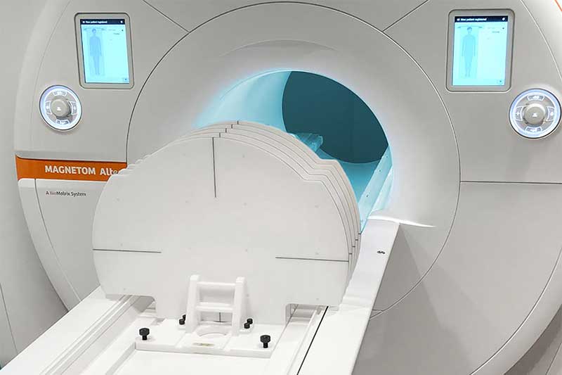
The use of Magnetic Resonance Imaging (MRI) simulation in radiation therapy leads to the need of acquiring images of high spatial accuracy and image integrity. This need has to be covered by additional radiation therapy (RT)-specific QA tasks and tools.
One of these tasks is detecting and monitoring the MR scanner’s image distortion. Covering this need, LAP and Siemens Healthineers developed the THETIS 3D MR Distortion Phantom.
This webinar introduces LAP’s THETIS 3D MR Distortion Phantom and describes ways of working with it. Together with a guest speaker from Siemens Healthineers we will show how THETIS integrates into the QA workflows of Siemens Healthineers MR scanners, covering the needs of MR imaging in radiotherapy.
SPEAKER: Torsten Hartmann is director of Product Management of the Business Unit Healthcare at LAP GmbH Laser Applikationen. He has more than 20 years’ healthcare experience, having held positions in software development, project and department leads. He joined LAP in 2006 and has since driven products in radiotherapy.
Arun Joseph
GUEST SPEAKER: Dr Arun Joseph serves as the global product marketing manager at Siemens Healthineers, overseeing the marketing of MR scanners dedicated for radiation therapy planning. With a background in biomedical engineering, Arun has maintained a profound connection with MR imaging since his days as a student and scientist. In his current role as product marketing manager, he passionately advocates for the pivotal role of MRI in radiation therapy planning.
At the Pius-Hospital Oldenburg in Germany, Dr Hui Looe and his team have used the LUNA 3D system pre-clinical on their ELEKTA Synergy LINAC and their CT since June 2023. After installing and establishing the LUNA 3D system, multiple use cases for a treatment have been tested and tried out leading to a broad user knowledge.
In this webinar, Dr Looe will present his pre-clinical experience with LUNA 3D while showing all the steps of implementation, first insights of the software and exciting (accuracy) measuring results of the system. Also, he we will focus on the combined use of SGRT on CT and LINAC.
LUNA 3D is the new SGRT system from LAP, which utilizes high-resolution stereoscopic cameras to provide precise and dose-free patient positioning and monitoring from CT simulation to treatment delivery, compatible with certain CT, bore -type and C-arm LINACs.
510(k) pending (K232031) – not available for sale in the US. Availability of products, features, and services may vary depending on your location.
Dr Hui Looe is the deputy head of medical physics at Pius-Hospital in Oldenburg, Germany, and a scientist in the Medical Radiation Physics group at the University of Oldenburg. As part of this working group, he undertakes clinical, teaching and research activities, leading the research group focusing on mathematical and computational methods in dosimetry. He also improves patient positioning using Surface Guided Radiation Therapy (SGRT). As part of these activities, he accompanies the validation and clinical implementation of the LUNA 3D system of LAP.
Tumours are prone to move relative to healthy tissue and organs-at-risk (OARs) as a cancer patient undergoes a course of radiotherapy – and can even change position during an individual treatment session. The ability of MR-guided radiotherapy (MRgRT) systems like the Elekta Unity MR-Linac to detect that target motion and adapt therapy accordingly – in effect, helping clinicians to “see what they treat” in real time – points the way to a more personalized radiation oncology tailored to the unique requirements of each patient. That end game, it seems, is edging ever closer with the clinical roll-out of Elekta’s Comprehensive Motion Management (CMM) upgrade for the Unity treatment system, with real-time tumour tracking and automatic gating as the underpinnings for online plan adaptation.
Among the early-adopting clinical customers for CMM is US-based University of Iowa Health Care. At its main radiation oncology clinic in Iowa City, this integrated cancer centre treats around 1600 patients each year using an all-Elekta suite of five external-beam radiotherapy systems: an Elekta Unity MR-Linac; three Versa HD machines (all with onboard imaging; one with HexaPOD robotic table); and a Leksell Gamma Knife Icon (for stereotactic radiosurgery of brain lesions). “We’ve been treating a range of indications with the Unity system over the past four-and-a-half years – mostly prostate, liver, pancreas as well as oligometastatic cancers,” explains Daniel Hyer, professor of radiation oncology and director of clinical physics at University of Iowa Health Care.
A catalyst for clinical innovation
Fast forward to September 2023 and the University of Iowa’s introduction of the CMM upgrade on its Elekta Unity MR-Linac – a clinical innovation that yielded immediate and significant impacts for Hyer and the multidisciplinary care team. “During our first week live with CMM,” he notes, “the system gated the beam during unexpected motion of a pelvic node. On another patient, we were able to perfectly track a target next to the heart despite cardiac and respiratory motion. Ultimately, we expect that CMM will enable us to treat many of our lung cases on the Elekta Unity system.”
It’s not hard to see why. Put simply, CMM’s motion-management features – tracking the tumour target automatically and responding to any movement in real-time – are fundamental to improving the accuracy of beam delivery and, in turn, enhancing therapeutic outcomes. “The core innovation with CMM,” notes Hyer, “is that the Unity system now actively tracks the tumour target on the live imaging and shuts the beam off automatically if the target moves outside its planned envelope of motion. These automated gating techniques can be free-breathing or when the patient is in breath-hold.”
Operationally, CMM supports three workflows to manage the treatment of tumour targets subject to periodic breathing motion. There are two free-breathing workflows (free-breathing exhale and free-breathing average) that avoid the need for the patient to hold their breath during radiation delivery – a challenging proposition for many – while predictive algorithms ensure precise motion management with virtually zero latency on target tracking.
In contrast, the breath-hold technique sees the patient coached to hold their breath while the Unity system acquires the daily 3D MR image in a single breath-hold (with automatic gating to ensure the treatment is delivered only during subsequent breath-holds). For unexpected non-respiratory motion – owing to rectal gas, say, or bladder-filling – the so-called exception gating strategy is used to track the tumour target in real-time, with the radiation being paused if the target moves out of tolerance.
In this way, CMM has already opened up new treatment pathways for the Iowa radiation oncology team, with five lung cancer cases treated on the upgraded Unity system in December alone (versus two lung patients on the MR-Linac in the preceding four years). “Previously with lung lesions,” notes Hyer, “we didn’t want to treat the entire motion envelope.” In the case of a lung tumour that moves 15–20 mm, for example, all of that motion had to be accommodated prior to CMM – which means a lot of healthy tissue being irradiated. “Now we can cut that volume down thanks to CMM,” Hyer adds. “If the patient is free-breathing, but we only want to treat a subset of that motion – say 5 mm – we can design the treatment plan accordingly and ensure the treatment beam turns on and off automatically as required.”
Another area of clinical innovation with the MR-Linac relates to the treatment of pelvic and prostate nodes. In this case, CMM enables Hyer and colleagues to rapidly shift the plan to account for systematic changes in target position that might occur during the treatment session – thereby circumventing the delay and workflow inefficiencies associated with reimaging and replanning.
“Before the introduction of CMM, we basically had to start over if the patient moved on the table,” notes Hyer. “Now, with CMM active tracking, we can do what’s called a baseline shift and recentre the treatment on the new target position.” This baseline shift plan typically takes around a minute, which means that clinicians are already becoming a lot less hesitant about tightening their margins on pelvic and prostate tumours in treatment planning.
Forward motion
So what does the CMM roadmap look like at University of Iowa Health Care through 2024? According to Hyer, preclinical testing is already underway using breath-hold sequences to enhance Elekta Unity’s anatomy-specific MR imaging of difficult-to-visualize structures like the pancreas. “We’re developing a whole strategy around breath-hold,” he says. “The imaging sequences so far have yielded exquisite visualizations of the duodenum, stomach and bowel with clear potential to help us with treatments in challenging locations like the pancreas.”
Target-tracking Breath-hold image for the pancreas generated with the Elekta Unity MR-Linac (18 s scan, balanced contrast). Automatic gating ensures radiation is delivered only during subsequent breath-holds. (Courtesy: University of Iowa Health Care)
Technical innovation is also in the pipeline, including plans to introduce real-time visual feedback to help the patient on the treatment couch see how the tumour target is lining up versus where it’s supposed to be. “Right now, we provide that feedback via audio coaching,” says Hyer, “so the next step is a visual representation on the inside of the MRI bore – an innovation that will effectively make the patient an active participant in their own treatment.”
Meanwhile, Hyer and the University of Iowa medical physics team are working closely with the two other sites involved in Elekta’s CMM pilot release: UMC Utrecht in the Netherlands and the IRCCS Ospedale Sacro Cuore Don Calabria in Negrar, Italy. “We’re collaborating on the physics side and comparing best practice on a range of issues including liver tracking,” Hyer concludes.