Researchers in Australia have detected and controlled changes to the shape of a single molecule in response to an applied mechanical force. The feat could enable the development of miniaturized implantable pressure sensors and accelerometers for the electronics industry.
The molecule in question, bullavene, is a hydrocarbon with the chemical formula C10H10. Importantly, it is piezoresistive, meaning that its electrical resistance changes in response to mechanical strain. In bullavene’s case, this strain occurs when the molecule changes between its different possible shapes, or isomers, altering the connectivity between its atoms and producing a measurable variation in its electrical resistance.
To measure bullavene’s changing resistance, the team used chemical attachments called diaryls to bind the molecule to gold contacts 7 to 15 angstroms apart. When these gold contacts move, the molecule remains bound to them, but the mechanical strain it experiences causes it to form a new isomer with a different shape. This shape change modifies the flow of electricity through the molecule, and the researchers were able to measure these changes using scanning tunnelling microscopy.
Miniature sensors and millisecond timescales
Piezoresistors are already widely deployed in a range of applications, including vibration detectors in electronic devices, pedometers in smartphones, triggers for car airbags and implantable medical sensors. Because bullavene molecules are so small, they could be used to create miniaturized versions of these conventional devices. A bullavene-based sensor could also detect the presence of other chemicals or biomolecules such as proteins or enzymes – something that might be important for detecting diseases, Darwish says.
The researchers, who detail their work in Nature Communications, say they can envisage creating devices as small as 3 to 100 nm2 that detect external forces and pressures simply by measuring changes in resistance. Another useful feature, Kosov adds, is that the piezoresistors can be made to oscillate at 800 Hz, which means they could be used to monitor processes that occurs on millisecond timescales.
The next stages in the team’s work will involve transferring the technology from an expensive microscopy experiment into a cheap sensing platform. “This will require us to develop nano-electrode sensors whose active elements are our shape-shifting molecules,” Darwish tells Physics World.
Changes to the operation of a planned Higgs factory could significantly improve its energy efficiency, but construction will have the biggest impact on the facility’s overall carbon footprint. That is the conclusion of an analysis of the potential environmental impact of the Cool Copper Collider (C3) – a proposed successor to CERN’s Large Hadron Collider (LHC).
Following the discovery of the Higgs boson in 2012 at the LHC, particle physicists are planning to build a so-called Higgs factory that would smash electrons with positrons to allow more detailed investigation of the properties of the Higgs boson and other particles.
The researchers found that, in general, linear accelerators are more environmentally friendly than circular designs. This is because their compact size simplifies construction and reduces the amount of material needed. Indeed, tunnel lengths for future circular accelerators approach 100 km in length, while the linear options are around 10 km long.
This 10-fold difference in length significantly reduces the use of concrete, which has a significant carbon footprint, but also allows for simpler construction methods. Overall, the team says that building the main tunnels for the FCC and CEPC would produce around 578 and 638 kilotonnes of carbon dioxide equivalents (CO2e), respectively, compared with 73 and 144 kilotonnes CO2e for the main tunnels of the CLIC and C3, respectively.
A new discussion
When it comes to the overall operating energy consumption of the proposed colliders, the CEPC was found to be the highest with CLIC the lowest and the other three remaining similar. This analysis was based on factors such as power consumption, expected years of operation and particle collisions per year. The researchers also estimate that dedicated renewable energy facilities, such as solar farms, could roughly half the carbon intensity of the electricity used by any future collider.
Yet SLAC particle physicist Caterina Vernieri and colleagues argue that it is also important that scientific impact is balanced against environmental cost and efforts (PRX Energy2 047001). While CLIC will have the lowest overall carbon footprint, they suggest that C3 would be the most environmentally friendly in terms of physics output. This is because it has a similar environmental footprint than CLIC but would carry out a precise determination of the Higgs bosons properties.
We need to think not only in terms of financial costs, but also environmental impact
Caterina Vernieri
The team found that the easiest way to lower the carbon impact of a future Higgs factory is to reduce its power usage. Their analysis shows that tweaks to the workings of the klystrons – responsible for creating the electromagnetic fields that drive the beams — as well as to the structure of the beam itself such as reducing bunch spacing, could reduce the energy consumption of the C3 beam, for example, by about 40%.
Vernieri says that given their huge costs and environmental impact studying the sustainability of physics projects is a new but necessary field. “We need to think not only in terms of financial costs, but also environmental impact,” she says, adding that at least there is now a “whole new discussion” that is examining the carbon footprint of particle physics.
The researchers also say it is worth noting that CERN plans to re-use the FCC tunnel once its work as a Higgs factory is complete as a high-energy hadron collider. Yet this would not necessarily be a clear-cut climate win as it would require new beamline infrastructure with its own carbon costs. The team states that a detailed lifecycle analysis would be required to determine the possible climate advantages of reusing the FCC tunnel, which is beyond the scope of this study.
Will robots take over the Earth? It’s a question that’s been asked by many sci-fi movies since the dawn of AI. We are simultaneously scared and excited by the prospect of creating robots to imitate – and do some of the work of – humans. These films follow a well-trodden path: we create the robots, they develop consciousness, they threaten to take over, we try to stop them (I, Robot, TheTerminator…). The Creator – the new film directed and co-written by Gareth Edwards – has a similar plot line, but it does add a new twist.
We find ourselves on an Earth where robots – or “synths” – have been created in our likeness and introduced into society some time before the story begins. But a nuclear-weapon detonation over Los Angeles put an end to this life of cohabitation. The West has called an all-out war on synths, which are still an integral part of the “New Asia” of the East. Our hero, Joshua (played by John David Washington), is a soldier on a mission to find and destroy the “Creator” (of the synths) and a new weapon that threatens the existence of humankind. Along the way, he finds himself in a struggle to identify who the real enemy is.
The synths are, in many ways, more human than most of the people in the movie. The “humanity” of the synths is reminiscent of the Blade Runner replicants, who became increasingly introspective as their end-date draws close. As they ask life’s big, universal questions, the synths turn to some very human concepts.
This is a big movie, with big special effects. Many of the beautiful scenes were filmed on location with effects added to them. The soundtrack is powerful and in places music is used in an unusual way to pull your focus to a particular part of the drama. Washington is hugely watchable throughout, as is newcomer Madeleine Yuna Voyles, who plays the synth Alphie.
The Creator is action-packed, edge-of-your seat stuff for most of its 133 minutes. But it’s a poignant film too, dealing with love and loss. It also argues against using a single act of violence as an excuse to commit genocide. Ultimately, though, The Creator suggests that we cannot blame technology for the problems we face in the world today – we must take full responsibility for those ourselves.
Earbuds equipped with biosensors can continuously measure the electrical activity of the brain and levels of the sweat secretion lactate. The device represents a potential new wearable sensing technology for detection and monitoring of neurogenerative diseases or long-term health monitoring.
Developed by a multidisciplinary team of engineers at the Center for Wearable Sensors at the UC San Diego Jacobs School of Engineering, the earbud sensors wirelessly transmit the recorded data to a smartphone or laptop computer for visual display and analysis. With this invention, the researchers foresee a future in which neuroimaging and health monitoring systems work with easily wearable sensors and mobile devices to track brain activity and levels of many health-related metabolites throughout the day.
The integration of individual brain and body signals into a single miniature platform represents a technological breakthrough for in-ear sensing technology. The researchers note that the sensors have a much smaller form factor, are less visually obtrusive and more comfortable to wear than a state-of-the-art scalp electroencephalogram (EEG) headset or commercial blood lactate meter, while offering similar performance.
“Being able to measure the dynamics of both brain cognitive activity and body metabolic state in one in-ear integrated device that doesn’t intrude on the comfort and mobility of the user opens up tremendous opportunities for advancing health and wellness of people of all ages, anytime and anywhere,” co-principal investigator Gert Cauwenberghs comments in a press statement.
When combined, EEG data, which reflect electrical activity in the brain, and measurements of sweat lactate, an organic acid produced by the body during exercise and normal metabolic activity, can be used to monitor effort during physical exercise, or to track levels of stress and focus. Such data can also be used for diagnosis of various seizures, including epileptic seizures.
Reporting their findings in Nature Biomedical Engineering, the researchers explain that in-ear electrophysiological sensing systems provide “elegant solutions to unobtrusive brain-state monitoring inside the ear canal”. The ear is located close to the central nervous system, major vasculature and the auditory cortex, providing access to physiological parameters such as EEG, pulse rate and oxygen saturation. It also has multiple exocrine sweat glands that enable analysis of vital metabolites.
The new device comprises two types of sensor screen-printed onto a 150 µm-thick flexible polymer substrate, which attaches around the earbuds. The sensors are designed to track daily activities using two principal sets of features that characterize brain–body health. The electrophysiological sensor implements a form of a wearable brain-computer interface in the ear to track brain state-related signals such as EEG and electrodermal activity.
The other sensor performs electrochemical analysis of metabolites in the ear (in this study, lactate in sweat). The electrochemical sensors are covered with a transparent, sponge-like hydrogel that acts as a mechanical cushion between the skin and sensors and improves sweat collection. They are spring-loaded to hold contact with the ear but adjust as the earbuds move.
To determine the optimal layout of the sensors, the researchers initially performed functional mapping inside the ear canal. Based on their findings, they oriented the electrophysiological electrodes towards the temporal lobe which has lower secretion of sweat, and the electrochemical electrodes towards the location with higher sweat secretion. This design minimizes potential crosstalk between the two sensors, which are separated by only 2 mm, and helps increase the signal-to-noise ratio. The team also customized the outline contour of the integrated ear sensors to match that of the earbud and one of three sizes of generic silicone tips.
The researchers validated the efficacy of their sensors by characterizing the electrode performance and the measured brain signal patterns. They also assessed the sensitivity, selectivity and long-term stability of the lactate sensors, verified the minimal crosstalk between sensors, and confirmed the mechanical and environmental stability of the integrated scanners.
The team also tested the tested the earbud sensors in healthy volunteers performing vigorous stationary cycling at a fixed level. The device detected elevated sweat lactate levels as well as changes in brain activity. Validating the collected data against results obtained from commercial dry-contact EEG headsets and lactate-containing blood samples revealed comparable data from both systems.
The lactate sensors currently require users to perform vigorous exercise or other activities that generate sweat. Without such exercise, sufficient lactate for analysis cannot be gathered, explains co-principal investigator Sheng Xu. The researchers plan to improve the design so that exercise will not be necessary for monitoring.
Fellow co-principal investigator Patrick Mercier advises that the team’s future plans also include creating a design to process the data on the earbuds themselves, with the goal of transmitting these processed data wirelessly to a computer or smartphone. The researchers also hope that the in-ear sensors could gather additional data, such as oxygen saturation and glucose levels.
The research could also lead to new therapies. “Auditory neurofeedback coupling the measured brain signals with sound played by the device in the ear may enable potentially far-reaching new therapeutic advances for active remediation of debilitating neurological disorders such as tinnitus,” says Cauwenberghs.
Complex interior: illustration showing the two different layers of the Martian mantle between the core and crust. Also shown is how the layers affect seismic waves detected by InSight. (Courtesy: IPGP-CNES)
Mars’ mantle is divided into a partially molten outer layer and a fully molten, silicon-rich layer that lies closer to the planet’s core. This discovery was made by two independent teams and challenges the previous view that the mantle – which lies between the Martian crust and core – has a uniform composition and structure. The new analyses used seismic data from NASA’s InSight Mars lander and could help shape our understanding of how the red planet formed and evolved.
Some of the seismic waves studied were created by meteorites impacting the planet. The waves will have travelled deep within Mars before they reached InSight’s seismometer, and studying them provides important information about the Martian interior.
“Such large epicentral distances allowed for the propagation of compressional waves that travelled in the lowermost Martian mantle as a diffracted wave,” explains Henri Samuel at CNRS in Paris, who headed one of the studies. “It was found that the propagation of these waves was too slow to be explained by a homogeneous mantle.”
Surprising abundance
The research has also provided further clues about the elemental composition of the Martian core. Previously, this had been calculated to contain a surprisingly high abundance of lighter elements, including carbon, oxygen, and hydrogen. However, these latest studies suggest that these lighter elements are not as common as had been predicted and the core is smaller and denser than previously thought.
The other study was led by Amir Khan at ETH Zurich, who explains, “This need for a large complement of these [lighter] elements posed serious cosmochemical problems, since it is difficult to imagine how Mars would have accreted such a large proportion of light elements, and sequestered them into its core”.
In their respective studies, Samuel and Khan’s teams both performed inversions of InSight’s seismic data – a mathematical technique that transforms the information into a subsurface models of a planetary interior.
Afterwards, each team took a slightly different approach to interpreting their inversions. For Khan and colleagues, this involved building up their calculations from first principles. “We computed the seismic wave speeds and density of iron–nickel light element alloys using quantum mechanics, which is completely novel for the conditions equivalent of Mars’s core,” Khan explains.
Attenuating structures
Samuel’s team went beyond considerations of density, composition, and seismic velocity and looked at how the interior structure of Mars attenuated seismic waves. “From this, we were able to infer the first attenuation structure model of Mars’s mantle based on seismological and other geophysical data,” he explains.
Yet even with these different methods, both teams came to a surprising conclusion. “Unlike the Earth, Mars appears to have a strongly stratified mantle with this enriched silicate layer above its core,” Samuel says. “The lower part of the layer is fully molten, while the thinner upper part is partially molten.”
Khan explains that his team reached a very similar conclusion. “The composition of the molten layer in our calculations is very close to that of the silicate mantle, which helps explain our finding of a slightly denser silicate layer relative to the mantle. The fact that the silicate is found to be slightly denser also explains why the layer remains stable at the bottom of the mantle.”
Despite the similarities in their results, the teams’ differing approaches allowed them to explore different implications of their discovery. For Samuel’s team, revealing the mantle’s structure in terms of attenuation allowed them to better explain the orbital path of Mars’ closest moon, Phobos.
Gravitational field
According to the team, a molten silicon layer could deform more easily under the moon’s tidal forces than would the colder, partially molten layer above. This would better explain the relationship between Mars’ gravitational field and Phobos’ orbit, while staying consistent with InSight’s measurements.
Through their own examination of Mars’ core, Khan’s team calculated that about 9–15% of its mass is made up of light elements. In terms of models of how Mars formed, this lower abundance seems more reasonable than the estimates of over 20% made in previous studies based on the assumption of a homogeneous mantle.
For both teams, the discovery marks a turning point in our understanding in how Mars first formed and evolved over the past 4.5 billion years. “With the presence of the stratification in the Martian mantle, we need to go back to re-analyse and re-interpret the roughly four year-long seismic record and all other geophysical observables in the light of this new paradigm,” Samuel says. “This could lead to additional discoveries on the deep structure of the Martian mantle and its core.”
Beyond improving our knowledge of Mars, the result could also help astronomers to gain a better understanding of rocky planets beyond the solar system. “Through acquisition of new data and new methods of analysis, we make new discoveries and keep refining and updating our current understanding of what the terrestrial planets are made of,” adds Khan. “Ultimately, this will be needed for understanding the origin and evolution of extrasolar planetary systems.”
The 2024 Rank Prize winners Left to right: Junzhong Liang, Donald Miller, Austin Roorda and David Williams. (Courtesy: Rank Prize)
Four scientists who pioneered the development of adaptive optics (AO) technologies for imaging the human retina have been awarded the 2024 Rank Prize for Optoelectronics. The winners – Junzhong Liang, Donald Miller, Austin Roorda and David Williams – invented instruments that use AO to capture high-resolution images of the living retina and provide new insight into the structure and function of the human eye.
AO was originally developed for use in astronomy, to eliminate atmosphere-induced blur in images from ground-based telescopes. It works by measuring distortions in a reflected wavefront using a wavefront sensor, and then compensating for these distortions with a wavefront corrector, which is often a deformable mirror.
In 1997, Liang, Williams and Miller demonstrated that AO can also be used to correct for distortions caused by imperfect optics within the human eye. Using AO, they created a retinal imaging camera with unprecedented resolution, enabling clear imaging of individual photoreceptor cells in the living human retina. Two years later, Roorda and Williams used this instrument to produce the first-ever images showing the distribution of the three types of cones in the human retina.
According to Donal Bradley, chair of the Rank Prize Optoelectronics Committee, the prize recognizes the winners “seminal contribution to imaging within the eye that opens new opportunities to understand this complex optical instrument and to improve eyesight through precise interventions”. Tami Freeman spoke to two of the winners to find out more.
Since its invention, how has AO impacted the field of eye imaging?
Donald Miller AO is the only technology that allows the visualization of individual retinal cells in a living eye. And because disease and pathology start at this cellular level, that’s the level we ultimately want clinicians to operate at, for earlier diagnosis and more effective treatments.
As one example from my own lab, we’ve recently been looking at the impact of glaucoma, one of the leading causes of irreversible blindness in the world, on retinal ganglion cells – the primary cell type that dies in this disease and which line the top of the retina. While effective treatments exist, the disease is unfortunately hard to diagnose early until significant damage has occurred. With AO, we can now, for the first time, monitor individual retinal ganglion cells and track them over time in these patients.
Using AO combined with optical coherence tomography (AO-OCT), we have found that, even in eyes under treatment, we see subclinical loss of cells. That’s important because clinicians can now use these cellular-level measurements to better establish whether or not their treatment is working. It also offers considerable potential for testing the efficacy and safety of new neuroprotective and regenerative strategies. The visualization of retinal ganglion cells in human subjects has only become possible within the last few years – we are entering a really exciting time.
Austin Roorda As treatments become available for the major blinding eye diseases, like diabetes, glaucoma and macular degeneration, we can now use AO to assess how effective they are. But there are other inherited retinal diseases due to gene mutations for which very little is known. In those rare diseases, previously the only way to see what was happening on a cellular scale was to wait for a donor eye and look at it under a microscope. AO has opened up the ability to examine the retina on a microscopic scale in these patients. Treatments such as gene therapy are on the horizon that could potentially cure or halt these inherited diseases. AO is poised to play a key role in that process – to understand how the mutation affects the retina, assess the state of the retina, predict the prognosis if the patient undergoes gene therapy, and then measure the effectiveness of that therapy.
How has AO technology progressed over the last 25 years?
AR AO was originally constrained by the technology that was available, which was largely developed for the field of astronomy. So the deformable mirror was big and wasn’t suited to the eye. Over the years, when companies started recognising the potential of AO in other fields, including ophthalmoscopy, they started to build wavefront sensing devices and wavefront correctors (the deformable mirror) that were a lot better suited to applications in the human eye.
DM When we first developed the AO system, we made a lot of guesses: what type of wavefront correction to use, what wavefront sensor, the loop speed and so on. In the next five to 10 years there were a lot of improvements in our understanding of the spatial properties and temporal dynamics of ocular aberrations. These then defined the AO components: how many actuators you need in your wavefront corrector, what the stroke [actuator displacement] should be, how many sampling points you need across the pupil, and how fast the AO system should go. Those have all been optimized over the years.
Step back in time David Williams, Donald Miller and Junzhong Liang around the first adaptive optics system for the eye at Rochester in 1997. (Courtesy: David Williams)
For example, the wavefront corrector we used in 1997 had 37 actuators that push and pull on the back surface of the mirror to warp its shape, and it would give four microns of stroke. The ones used today have close to 100 actuators and give an order of magnitude more stroke, which is important because the eyes have severe aberrations; that’s made a big difference.
AR Now, when you use AO, you push a button and it runs automatically at anywhere from tens to hundreds of hertz. Before, we had to take a picture, a map of the of the eye’s aberrations, and scrutinize it to make sure there weren’t any errors in the initial image analysis. Then you would push the next button to apply that shape to the mirror. So the user was an integral part of the closed-loop AO system. It was fun, but it was slow.
Initially, Don, David and Junzhong built a standard flood-illumination camera that would look at the retina through an AO system to reveal the microscopic structure. Later, I incorporated AO into a scanning system to create an AO scanning laser ophthalmoscope (AOSLO) that can record video of the retina and perform depth sectioning. That’s an entirely new AO imaging platform. Other researchers have incorporated a type of phase contrast imaging that can visualize otherwise transparent cells in the retina, and in David’s group they are performing fluorescence imaging in animal eyes.
What’s your current main area of research?
AR If there was a theme for what I’ve been doing for the last 15 years or so, it’s structure and function. It turns out that our AOSLO imager is also the world’s best eye tracker. You can track eye motion very quickly and accurately because you can see the movement of single cells in the back of the eye. We took this a step further, using the scanning laser system not only to image the retina, but to control the placement of images onto the retina on the scale of a single cone.
Structure and function Austin Roorda working on the adaptive optics scanning light ophthalmoscope (AOSLO) at UC Berkeley. (Courtesy: Austin Roorda)
We’ve been measuring functional properties in living humans. If you were in the device, I could deliver flashes of light into individual cones and ask whether you could see them or what colour you see. Early on, we mapped the cone mosaic, that was one of the big AO-enabled discoveries. Now we can take that cone mosaic and start asking questions about basic retinal circuits or the fundamental properties of human colour vision. We’re doing the same in eye disease. If we look at an array of cells in a patient and it doesn’t look normal, we’re interested in the functional consequences – not just seeing the structure of that diseased retina but asking about the visual outcomes.
DM We’re also focused on structure and function, but using AO-OCT. The big advantage of OCT is its axial resolution, which lets you section out whatever depth in the retina layer you want to visualize. Cones are very bright and high in contrast, but other cells tend to be much harder to image as they reflect a lot less light back. We’ve made quite a bit of headway using AO-OCT to image these other neurons in the retina at different depths. It was a big step to be able to image retinal ganglion cells, as they are highly transparent and have very low contrast.
We’ve also been using AO-OCT to look at function within photoreceptors. In 2000, Austin and David had developed their pioneering AO retinal densitometry method for cone classification. Twenty years later, we can use the phase information provided by AO-OCT to measure subtle changes in the elongation of these photoreceptor cells when stimulated by different colours of light. That turned out to be a much more accurate and far less time-consuming way to do cone classification and is a good example of the evolution of AO imaging technology.
How do you see the field of AO evolving in the future?
AR In my lab, we focus a lot on subjective measures of function, such as eye movements, acuity and colour vision. But I would envision that as AO techniques evolve, we’ll be able to measure functional properties of most cell classes in the retina. Right now, Don has generated beautiful images of ganglion cells using AO-OCT. These are the last cells before the signals from the retina reach the brain, so it’s a class of neurons whose function we’re very interested in. Using phase methods, or methods we can’t even conceive of right now, we may be able to measure the functional properties of those and other neurons in the retina.
David, Don and I are immersed in basic research, but there are a lot of other people thinking about how to get these systems into the clinic. AO is not easy and it’s not cheap, it’s a complicated technology so the path to the clinic is not easy. There are a few companies now that will sell AO imaging devices, but they’re not used routinely by any stretch.
DM The field of AO waxes and wanes between trying to improve AO performance versus making AO more accessible and commercially viable. In our labs, we’re trying to achieve the very best performance, correcting aberrations and getting sharper images for research or clinical purposes. But there’s a whole other side pushing this technology to make it more compact, cheaper and more automated. The real potential is marrying AO with SLO and OCT for commercial use. I think that it’s just a matter of time.
Founded in 1972 by the British industrialist and philanthropist Lord J Arthur Rank, the Rank Prize is awarded biennially in the fields of nutrition and optoelectronics. The Prize will be awarded formally on 1 July 2024.
A version of this article was first published as the foreword to the second edition of Andrew Robinson’s The Last Man Who Knew Everything, to mark the 250th anniversary of Thomas Young’s birth. It is published under a CC BY 4.0 licence
“The experiment I am about to relate…may be repeated with great ease, wherever the sun shines and without any other apparatus than is at hand to every one.” That was how British scientist Thomas Young (1773–1829) described his newly devised double-slit experiment to the members of the Royal Society in November 1803. His experiment revealed a key aspect of the true nature of light, and today it forms one of the main pillars of quantum mechanics.
Physics students across the globe are familiar with this classic optical experiment (Young’s slits), as well as his definition of elasticity (Young’s modulus). But not everyone may be as aware of Young’s many other astonishing achievements across the sciences – covering everything from fluids to human vision – or the fact that Young was a trained physician and self-trained linguist. Not only did he understand many ancient and modern languages, he also analysed the vocabulary and grammar of some four hundred languages, and is especially celebrated for his role in deciphering the scripts on the Rosetta Stone.
“‘Physicist, physician and Egyptologist’ is how encyclopaedias struggle to summarize Young. Physics and physiology were his forte, physic his profession, Egyptology his penchant. But his expertise extended well beyond these vast (even in his day) fields of knowledge,” writes Andrew Robinson in the introduction of his book The Last Man Who Knew Everything. Indeed, Young’s writings were literally encyclopaedic, and he ranks as one of the most prolific polymaths in history.
Young was remarkable from his early childhood in rural England in the 1770s. The first chapter of Robinson’s book outlines how Young was exceptionally precocious in languages and in mathematics. Some “child geniuses” burn out in adulthood, but Young emphatically did not. His youthful accomplishments were a precursor to the brilliance and breadth he displayed throughout his life – which ended in 1829 when he was only 55.
At a glance: Thomas Young
Thomas Young is best known for demonstrating the phenomenon of interference, which led him to promote the wave theory of light in opposition to Newton’s then-dominant idea that light was composed of particles
A polymathic mind, Young gave a brilliant series of lectures to the Royal Institution in 1802–1803 containing insights into mechanics and heat that were only fully appreciated years later
He was the first person to use the term “energy” in its modern scientific sense as a measure of a system’s ability to do work
Young was also the first physicist to estimate the diameter of a molecule
He even linked heat and light as one phenomenon, and proposed the modern concept of a continuous spectrum of radiation, in which wavelength rises as frequency falls
In addition to his work in physics, Young was a celebrated physiologist, physician and linguist, who inaugurated the decipherment of Egyptian hieroglyphs
Societies and the spirit of enquiry
An important advantage for Young was that London, at the turn of the 18th century, offered a culturally vibrant and stimulating atmosphere. As described in The Age of Wonder, Richard Holmes’ fascinating book about science in this Romantic period, there was an intermingling between the arts and sciences. The fruits of discovery by explorers and naturalists such as James Cook and Joseph Banks, melded with the creativity of poets like Samuel Taylor Coleridge and Percy Bysshe Shelley. There was no split between “two cultures” but instead boisterous interactions between scientists, literati and explorers.
This spirit of enquiry dated back at least to the beginnings of the Royal Society in 1660. The society’s founding members – Christopher Wren, Robert Hooke, Samuel Pepys and other “ingenious and curious gentlemen” (as they described themselves) – met regularly. Their motto was to accept nothing on authority. They did experiments, dissected weird animals and peered through newly invented telescopes and microscopes. One experiment even involved the transfusion of blood from a sheep to a man (who survived the procedure).
However, as well as indulging their curiosity, these pioneering scientists immersed themselves in the practical agenda of their era: improving navigation, exploring the New World, and rebuilding London after the Great Fire. Some of them were deeply religious, but their scientific inspiration was Francis Bacon, who envisioned two goals to which scientists should aspire: to be “merchants of light”, and to promote “the relief of man’s estate”. A century or so later, the American Philosophical Society was founded in Philadelphia for the “promotion of useful knowledge”, with the polymathic Benjamin Franklin as its first president.
The 18th-century Royal Society encouraged young talent. Young was elected a fellow in 1794 after presenting a paper on the structure of the human eye, and how it “accommodates itself to the perception of objects at different distances”. He was only 21 years old at the time, but such early admission to fellowship – and on the basis of just one paper – was less exceptional then than it would be today. Young remained active in the society for the rest of his life, but it is unlikely to have offered him great stimulus. Indeed, many of the fellows at the time were well-heeled amateurs with zero pretensions to scientific achievement.
Visualizing science This plate from the 1807 publication of Thomas Young’s lectures at the Royal Institution in 1802 illustrates several optical phenomena. These include ocular anatomy and the double-slit phenomenon. (Public domain)
By the late 18th century, the Royal Society – much like the universities of Oxford and Cambridge – was far from vibrant. Instead, the lively interest in science in London led to the foundation of other learned societies in the early 19th century. Some were specialized – such as the Linnean Society and the Royal Astronomical Society – but one of them, the Royal Institution (RI) founded in 1799, genuinely rivalled the breadth of the Royal Society.
The RI was bankrolled by a hyper-talented but roguish adventurer, Benjamin Thompson, Count Rumford, who donated sufficient funds to provide a fine building on Albemarle Street in central London. Rumford’s most famous scientific contribution was his theory of heat, thanks to his experiments on gunnery and explosives. Rather than heat being a substance, “caloric”, he realized – by studying the process of boring a metal cannon – that heat was generated by the agitation of atoms and molecules.
Rumford envisaged the RI’s mission as not only research and experimentation, but also as dissemination of scientific understanding among the wider population. Indeed, the RI was fortunate in the calibre of its first two directors, Humphry Davy and Michael Faraday. Both were outstanding scientists but also promoted outreach, mainly via weekly “discourses”. These were lectures that attracted many of the London elite and continue today, albeit with less allure. Young was one of the first to hold a professorship there, from 1801. Though not a charismatic lecturer like Faraday, his talks were comprehensive, and their published versions remain an important source for understanding the state of knowledge in that era.
Beyond the “gentleman scientist”
By this time, Young had also begun to establish himself as a professional medic. Though cushioned by a modest inheritance, he was not wealthy enough to be a lifelong gentleman scientist. He had studied medicine in London and Edinburgh, and pursued further studies in Göttingen and Cambridge during the 1790s. His training allowed him to support himself as a physician, but medicine’s time-consuming professional commitments render his scientific achievements all the more remarkable.
Throughout his medical work, Young retained contact with the Royal Society and became its treasurer, and then its foreign secretary in 1804. In his later years, he was sounded out about taking the presidency, but declined because he did not relish committee work and official activity.
Nevertheless, he undertook important administrative roles. Pre-eminent among them was secretary of the Board of Longitude and superintendent of its Nautical Almanac from 1819. This body had, famously, been established in 1714 to offer a prize of £20,000 to the person who could devise methods of determining (within a specified precision) the longitude of a vessel at sea. Following the success of John Harrison, a carpenter and clockmaker from Yorkshire, the board remained in existence for more than a century, funding expeditions and novel scientific instruments. It was, in essence, the first research council.
The present-day frontiers of our understanding are more extended and harder to reach, and that is why we can never expect modern scientific polymaths who can range as widely across frontiers as Young
Meanwhile, its annual Nautical Almanac tabulated the positions of heavenly bodies. High-precision data were of course crucial for astronomers, but navigators at sea wanted something simpler and more robust. Their inevitable divergence of view proved deeply contentious and led to the dissolution of the Board of Longitude in 1828, which proved to be a stressful diversion in Young’s later years.
This split between experts was an early instance of the balkanization of the map of learning: a trend that has continued to the present day. Scientific understanding has vastly increased, and most research involves elaborate equipment and team efforts. The frontiers of our understanding are more extended and harder to reach, and that is why we can never expect modern scientific polymaths who can range as widely across frontiers as Young.
Buried and forgotten
As Robinson notes in his book, quoting the words of the great 19th-century physicist and physiologist Hermann Helmholtz (with whom Young shares posthumous credit for the three-colour theory of colour vision), Young was “one of the most acute men who ever lived, but had the misfortune to be too far in advance of his contemporaries. They looked on him with astonishment, but could not follow his bold speculations, and thus a mass of his important thoughts remained buried and forgotten in the Transactions of the Royal Society until a later generation by slow degrees arrived at the rediscovery of his discoveries, and came to appreciate the force of his arguments and the accuracy of his conclusions.”
Of course, in the early 1800s some technologies and crafts were already professionalized and had incrementally advanced over several centuries: cathedrals, ships and bridges were built with a sophistication still amazing us today. Steam engines were improved without formal input from the subject of thermodynamics. But far less intellectual effort was deployed in what we would now call “science” – understanding the physical world and its governing principles – than in the “useful arts”.
Performative science This satirical illustration by British caricaturist James Gillray published in 1802 portrays Thomas Young performing a demonstration of “the powers of air” at the Royal Institution, assisted by Humphry Davy. (Public domain)
Indeed the word “scientist” did not exist in the time of Young. It was introduced in 1833 by William Whewell: a polymath and a scholarly grandee at Cambridge, who nevertheless opposed the formal teaching of science, averring that the young should focus on the eternal verities of mathematics and theology. Only later did science become an established profession – and a proper part of the Cambridge curriculum, leading to the symbiosis between “pure” and “applied” science whose consequences have shaped the modern world.
For all his formidable talent, erudition and prescience, Young was a modest and genial human being. One can imagine him being genuinely supportive and comforting to his medical patients, even though he would have known better than anyone how little could be achieved by the medicine of his time. In an era before the invention of anaesthetics or the emergence of the “germ theory” of disease, much of medicine was based on the anatomical knowledge and dexterity of surgeons, and on the potions of apothecaries.
Being a polymath, Young has tended to be underappreciated by posterity, both for his achievements and for his personality. Young himself was dubious about polymathy, writing not long before his death, “It is probably best for mankind that the researches of some investigators should be conceived within a narrow compass, while others pass more rapidly through a more extensive sphere of research.”
As a contemporary Oxford historian with an interest in polymathy, Alexander Murray explains, “History is unkind to polymaths. No biographer will readily tackle a subject whose range of skills far exceeds his own, while the rest of us, with or without biographies to read, have no mental ‘slot’ in which to keep a polymath’s memory fresh. So the polymath gets forgotten, or, at best, squashed into a category we can recognize, in the way Goethe is remembered as a poet, despite his claim to have been a scientist, or Hume as a philosopher, for all the six dumpy volumes of his History of England.”
We should be grateful then, that Robinson has surmounted these challenges. By portraying Young from a broad and engaging perspective, he brings a great polymath to life in his biography.
The Last Man Who Knew Everything: Thomas Young, the Anonymous Polymath Who Proved Newton Wrong, Explained How We See, Cured the Sick, and Deciphered the Rosetta Stone, Among Other Feats of Genius by Andrew Robinson is published by Open Book, DOI:10.11647/OBP.0344
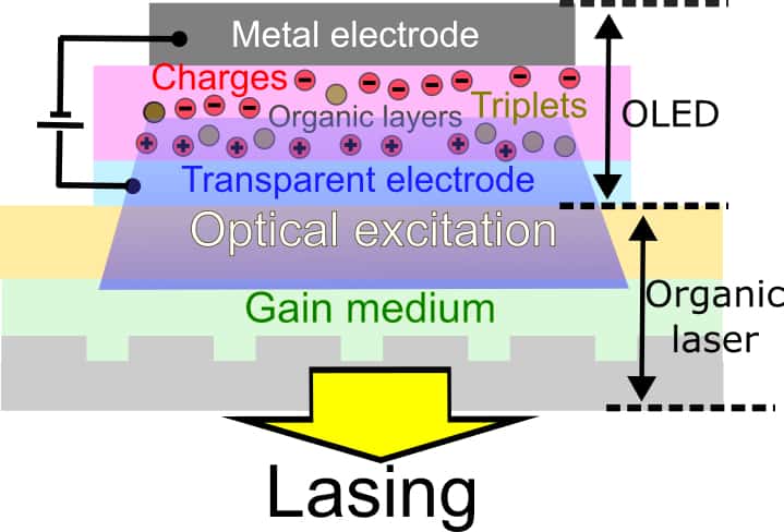
A schematic of the device. The organic LED is at the top – organic layers in between contacts. A voltage is applied to it, injecting charges and generating light that then excites the organic laser (lower half of figure). The organic laser contains a grating, applies feedback and also diffracts some of the laser light out of the structure – the yellow arrow in the figure. (Courtesy: Kou Yoshida)
Researchers at St Andrews University in Scotland have made the first organic semiconductor laser that does not require a separate light source to operate – something that had proved extremely challenging. The new all-electrically-driven laser is more compact than previous devices and operates in the visible region of the electromagnetic spectrum. As such, its developers say it might find use in applications such as sensing and spectroscopy.
Lasers work by bouncing light back and forth many times in an optical cavity comprising a gain medium sandwiched between two mirrors. As the light reflects back and forth between the mirrors, the gain medium amplifies it, stimulating the emission of more light and creating a coherent beam with a very narrow spectral range.
The first organic laser – that is, one made from a carbon-based material – was created in 1992. That laser, however, used a separate light source to drive its gain medium, which complicated its design and limited its applications. Ever since, researchers have tried to find a way to make an organic laser that works using only an electrical field to drive it, but without success. “This has therefore been a grand challenge in the field for the last 30 years,” explains physicist Ifor Samuel, who co-led the new study with his St Andrews colleague Graham Turnbull.
First, break a world record
There are two main strategies for designing an electrically-driven organic laser, Samuel explains. The first is to place electrical contacts on the organic laser gain medium and inject charges through them. It is difficult to make a laser in this way, however, because the injected charges absorb light across the luminescence spectrum of the material via so-called triplet states. The contacts themselves also absorb light. “Since a laser needs gain (optical amplification) to exceed losses, this light absorption is a huge barrier,” says Samuel.
In the new work, which is detailed in Nature, the researchers tackled this problem in the second way: by spatially distancing the charges, triplets and contacts from the laser gain medium. Doing this was no easy task either, however, as it meant they needed to make a pulsed blue organic light-emitting diode (OLED) with world-record light-output intensity to drive the gain medium. They then needed to figure out a way to couple all of this OLED’s light into the laser, which they made from a thin layer of semiconductor polymer that emits green light.
“To make the device, we initially fabricated the OLED and laser cavity separately before transferring the OLED, on a substrate of only a few microns thickness, onto the surface of the laser waveguide,” he says. “Careful integration of the two sections was crucial for the gain medium to access the intense electroluminescence generated internally in the OLED.”
To complete the design, the team used a diffraction grating in the thin-film laser to provide a distributed feedback of stimulated light emission in the plane of the film, while diffracting an output laser beam from the surface.
A slow technology speeds up
Organic semiconductor devices are widely thought of as a “slow” technology because the charge mobility in organic materials is typically orders of magnitude lower than it is for silicon or III-V crystalline semiconductors. Turnbull, however, thinks the team’s innovations could start to change that perception. “Our work is pushing these materials into a very fast and intense operating scheme,” he tells Physics World.
As for applications, the researchers say the new all-electric organic semiconductor lasers would be straightforward to integrate into point-of-care medical devices that use light-based sensing and spectroscopy to diagnose illness or monitor symptoms. “Electrical driving removes the need for a separate light source to pump them, which should widen potential applications,” Turnbull says.
There is further work to do, however, to optimize the new laser’s output power and efficiency and to broaden its light output across the visible spectrum. “The next big challenge in the field will be to make continuous-wave organic semiconductor lasers, which will need further control of the troublesome triplet population,” Turnbull concludes.
Magnetic levitation is already employed in systems such as Maglev trains, flywheels and high-speed machinery. Now researchers at the Technical University of Denmark have given this old technology a fresh twist by explaining how a magnet can be levitated simply by rotating another magnet of a similar size near it. This unusual effect was first demonstrated by an electronics/computer and software engineer, Hamdi Ucar, in 2021, and the TU-Denmark team say it could be exploited in the contactless handling of objects or used to trap and manipulate ferromagnetic microparticles.
There are three main types of magnetic levitation. In the first, known as active magnetic stabilization, a control system supplies the magnetic force required keep the levitating object balanced. The second, termed electrodynamic suspension, is used in Maglev trains. Here, a moving magnet induces a current in a stationary conductor, producing a repulsive force that increases with the speed of the moving magnet. The last category, called spin-stabilized levitation, features a levitating magnet that spins at around 500 revolutions per minute (rpm) and remains stable thanks to the gyroscopic effect.
A rotor and a floater
The new type of levitation involves two magnets. The first, dubbed the “rotor”, is mounted on a motor with its north and south poles oriented perpendicular to its rotation axis and made to rotate at velocities of around 10 000 rpm. In the TU-Denmark team’s experiments, this magnet was spherical, 19 mm in diameter and made from neodymium-iron-boron.
The second magnet – the “floater” – is placed near the rotor, begins to spin automatically, and then moves towards the rotor until it hovers in space a few centimetres below it. The floater precesses with the same frequency as the rotor and its magnetization is oriented close to the axis of rotation and towards the like pole of the rotor magnet. If disturbed, a restoring force moves it back to its equilibrium position.
Using computer simulations that take into account the magnetostatic interactions between the two magnets, the team found that the new type of levitation appears to be caused by a combination of the gyroscopic effect and magnetostatic dipole-dipole coupling.
“It is intuitive to all that the magnetostatic force that one magnet exerts on another can be both attractive and repulsive,” explains Durhuus. “Without rotation, the free magnet will rotate so the force becomes purely attractive and then the magnets will collide into each other. What makes the new magnetic levitation system so special is that the rotation itself allows the floater to remain in a counterintuitive configuration, nearly perpendicular to the rotor’s field, where the magnetostatic force both attracts and repels it.”
This process then leads to a mid-air energy minimum in the dipole interaction potential, he tells Physics World. This minimum was revealed using the team’s computer modelling, and it is here that the floater can levitate stably.
While applications of the new type of magnetic levitation are difficult to predict at this stage, the researchers say that contactless handing of objects (by robots, for example) is one obvious possibility. “Other possibilities will depend on the extent to which the phenomenon can be up- or down-scaled and how low the energy cost will be,” Durhuus says. “This will require further investigation.”
The first direct measurements of thin, supposedly insulating deposits that form in ageing rechargeable lithium-ion batteries have turned up a surprising result: the deposits are not a perfect insulator after all. According to researchers at the US Department of Energy’s Pacific Northwest National Laboratory (PNNL), the solid electrolyte interphase (SEI) layer, as it is known, is instead prone to “leaking” electrons – a finding that may help scientists develop longer-lasting batteries.
The SEI layer begins to form the very first time a rechargeable battery goes through a charging cycle. Over time, the layer gradually thickens, and its condition is thought to be related to the stability of a battery’s performance. Previously, scientists hypothesized that the SEI was electrically insulating, but its exact physical properties – including its electrical behaviour – were unknown because there was no direct way of characterizing them.
Researchers led by battery technologists Chongmin Wang and Wu Xu have now addressed this knowledge gap by embedding a nanoscale copper probe directly into a battery cell to grow a SEI film on copper or lithium. They then transferred the film to an in situ biasing transmission electron microscope and measured its electrical properties.
Leaking electrons reduce battery lifetime
The results show that as the voltage in the battery increases, the SEI layer “leaks” electrons, so reducing battery lifetime. The researchers say this hitherto unobserved semiconductor-like behaviour is mainly due to the carbon-containing components of the SEI layer. This finding suggests that minimizing these components should mean that batteries will last longer.
To back up these findings, the PNNL team’s collaborators at Texas A&M University (TAMU) created a theoretical model of an electrolyte on the surface of a lithium metal electrode. Led by Jorge Seminario and Perla Balbuena, the TAMU team members then used computational simulations to evaluate how the electrolyte evolved chemically, focusing on how chemical and physical interactions between the electrolyte and the electrode cause the SEI to form.
“Using extensive quantum physics techniques, we tested the electrolyte formulations, evaluated their chemistries and morphologies of their SEI growth, and calculated their current-voltage characteristics,” explains Seminario. “The impressive outcomes, where both the theoretical and experimental results followed the same trends, provide a way to determine the best electrolytes to use for lithium-metal batteries.”
A fruitful exchange of ideas
Balbuena adds that their results allow them to suggest possible candidate electrolytes to their experimentalist colleagues based on first principles. Conversely, they can also investigate possible alternative materials using their model before the experimental team tests them in the lab. Thanks to what Balbuena calls “the synergy of experimental chemistry with fundamental theory in the study of materials”, the team’s findings should have applications for research on batteries, sensors, biomedical devices and nano- and molecular electronics, as well as neuromorphic computing.
Seminario and Balbuena plan to expand their ab initio studies to include all components of lithium-ion batteries, and will also extend their analyses to other chemistries so they can fully untangle the degradation effects that arise during battery cycling.
Wang and colleagues, for their part, will be looking into how the different materials in an SEI layer are spatially distributed and correlated, and how this influences the layer’s physical properties. “We will also establish a direct correlation between the properties of the SEI with the chemistry of a liquid electrolyte and aim to tailor the SEI by optimizing the composition of the electrolyte for making better batteries,” Wang tells Physics World.