Ultrasound waves in air have been used to manipulate powerful laser beams – in a first claimed by researchers in Germany. The team’s acousto-optic Bragg grating could lead to new and useful ways to manipulate light.
From gravitational wave detection to semiconductor fabrication, much of modern science and technology relies on the precise control of laser light.
“Optical elements like gratings, lenses or modulators have always formed the basic ingredients behind optical devices including lasers, microscopes and atomic clocks, which have enabled many breakthroughs in various scientific fields,” explains Christoph Heyl at DESY, who led the research.
However, demands for higher power, shorter pulses and tighter control over the properties of laser light are pushing even the most advanced optical elements beyond their limits. Today, researchers are having to adapt their methods to avoid light-induced damage to optical components, and mitigate against unwanted absorption and nonlinear effects that degrade the quality of laser light.
Density manipulation
Now Heyl and colleagues have taken novel approach to controlling light that promises to avoid some of the problems associated with conventional optical components. Their technique involves manipulating the density of air at length scales on par with the wavelength of light.
“We employ highly intense ultrasound fields to control and redirect laser beams under a small angle directly in ambient air, employing the principle of acousto-optic modulation,“ Heyl explains.
In their experiment, the researchers mounted an ultrasound transducer opposite a planar sound reflector. This establishes a high-pressure standing ultrasound wave in the air gap – a wave that features sharp, periodic variations in air density. The index of refraction of air increases with density, so the standing wave acts as a Bragg grating that can deflect light using optical diffraction. While this technique is used to create gratings in solid media such as glass, the team says that this is the first time it has been done using air.
To use their grating, Heyl and colleagues placed a pair of opposite-facing mirrors perpendicular to the standing ultrasound wave. A beam of light enters the device and is reflected back and forth many times before exiting the device. This increases the distance that the light travels through the Bragg grating, enhancing the diffraction effect.
High-power handling
The team found that about 50% of the incident light was deflected and the rest was transmitted – with the quality of the incident laser light preserved. The team says that numerical simulations suggest that this percentage could be increased significantly in the future. What is more, the grating can handle gigawatt laser pulses that are about one thousand times more intense than the upper limit of devices that employ the acousto-optical modulation of solid materials.
“Our approach provides a bypass to the restrictions that solid media usually impose: including orders of magnitude lower dispersion, higher peak powers, and wider wavelength ranges,” explains team member Yannick Schrödel, who is a PhD student at DESY.
Based on these results, the team predicts a diverse array of future applications for their acoustic optical Bragg grating. “Our method provides direct routes to novel optical amplitude and phase modulators, switches, beam splitters and many more elements, directly implemented using gas-based gratings,” says Schrödel.
The team is also looking forward to the development of other new technologies for manipulating light. “In addition, more advanced optical elements could be realised,” Schrödel continues. “This could enable exciting new directions for ultrafast optics, and other fields which face limits in optical power and spectral coverage.”
The acousto-optic Bragg grating is described in Nature Photonics.
Most physicists are drawn to the subject by a simple desire to learn how the world works. But physics is also vital for the economy, supporting high-value jobs, generating profits, and spawning innovation in sectors ranging from renewable energy and medicine to communications and computing. It was to celebrate such firms that in 2012 the Institute of Physics (IOP), which publishes Physics World, launched its business awards to honour companies in the UK and Ireland for their innovative application of physics.
The awards have grown from strength to strength over the last dozen years (full disclosure: I was a member of the judging panel this year). In 2018 a new category was launched to honour promising start-up firms, while in 2020 the Lee Lucas award was also introduced for early-stage medical and healthcare companies thanks to a generous donation by Mike and Ann Lee (née Lucas) funding the £5000 cash prize in perpetuity. This year nine firms have won IOP business innovation awards, bringing the total number recognized to 83.
Given the size, breadth and depth of the medical-physics market, it is no surprise that five of the nine award-winners are businesses in medical physics.
Given the size, breadth and depth of the medical-physics market, it should come as no surprise that five of the nine award-winners in 2023 are businesses operating in medical physics and allied areas. Fortune Business Insights has estimated that the market for medical devices – from dentistry and surgery to diagnostics and cancer treatment – was worth $489bn in 2021 and will grow to $719bn by 2029. The market is even bigger if applications in animal welfare and food production are taken into account.
Drug delivery
The first 2023 winner I’d like to mention is the Glasgow-based firm Nebu~Flow, which bags an IOP business start-up award for its devices that deliver drugs into a patient’s lungs in aerosol form. Having previously won a Lee Lucas award in 2020, Nebu~Flow has built an acoustic nebulizer based on surface-acoustic-waves. Its device helps the medication to reach the right part of a patient’s lungs, which isn’t always possible with existing devices.
Nebu~Flow says its technology has performed exceptionally well in pre-clinical trials, with the device, for example, boosting the efficiency with which the body absorbs salbutamol – a common asthma drug – from 60% to 90%. It does this using the mechanical energy of the acoustic waves to control the aerosol droplets to be less than 5 microns in size. The technology could also allow patients to inhale drugs for treating lung hypertension and cystic fibrosis.
As for this year’s Lee Lucas award, it was won by ArtioSense, which has built a device to help surgeons position implants precisely, especially in patients having replacement hips fitted. The core technology was developed by Sohini Kar-Narayan, a materials scientist at the University of Cambridge, who co-founded the firm with the consultant orthopaedic surgeon Vikas Khanduja. Using microfluidic sensors that provide real-time force measurements, the device should make operations more successful.
Another winner this year is Bristol-based Siloton, which picks up a start-up award for developing a personal optical coherence tomography (OCT) system. It contains a specially developed photonic chip called Akepa that can image into body tissue, such as the back of the eye. Siloton’s device will be particularly useful for patients with sight loss caused by age-related macular degeneration (AMD). The condition affects a quarter of people over 60 with estimates suggesting there could be as many as 300 million AMD patients globally by 2040.
Current OCT systems for monitoring AMD are large, fragile and expensive, meaning they are mostly the preserve of hospitals and optometrists. But as Siloton’s devices are small and low cost, the company believes that patients can simply be prescribed one of the devices, which they can use themselves a couple of times a week wherever they are. It would therefore enable highly precise and personalized treatment to be devised, reducing unnecessary sight loss, and freeing up hospitals to focus on diagnosis and treatment.
Another start-up award has gone to Occuity in Reading, which has developed a hand-held device that can quickly screen and monitor ophthalmic and chronic diseases using the eye as “a window to health”. The company also hopes to start selling the world’s first truly non-contact blood glucose monitoring system for people with diabetes. Co-founded by the physicist Dan Daly, the device is non-contact, non-invasive, disposable-free and could even be used to diagnose Alzheimer’s disease, vascular dementia and sepsis.
The final company I want to mention is Dyneval, which is based in Edinburgh and has won a 2023 business start-up award for developing equipment to measure the quality of semen in livestock. Dyneval’s kit does this by monitoring the tiny fluctuations in the intensity of light passing through a sample of semen, which provides information on how well the sperm are moving. Marketed as Dynescan, the equipment allows lab-quality measurements to be carried out on farms, allowing farmers to save money by boosting conception rates among their animals.
Challenging times
One of the challenges that all physics-based firms face is the time it takes to develop products or services and to become globally significant. Medical-physics companies face the added difficulty of having to gain regulatory approval in each market, which means it can even longer to start selling their tech. There’s also the age-old difficulty of explaining a technical product idea to potential investors who have little or no science background.
That’s why the IOP’s start-up awards are great. They give small firms evidence that their technology has been reviewed by an experienced judging panel with solid physics and business experience. I hope this year’s winners will inspire your company to apply – and it may even encourage you to set up a firm of your own. Award entries open soon so watch this space and IOP announcements. In next month’s column, I’ll look at the four other companies to have won awards.
Mitigating skin tone bias Conventional (top rows) and SLSC (bottom rows) photoacoustic images of volunteer 1 (light skin tone) and volunteer 18 (dark skin tone) at 750, 810 and 870 nm. The green arrows indicate the radial artery location; the white arrows show a smaller blood vessel enhanced with SLSC imaging. (Courtesy: CC BY 4.0/G S P Fernandes et al Photoacoustics 10.1016/j.pacs.2023.100555)
Photoacoustic (PA) imaging is a non-invasive diagnostic technique that can visualize anatomic structures – such as arteries and blood vessels – up to several centimetres deep into tissue. But while this imaging modality works well for people with lighter skin tones, it cannot create as clear images from patients with darker skin.
PA imaging works by illuminating tissue with near-infrared light. The tissue absorbs the optical energy, causing it to expand and generate acoustic waves, which can be detected by conventional ultrasound transducers. When imaging darker skin, however, the increased melanin absorbs more of the incident light, reducing the illumination efficiency. This absorption also generates PA waves at the skin surface, creating image artefacts known as acoustic clutter.
A research collaboration between Johns Hopkins University and the University of São Paulo has found a way to address this problem, using a method called short-lag spatial coherence (SLSC) beamforming, which effectively removes the unwanted clutter signals. The team demonstrated that the SLSC beamforming algorithm significantly increased image quality in all participants – and worked particularly well in those with darker skin tones – reporting their findings in Photoacoustics.
Muyinatu Bell “We’re aiming to mitigate, and ideally eliminate, bias in imaging technologies.” (Courtesy: Johns Hopkins University)
“When you’re imaging through skin with light, it’s kind of like the elephant in the room that there are important biases and challenges for people with darker skin compared to those with lighter skin tones,” says co-senior author and SLSC inventor Muyinatu Bell in a press statement. “Our work demonstrates that equitable imaging technology is possible.”
To test this approach, the team recorded PA data from the forearms of 18 volunteers with differing skin tones, using 750, 810 and 870 nm excitation. To determine the effect of melanin content on conventional amplitude-based PA images, as well as the impact of SLSC beamforming, they calculated the signal-to-noise ratio (SNR) of the radial artery running through the forearm and the level of surrounding clutter artefacts for both conventional and SLSC PA images.
Rather than classifying skin tone with the commonly used, but subjective, Fitzpatrick skin type scale, the researchers used a quantitative method. By measuring the individual typology angle, which correlates with epidermal melanin content, they classified each participant’s skin tone as very light, light, intermediate, tan, brown or dark.
In conventional amplitude-based PA images acquired at 810 nm, the image of volunteer 1 (who had the lightest skin tone) provided good visualization of the radial artery and surrounding small blood vessels. The PA image of volunteer 18, who had the darkest skin tone, contained strong clutter artefacts that compromised radial artery visualization.
Examining the skin PA signal level as a function of skin tone for each volunteer showed that the amplitude of this signal increased with increasing epidermal melanin content. The skin PA signal was also dependent on the optical illumination wavelength, with lower signals observed at higher wavelength.
Conventional PA images of volunteer 1 had minimal acoustic clutter at all three wavelengths. For volunteer 18, however, strong clutter artefacts were seen in the images, particularly at 750 nm, where the radial artery could not be distinguished from the background.
The SLSC PA images, on the other hand, contained far less clutter artefacts, for both light and darker skinned volunteers. For light skin tones, SLSC PA imaging reduced the median clutter level by 0.7 dB, from −16.2 dB with amplitude-based PA imaging to −17.0 dB. The effect was more pronounced for dark skin tones, reducing clutter by 6.1 dB, from −7.9 to −14.0 dB.
University of Sao Paulo team From left to right: João H Uliana, Adilton Carneiro, Luciano Bachmann, Theo Pavan and Guilherme Fernandes. (Courtesy: University of São Paulo)
This reduced level of clutter led to better visualization of the radial artery. In addition, SLSC PA images from volunteer 18 revealed a small blood vessel that was previously masked by clutter in the conventional PA images.
Calculating SNR values for the radial artery (for the three wavelengths combined) showed that for all volunteers, the SNR decreased for darker skin tones in the conventional PA images. SLSC beamforming improved this SNR by a median of 3.8 dB across all skin tones, enhancing radial artery visualization. The team notes that the SLSC SNR achieved with the darkest skin tone was comparable to the conventional SNR for the lightest skin tone.
The researchers conclude that the quantifiable bias introduced by skin tone variations was successfully mitigated with SLSC beamforming. They are now working to apply these findings to breast cancer imaging, to image the blood vessels that accumulate in and around tumours.
“We’re aiming to mitigate, and ideally eliminate, bias in imaging technologies by considering a wider diversity of people, whether it’s darker skin tones, higher breast densities or greater body mass indexes – these are currently outliers for standard imaging techniques,” says Bell, who recently received a Science Diversity Leadership Award for her research into inclusive imaging technologies. “Our goal is to maximize the capabilities of our imaging systems for a wider range of our patient population.”
“To the best of our knowledge, our study represents the first quantitative analysis of the relationships between skin tone, clutter level and image quality in photoacoustic imaging,” co-senior author Theo Pavan tells Physics World. “Our findings suggest that it would be beneficial for future clinical studies utilizing photoacoustic imaging to incorporate participant skin tone information in their research.”
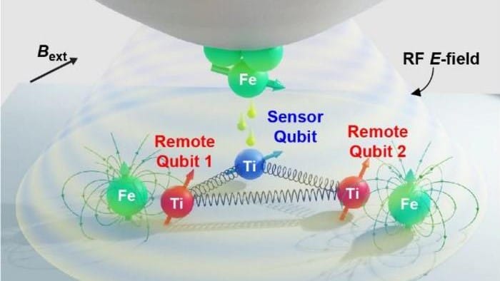
Multiple qubit platform: In this diagram, an STM tip coated with iron (top) operates the sensor spin qubit. Also shown are the remote spin qubits, which are aligned by the magnetic fields of nearby iron atoms. (Courtesy: Institute for Basic Science)
A quantum computing platform that is capable of the simultaneous operation of multiple spin-based quantum bits (qubits) has been created by researchers in South Korea. Designed by Yujeong Bae, Soo-hyon Phark, Andreas Heinrich and colleagues at the Institute for Basic Science in Seoul, the system is assembled atom-by-atom using a scanning tunnelling microscope (STM).
While quantum computers of the future should be able to outperform conventional computers at certain tasks, today’s nascent quantum processors are still too small and noisy to do practical calculations. Much more must be done to create viable qubit platforms that can retain information for long enough for quantum computers to be viable.
Qubits have already been developed using several different technologies, including supercomputing circuits and trapped ions. Some physicists are also keen on creating qubits using the spins of individual electrons – but such qubits are not as advanced as some of their counterparts. However, that does not mean that spin-based qubits are out of the running.
“At this point, all existing platforms for quantum computing have major drawbacks, so it is imperative to investigate new approaches,” explains Heinrich.
Precise assembly
To create a viable spin-based processor, qubits must be assembled precisely, coupled together reliably, and operated in a quantum-coherent manner, all on the same platform. This is something that has so far eluded researchers, until now – according to the Seoul-based team.
The researchers created their multi-qubit platform with the help of an STM, which is a powerful tool for imaging and manipulating matter on atomic scales. When the conducting tip of an STM is brought very close to a sample surface, electrons are able to quantum-mechanically tunnel between the tip and the sample surface.
Since the probability of tunnelling strongly depends on the distance between tip and surface, an STM can map out the sample’s nanoscale topography by measuring the current of these tunnelling electrons. Individual atoms on surface can also be manipulated and assembled by pushing them around by the nanoscale forces applied by the tip.
Using these capabilities the team has “demonstrated the first qubit platform with atomic scale precision,” according to Heinrich. “It is based on electron spins on surfaces, which can be placed at atomically precise distances from each other.”
Sensor qubit
Using STM, the researchers assembled their system on the pristine surface of a magnesium oxide bilayer film. The system includes a “sensor” qubit, which is a spin-1/2 titanium atom that is located directly below the STM tip. The tip is coated in iron atoms, which means that it can be used to apply a local magnetic field (see figure).
To either side of the tip are a pair of “remote” qubits – also spin-1/2 titanium atoms. These are placed at precise distances from the sensor qubit, outside the region where electron tunnelling between atoms can occur.
To control the remote qubits simultaneously with the sensor qubit, the team created a magnetic field gradient by placing iron atoms nearby. The iron atoms behave as single-atom magnets because their spin relaxation times far exceed the operation times of individual qubits.
In this way, the iron atoms each act as a substitute for the STM tip in providing a static, local magnetic field for aligning the spins of each remote qubit. Transitions between the spin states of the qubits are done by using the STM tip to apply radio-frequency pulses to the system – a technique called electron spin resonance.
Addressed and manipulated
The team initialised their qubits by cooling them to 0.4 K, then applying an external magnetic field to bring them into the same spin state and coupling them together. Afterwards, the state of the sensor qubit depended reliably on the states of both remote qubits, but could still be addressed and manipulated individually by the STM tip.
The overall result was entirely new qubit platform that allowed multiple qubits to be operated simultaneously. “Our study has achieved single qubit, two qubit, and three qubit gates with good quantum coherence,” Heinrich says.
He adds that, “the platform has its pros and cons. On the pros, it is atomically precise and hence can be easily duplicated. On the cons, the quantum coherence is good but needs to be improved further.”
If these challenges can be overcome, Heinrich and colleagues see a bright future for their system.
“We believe that this approach can relatively easily be scaled to tens of electron qubits,” Heinrich says. “Those electron spins can also be controllably coupled to nuclear spins which might enable efficient quantum error correction and increase the available Hilbert space for quantum operations. We have just scratched the surface!”
Earlier this year, the Institute of Physics (IOP), which publishes Physics World, launched a campaign to persuade journalists to stop using the outdated slang term “boffin” when referring to scientists. The initiative was aimed at the red-top tabloids such as The Sun as well as The Daily Star, who have a particular soft spot for the word.
The IOP says that using the term in their coverage has a negative impact and puts people off from studying science. This is because boffin conjures up the image of a scientist being a slightly dishevelled, elderly white man. In this new video from Sixty Symbols, physicist Philip Moriarty at the University of Nottingham, broadly agrees as he discusses the pros and cons of the campaign. You can watch the video above.
Winter is approaching in Austria, and soon the country’s famous alpine resorts will be thronging with skiers and snowboarders. But in the alpine spa town of Bad Gastein, some visitors are going deep under the mountains rather than up on ski lifts. Guests of the Gasteiner Heilstollen spa can don their swimsuits and hop on a small train that takes them more than 2 km underground to caverns where they can “bathe” in radiation given off by naturally occurring radon gas.
No proven benefits
According to an article in The Guardian by Colin Nicholson, radon bathers receive a radiation dose that’s about 25% of a medical X-ray. Although Nicholson points out that there are no proven health benefits from the radiation, he says that some Austrians have their visits covered by their health insurance.
Nine years ago Symmetry published a list of ten physics themed Halloween costumes. And now, just as physicist trick-or-treaters have exhausted those ideas, the magazine’s Aspen Stuart-Cunningham has come up with ten more. The latest suggestions include going as a neutrino by continuously changing between three different costumes – to mimic how the subatomic particles oscillate between three different flavours.
Or maybe you fancy brightening up a party by dressing up as the High-Luminosity Large Hadron Collider – which should come on line at CERN in 2029. Stuart-Cunningham’s suggests wrapping yourself with a hula hoop and just about every portable source of illumination you have around the house including Christmas lights and bike lights.
Duelling dark sector
I think my favourite suggestion is teaming up with a friend to be dark matter and dark energy. You could pretend to be in conflict all evening, with dark energy expanding the universe and dark matter’s gravity trying to pull it in the opposite direction.
We will delve into the world of MRI-guided radiotherapy systems and the critical role of quality assurance in delivering precise, personalized cancer treatments.
This webinar will address the unique challenges and considerations that MRgRT QA presents. From image distortion and system calibration to complex workflows, understanding and mitigating these challenges is vital for every healthcare professional working with MRgRT systems. Moreover, the webinar will delve into well-established quality assurance protocols and procedures that are tailored specifically to MRI-guided radiotherapy systems.
All facets of quality assurance, spanning machine QA, patient-specific QA, and real-time adaptive QA, will come under thorough scrutiny and discussion throughout the session.
Stephanie Tanadini-Langis co-vice chair of the department of radiation oncology at the University Hospital Zurich in Switzerland. She is also head of medical physics at the hospital, where she is responsible for all dosimetric aspects of 1700 patients per year in the radiation oncology department, and is group leader of the radiomics and modelling research team. After studying for a MPhys at the University of Constance in Germany, Stephanie obtained a MAS in medical physics from ETH Zurich and a PhD in medical physics from the University of Zurich. Her research to date has included studies of quality assurance for complex radiosurgery, radiomics for non-small cell lung cancer, quantitative image analysis in glioblastoma, deep learning algorithms and synthetic CT images.
This is the first in the Physics World webinar series on radiotherapy quality assurance. Even if you’re not able to join the live event, registering now enables you to access the recording as soon as it’s available.
In December 1942 US president Franklin D Roosevelt signed the Manhattan Project into existence. A scientific endeavour that culminated in the dropping of the Little Boy and Fat Man bombs three years later, the project was – for better or worse – the most significant development in the long history of nuclear physics. What is perhaps surprising, though, is that this pioneering field of discovery is captured forever through the medium of postage stamps.
Marie Curie has appeared on more than 600 postage stamps and holds the record as the physicist with the most stamps ever issued in their name
Our story begins with Marie Curie, who shared the 1903 Nobel Prize for Physics with Pierre Curie for their studies of radioactivity. This phenomenon had been discovered in 1896 by Henri Becquerel, who won the other half of that year’s prize, but it is Marie Curie who is easily the most famous of the three scientists. She has appeared on more than 600 postage stamps and therefore holds the record as the physicist with the most stamps ever issued in their name. My favourite is the 1938 Afghanistan 15 pul stamp, which is the only one featuring Curie with her electrometer and was also the first stamp to depict a female scientist.
From her lab in Paris, Curie famously studied the radiation emitted by pitchblende – a glowing mix of uranium oxide and lead, which hailed from the Jáchymov mine in Bohemia, now part of Czechia. Known for its production of silver, the ore was delivered to Curie, who also used it to discover the elements polonium and radium. The mine’s fame as the birthplace of nuclear science was commemorated by the former Czechoslovakia in 1966 with a 60 haléř stamp (click here to view).
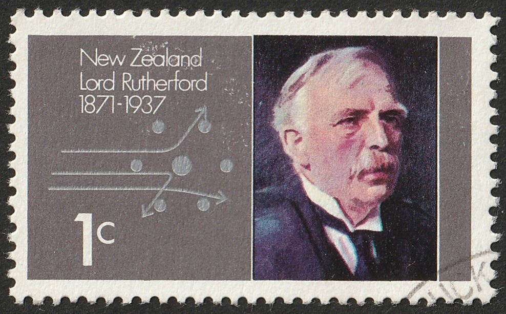
Ernest Rutherford – the New-Zealand-born physicist who discovered the atomic nucleus – is also commemorated on several stamps. One I particularly like was issued by New Zealand in 1971 to commemorate the centenary of his birth. The 1 cent stamp of the set includes a portrait of Rutherford along with a diagram of the Rutherford atomic model, which – correctly – envisaged electrons surrounding a dense central nucleus. The stamp nicely shows alpha particles being scattered back from the nucleus – the famous “gold-foil” experiment found in every school physics syllabus.
Marked man This 1 cent New Zealand stamp was issued in 1971 to commemorate the centenary of Ernest Rutherford’s birth. (Crown Copyright New Zealand. Reproduced with permission. Photo courtesy: Ian Briggs)
Rutherford could – and perhaps should – have won a Nobel prize for his discovery of the nucleus but he of course won the Nobel Prize for Chemistry in 1908 for his work on the decay of radium. The Nobel committee obviously viewed radioactivity as chemistry, not physics, prompting Rutherford to famously remark that he had dealt with many different transformations, but that the quickest was his “own transformation in one moment from a physicist to a chemist”. Be that as it may, winning a Nobel prize is a sure-fire way to philatelic fame.
The Danish physicist Niels Bohr – who won the 1922 Nobel Prize for Physics for his work on the structure of atoms – has appeared on several Swedish stamps but my favourite is actually a Greenland 1963 issue, celebrating 50 years of “Bohr theory”, which describes how electrons exist in discrete orbits and can jump between them. I like this stamp because rather than containing just a visual portrait of the scientist, as was the trend until then, it also depicts Bohr’s work in the form of an equation (hν = E2–E1) and a diagram of orbiting electrons.
As the 1920s turned into the 1930s, the pace of research in nuclear physics picked up. In 1932 James Chadwick discovered the neutron. In 1938 Otto Hahn and Fritz Strassman, along with Lise Meitner and Otto Frisch (working under Bohr), discovered atomic fission. In 1939 Frédéric Joliot-Curie, Enrico Fermi and Leo Szilard confirmed the chain reaction experimentally. The final pieces of the bomb jigsaw were provided by Francis Perrin, who calculated the critical mass of uranium needed for a self-sustaining reaction, along with further work from Rudolf Peierls in Birmingham, UK.
Images on postage stamps are a great reminder of the role of science in the world around us and yet, they can also entrench inequities
Discovery in science is a bit like a self-sustaining reaction, in which new ideas are built on old ones and researchers stand on the shoulders of the giants who went before. Images of postage stamps are a great reminder of the role of science in the world around us and yet, they can also entrench inequities. The beautiful 60 pfennig German stamp first issued in 1979 (click here to view), for example, shows the splitting of a uranium nucleus but it mentions only Hahn, who was awarded the 1944 Nobel Prize for Chemistry. His co-discoverers – Meitner, Strassman and Frisch – who were left empty-handed are, once again, omitted from history.
Stamps don’t just reflect history but can shape it too.
This episode of the Physics World Weekly podcast features two pioneers in their fields.
Margaret Gardel is a biophysicist who is setting up a new National Science Foundation Physics Frontier Center at the University of Chicago. The Center for Living Systems will focus on the physics of adaptation, a new field that looks at how living matter stores, retrieves, and processes information as it adapts to change. Gardel explains how physics-inspired theory and experiments are providing fresh insights into biological systems.
Our second pioneer is Susannah Glickman who has just completed what is probably the first scholarly history of quantum computing. A historian based at Stony Brook University in the US, Glickman explains why there has been so much enthusiasm for quantum computers, despite the fact that that the technology is far from settled. She also talks about the process of writing her history and the generosity of some of the quantum-computing experts who provided her with crucial information about how the field has developed.
Stereotactic ablative radiotherapy (SABR) is a precision cancer treatment that delivers highly focused, intense radiation doses over just a few treatment sessions. Also known as stereotactic body radiation therapy, SABR is the standard-of-care for inoperable early-stage non-small cell lung cancer (NSCLC). However, it can confer a higher risk of severe-to-potentially fatal toxicities than more conventionally delivered radiotherapy
To minimize such risks, researchers at Stanford University have developed an individualized approach for lung SABR, using dose and fractionation regimens based on tumour volume, location and histologic findings. Their strategy and clinical trial, described in JAMA Oncology, produced excellent local control with few toxic effects for its 217 participants.
“The iSABR [individualized SABR] trial demonstrated that we can personalize treatment to the characteristics of individual patients to optimize the clinical outcomes by balancing effective control of lung tumours with minimizing side effects,” explains Billy Loo Jr, co-principal investigator of the trial along with Maximilian Diehn. “In prior studies, these factors impacted the likelihood of the tumours being controlled by SABR, or the risk of serious normal organ injury by SABR. By individualizing treatment according to these factors, we hypothesized the impact of these factors could be overcome.”
In their phase 2 non-randomized trial, the researchers enrolled 214 patients at Stanford and three at Hokkaido University in Sapporo, Japan. Patients were categorized into three groups: those with a single NSCLC (group 1); those having a new primary NSCLC with a history of prior NSCLC or having multiple NSCLCs (group 2); and those with lung metastases from NSCLC or another solid tumour (group 3).
Based on tumour volume and location, the researchers designed five dose-and-fractionation schedules, with up to four tumours treated with once-daily SABR. Doses ranged from 25 Gy in one fraction (for 55% of the patients) to 60 Gy in eight fractions. Large tumours received higher doses, while central tumours generally received a lower dose per fraction.
For smaller tumours, with a volume of 10 cm3 or less, the researchers prescribed a biologically effective dose (BED10) of 80–87.5 Gy, lower than the standard dose of 100 Gy BED10. All other regimens had BED10 greater than 100 Gy. Patients with colorectal metastases (which exhibit radioresistance) received at least 50 Gy in four fractions.
The researchers followed the patients for a median of 33 months. Of the 285 treated tumours, 26 (or 9%) had a local recurrence. Freedom from local recurrence at one year was 97%, 94% and 96% for patients in groups 1, 2 and 3, respectively, confirming the hypothesis that the one-year local recurrence risk was less than 20% for each of the groups. At two years, freedom from local recurrence ranged from 90% in group 1 to 95% in group 3, and at five years it ranged from 83% in group 1 to 93% in group 2. The cumulative incidence of treated-tumour recurrence for all participants was 3% at one year, 5% at two years and 7% at five years.
Compared with published clinical trials of patients prescribed standard doses without individualization, fewer patients in this study experienced side effects, and these were much less severe than those previously reported. Sixteen patients developed grade 2 or higher pneumonitis, 13 had noncardiac chest pain and five had pleural effusion. Only four patients experienced grade 4 toxicities and one patient had a grade 5 adverse event. The researchers report that the risk of more severe toxic effects was higher in patients with ultracentral tumours.
Future prospects
In an accompanying invited commentary in JAMA Oncology, Vivek Verma from the University of Texas MD Anderson Cancer Center writes that “very few prospective trials have attempted to reduce BEDs”. He notes that “among the most common misconceptions of lung SABR is that virtually all patients experience high local control”. As a result, many radiation oncologists reflexively prescribe 100 Gy10 (50 Gy in five fractions) for virtually all lung SABR cases. “In reality, the dogmatic 100 Gy10 BED threshold is merely based on retrospective data. It is essential to understand there is an interplay between tumour size/histologic type and the BED required for durable local control,” Verma writes.
The researchers are currently conducting a clinical trial (ADAPT-E) in which patients with early-stage lung cancer are selected for adjuvant treatment with immunotherapy following their initial treatment with SABR or surgery, based on whether there is evidence of microscopic residual cancer detected by an ultrasensitive assay for circulating tumour DNA in the blood.
“Diehn’s laboratory is developing ultrasensitive novel assays for circulating tumour DNA and also identified the KEAP1/NFE2L2 pathway as a radioresistance mechanism,” Loo tells Physics World. “We know that the genetic characteristics of tumours can also determine their response to therapy. Prior work has demonstrated that approximately half of the local tumour recurrences following radiotherapy might be explained by mutations in the KEAP1/NFE2L2 gene pathway that lead to ramping up the tumour cells’ antioxidant defences against radiation damage.”
Loo adds that his team is also studying the biological effects of FLASH therapy, the delivery of ultrahigh dose rates in a fraction of a second. “FLASH therapy has been shown in preclinical research to reduce damage to normal tissues without compromising tumour control. My colleagues and I are developing new technology to translate FLASH treatment to clinical use and to be able to optimize clinical outcomes even more in the future.”
In today’s hyperconnected world, where Zoom meetings and remote working have become the norm, it can be easy to overlook the pivotal role that scientific meetings play in driving research progress. For Stanley Whittingham, who shared the 2019 Nobel Prize in Chemistry for his pioneering work on lithium-ion batteries in the 1970s, the meetings of The Electrochemical Society (ECS) were the quickest and most effective way to share and discuss the latest research results.
“There were two main groups working on lithium-ion batteries, my group at Esso [now Exxon] and the other at Bell Labs about 10 miles away,” says Whittingham, who joined the ECS as a post-doc at Stanford University in 1970 and now continues to investigate battery chemistry at Binghamton University, part of the State University of New York. “Everyone working in the field knew everyone else, and the ECS meetings were the logical place for us to meet, discuss our latest findings, and exchange ideas.”
Stanley Whittingham, who shared the 2019 Nobel Prize for Chemistry for his pioneering work on lithium-ion batteries, has played an active role in the ECS meetings since the 1970s. (Courtesy: ECS)
In those days, recalls Whittingham, it could take months for an article to appear in the Journal of the Electrochemical Society, the only publication at the time that would accept battery research. “All the correspondence with the editors and referees had to be sent by post, and once the article had been published it could take a while for the journal to reach people in remote parts of the world,” he says. “The meetings provided a faster way to share new research results and to find out what other people were working on.”
Even today, when online journals have allowed emerging research to be shared in a fraction of the time, the meetings offer a valuable opportunity for attendees to immerse themselves in the science and the progress being made. “At a meeting I can really pay attention to the presentations and think about the new work that is being reported,” says Iryna Zenyuk, who leads a group developing fuel-cell and hydrogen technologies at the University of California, Irvine. “Our days are so full that it’s easy to get distracted or interrupted, but a meeting allows me to absorb more information because I am fully present in the moment.”
Both Whittingham and Zenyuk play an active role in presenting work and organizing symposia at the ECS meetings, which are convened twice a year in the spring and fall. “The ECS is still the major society for battery scientists, and you can almost guarantee that you will find some interesting new research and meet some new people,” says Whittingham. “The field has grown so much, and many other conferences now cover battery research, but the core people still go to the ECS meetings.”
For Zenyuk, one unique aspect of the ECS events is the ability to connect with scientists and engineers from across the electrochemical community. “What I like about ECS meetings is the presence of people from industry, the national labs and the academic sector,” she says. “The meetings provide a focal point for people from different disciplines to come together, and for fundamental research scientists to connect with engineers and technologists who are more focused on applications. The field is defined by all these different sectors, and it’s very important to get a perspective from all of these key stakeholders.”
Hydrogen scientist Iryna Zenchuk has established several industrial collaborations by showcasing her research at ECS meetings. (Courtesy: ECS)
From her own personal experience, Zenyuk has found the meetings particularly useful for establishing new connections with industrial partners. “Several of our industrial collaborations have started through ECS meetings, since someone from the company might have seen my presentation and discovered that we have a capability that could be useful to them,” she says. “Among academics we already know what other people are working on, and we can approach each other if there is an opportunity to collaborate. But we don’t know if people from industry have a specific need because they are more inclined to keep their activities to themselves. We need to wait for them to come to us.”
Making new connections across the wider electrochemistry community is particularly important for younger scientists and engineers, with both Whittingham and Zenyuk encouraging their students to attend the meetings and present their work. “The younger people need to build links, which is easier to do in person than online,” says Whittingham. “Most research these days is done in teams, and the ECS provides a place for students and early-career researchers to get to know each other, find out what other people are working on, and establish collaborations and research projects.”
The meetings also provide a valuable opportunity for young electrochemists to share their experiences and understand how their work is contributing to the wider scientific endeavour. “Students work hard on their experiments, and sometimes it can feel tedious or difficult,” says Zenyuk. “Going to a meeting is really special for them because they realize that other people are struggling with the same problems. It helps them to feel part of something bigger and to gain more confidence in their own abilities.”
The ECS also provides plenty of incentives for students to get involved. Many of the society’s divisions and sections offer travel grants to undergraduates, graduate students and early-career researchers who are presenting their work, plus they support a number of student awards for poster presentations. Zenyuk, who won an award as a PhD student in 2013, is now responsible for organizing the poster session for the symposium on fuel cells and electrolysers, typically the largest one at the meeting.
“The symposium has 20 organizers who work together to arrange plenary sessions and invited talks, and to decide which of the hundreds of submissions will be offered a presentation slot,” she explains. “My focus is to arrange the poster session, including the judging of the student awards. That’s the fun part, because I received an award when I was a student and now I’m responsible for organizing the student competition.”
Presenting a poster brings plenty of other benefits too, as Zenyuk discovered from her experiences as a PhD student. “At the time I had two offers for post-doc positions,” she recalls. “I would tell people who people came to my poster about the offers, and they offered me advice that helped me to accept a position at the Lawrence Berkeley National Lab. My poster was an opportunity not just to showcase my work, but also to meet the community and learn from them.”
Building collaborations The ECS meetings enable scientists and engineers from all over the world to discuss their latest work and identify opportunities for joint research projects. (Courtesy: ECS)
For Zenyuk, it is that human component of scientific meetings that sets them apart from other forms of scholarly communication. “Most of us are friends, we are part of the same community, and it’s good to catch up,” she says. “That face-to-face interaction makes it easier to meet new people as well as to reconnect with friends and colleagues.”
While Whittingham has witnessed a surge in battery research since his seminal work in the 1970s, he agrees that the ECS meetings continue to provide a vital focal point for the community to share results and build collaborations. “In the old days everyone working in the field could sit around a table for lunch, but now there might be three or four parallel sessions focused on batteries,” he says. “Even as the meetings have got larger the younger folks still get to know each other outside of the sessions, and make the connections that will support them throughout their career.”
The 245th biannual meeting of The Electrochemical Society will be held on 26–30 May 2024 in San Francisco, US, while its 246th meeting will form part of PRiME 2024, a joint international meeting with the Electrochemical Society of Japan and the Korean Electrochemical Society that will be held in Honolulu, US, on 6–11 October 2024.