What’s the best way to recognize the death of an eminent scientist? The Royal Society memorializes its late members – as do many other national scientific academies – by commissioning colleagues to write what are known as “biographical memoirs”. Each article, which aims to provide a “definitive account” of that person’s life and work, is peer-reviewed and published in a journal called the Biographical Memoirs of Fellows of the Royal Society.
Free to read online, the memoirs are supposed to focus on “science and scientific endeavour, while also shining a light on the human side of scientific success [and] offering a fascinating insight into the character and personalities of the individuals involved”. According to the Royal Society, this remit makes the memoirs “a valuable resource for both scientists and historians of science”. The University of Cambridge astrophysicist Malcolm Longair – current editor-in-chief of the Biographical Memoirs – says they contain “the DNA of the society”.
In a lecture on the Royal Society’s website, Longair set outs his philosophy of what the memoirs are trying to achieve. Unlike the obituaries you might find in national newspapers, which are aimed at the non-specialist, the memoirs are “written by scientists for scientists”. They focus on the deceased’s scientific contributions and personality but steer clear of any philosophical and sociological concerns. If an article can capture the scientist’s individual genius, Longair adds, it might inspire young people to enter their field.
I’ve never written a biographical memoir for the Royal Society, but I have done three equivalent articles for the US National Academy of Sciences (NAS), which adopts a similar approach. Natalie Shanklin, who works in the NAS communications team, told me its biographical memoirs are meant to offer an “in-depth and scholarly lens on the research and career of the NAS member”. Being written by someone who knows the subject well, they add colour by including “more sentimentality and sometimes anecdotal asides”. Overall, the NAS memoirs aim to provide “a biographical history of science in America”.
Technically, I wasn’t a colleague of the three people I’ve written about but in each case I did have a connection with them. For the nuclear physicist Robert Serber (1909–1997), who had worked on the Manhattan atomic-bomb project, I had helped him write and publish his recollections. For the physicist and historian Abraham Pais (1918–2000), I had been recruited to complete his unfinished biography of Robert Oppenheimer. As for Maurice Goldhaber (1911–2011), who is best known for discovering that neutrinos have a left-handed helicity, I had interviewed him extensively for other projects.
A long history
Over in France, the Paris Academy of Sciences began publishing éloges or eulogies about recently deceased members as long ago as 1699. In his book Science and Immortality, the US historian Charles B Paul examines the first century of these articles, which were initiated by the French essayist and scholar Bernard Le Bovier de Fontenelle, who had towering ambition. He conceived the memoirs not as storehouses of historical and biographical information, but as a means to extoll “the moral virtues of the post-Renaissance sciences and their practitioners”. He wanted to elevate scientists to the status of the “political, military and religious heroes memorialized since the beginning of time”.
Tensions persist in biographical memoirs, for they have incohesive, almost oxymoronic goals – “biographical” suggests something third-person and objective, while “memoirs” suggests first-person and subjective
If today’s biographical memoirs are less daring, that’s largely due to the success of the tradition started by Fontenelle. However, tensions persist in biographical memoirs, for they have incohesive, almost oxymoronic goals. The word “biographical” suggests something third-person and objective, while “memoirs” suggests first-person and subjective. They are also aimed simultaneously at non-scientists, scientists, biographers and historians alike.
I have recently been asked to write two further biographical memoirs. One is for the Royal Society to honour the British experimental physicist Francis Farley (1920–2018), who worked on the first four g-2 experiments, which were designed to test the soundness of quantum electrodynamics via a measure of the muon spin. The other will be for the NAS on the theoretical physicist Toichiro Kinoshita (1925–2023), who pioneered the theory of g-2 calculations. I knew Farley and had interviewed him at length, while Kinoshita was a personal friend, as I mentioned in a recent column. Still, I will be writing the memoir of each person with one of their scientific collaborators.
But despite my personal connections with both people, I find composing these memoirs no easier than others I have written. As well as trying to address the seemingly competing aims for biographical memoirs, I recognize what I think is yet another purpose. The most important goal of a biographical memoir, in my opinion, is not just to be a “valuable resource” but also to memorialize the person – to craft something that lives after that person that prompts us to remember them as a full human being.
The critical point
When I write a biographical memoir, my goal is to make readers wish that they had known, learned from and conversed with the subject about both personal and scientific matters. I try to make readers wish they’d known the person as a friend, and even have invited them over for, say, drinks or a family dinner. I want to show that who that person was and what they did belong together – and that whatever honours that person received were the natural thing to bestow on them.
Doing this is of value for a national academy, I think, because it addresses a recognition that its members have of themselves. They are not just people who sit on committees and work on problems but communities of diverse, highly skilled and carefully chosen scientists joining forces on valuable, shared goals. The aim of memorializing someone might not be explicitly acknowledged in the instructions for writing a memoir, and it’s a hard piece of advice to put into words. Still, it’s why writing a good biographical memoir is both worthwhile and not as easy as it sounds.
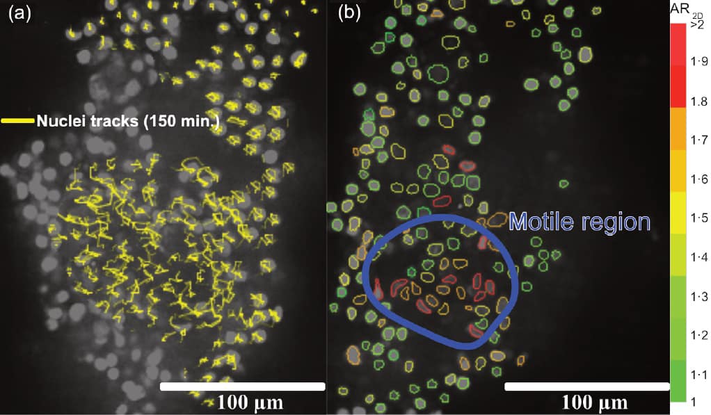
Vital cell tracking in a breast tumour explant (a) Yellow lines show the tracks of cell nuclei movement over 150 min; an unjammed region of cell motion is seen in the lower middle of the sample, surrounded by jammed cells. (b) Cell nuclei colour-coded for aspect ratio (AR), with red indicating elongated (high AR) cells and green showing rounder (low AR) cells. The region of motile cells exhibits more elongated nuclei than the surrounding cells. (Courtesy: CC BY 4.0/P Gottheil et al Phys. Rev. X 10.1103/PhysRevX.13.031003)
The physical concept of jamming – originally developed to understand the behaviour of glasses and granules – could improve predictions of whether cancers are likely to spread.
Research carried out by doctoral student Pablo Gottheil and colleagues from the Soft Matter Physics Division of Leipzig University has shown that the prognostic accuracy for cancerous tumours spreading increases by 26% when the detection of cancer cell unjamming is employed alongside existing prognostic methods.
Cancers spread when cells from a primary tumour travel through the body via the bloodstream or lymphatic system, then seed further tumours known as metastases. This involves cells that were packed together in the primary tumour becoming mobile, in a similar manner to the state transition between jammed solid-like and unjammed liquid-like behaviour within amorphous materials and granules: when unjammed, particles can flow like table salt onto your dinner, but being jammed is akin to the salt flow stopping when it gets into a configuration that blocks the tiny salt cellar hole.
Since living human cells within closely packed cell aggregates and tissues exhibit similar jamming and unjamming, similar concepts can be used to study the process. “To understand cancer you can’t just look at the genome, you also have to look at physics,” says Josef Käs, head of the research team and Gottheil’s PhD supervisor. “Metastasis has emergent collective effects which can be described by mechanics and soft-matter physics.”
In a first clinical study, reported in Physical Review X, the researchers analysed clinical data and histological samples from 1380 patients with breast cancer. Unlike previous work by other groups that characterized cell unjamming in terms of either the cell’s shape or its density, the Leipzig researchers reconciled this conflict by taking shape and nucleus packing into consideration.
Their research quickly confirmed that both cell and nucleus shape, as well as nucleus number density were involved: specifically, when a tumour is unjamming, its cells and nuclei elongate and the cell nuclei are more loosely packed. This enables the cells to move more readily than when they are rounder, as it is easier for them to squeeze between other cells and exchange places with them. Furthermore, the team found that an increased prediction of unjamming occurring for a given tumour correlated well with the actual development of secondary cancer in that patient any time up to a decade later.
The researchers represented the cancer cell and nucleus shape, and the cell area as a measure of nuclei packing, in a comprehensive state diagram of cancer cell unjamming that indicates patients at high and low risk of developing metastases. This simple diagram could be used to assess prognoses in 92% of cancers.
“It is intriguing that the static geometric signature of the primary tumour cells can serve as a proxy for cell motility, which then actually correlates with tumour progression,” Gottheil tells Physics World. “Established markers, such as the status of affected lymph nodes, already show that cancer cells left the primary tumour, whereas our marker predicts that if classified as motile and unjammed, the tumour should have a higher likelihood of distant metastasis formation.”
“Currently one of the biggest problems in all cancers is over- and under-treatment because we don’t have a precise prognosis in cancer diagnosis,” adds Käs. He hopes that their technique – which can be applied to histological samples already routinely collected – could reduce that problem. His team will shortly embark on a retrospective clinical trial working with 25,000 breast cancer patients and Pathology Hamburg-West, and strive to establish prospective multi-centre clinical trials for breast and colon cancer.
Käs intends keeping the work in the public domain so as many patients as possible can benefit. “It is so horrible for people not knowing from one year to the next if their cancer will relapse. Anything I can do to alleviate that situation makes me happy,” he says.
I grew up in the Canadian province of Ontario, so I spent a lot of time around lakes and streams when I was young. I remember being intrigued by water striders, which are long-legged insects that literally walk on water. Now that I live in England, we even have our own water striders living in our small garden pond.
Water striders’ legs are hydrophobic, so they float by repelling water, and under each leg is a dimple in the water – called a meniscus. One thing that I didn’t know about water striders is that they can jump very rapidly from the surface of the water when attacked from below by predators. Scientists have known for some time that the insect does this by pushing down on the water, making the dimple larger. Then, they use the upward recoil of the water’s surface to aid in their propulsion.
But now, an international team of researchers has discovered a new jumping mechanism that is used by larger water striders that weigh more than about 80 mg. When these behemoths push down on the water, their legs breakthrough the surface – so they cannot take advantage of the springy dimples.
Hairy legs
The researchers found that a layer of air becomes to attached to the hairy legs as they plunge into the water. This air increases the resistance that the legs encounter as they move down through the water, giving the insects the extra purchase needed to jump out of the water.
The observations were made during an expedition to Vietnam to study that country’s giant water strider and the layer-of-air hypothesis was quantified by creating a mathematical model. The team says that their research could help in the developing of robots that walk on water and could also shed light on the evolutionary development of water striders.
Speaking of air–liquid interfaces, physicists have studied the physics of “beer-dancing peanuts”, which I am told are all the rage in Argentina. To see the effect for yourself, drop a peanut into a glass of beer. Being denser than the liquid, the peanut will first sink to the bottom of the glass. However, after a few moments the peanut will float to the surface of the beer, where it will remain for a few moments before it sinks and repeats the process again.
Now, you are probably thinking that this has something to do with bubbles from the beer accumulating on the surface of the peanut until it is buoyant and floats to the surface. There, the bubbles probably burst, causing the nut to sink back down. And that’s what researchers in Germany have observed when they dropped peanuts into a litre of lager-style beer – with the added detail that the rotation of peanuts on the surface causes the bubbles to burst. What is more, they found that the process repeated itself for 150 min until the peanut came to rest at the bottom of the vessel. Something that only very slow tipplers would notice.
If you want to read more about this study, check out this article in Physics, which also explains how the study could shed light on the behaviour of magma under the Earth’s surface
Theoretical physicists do a good job when peer reviewing scientific papers, but tend to be more impressed by the significance of new research rather than the rigour with which it was carried out. That is the provocative suggestion of a new study carried out by researchers in the UK and Poland that examines the reliability of peer review in theoretical physics.
Peer review involves sending research papers to independent, external referees to decide if an article is scientifically credible and appropriate for the journal to which it has been submitted. Despite being a central part of science for hundreds of years, few academics are trained in peer review, and anecdotal evidence suggests that disagreements between individual reviewers about a paper are common.
“In an ideal world, all reviewers would evaluate each submission with perfect accuracy and render a perfect decision,” says Mike Thelwall, a data scientist from the University of Sheffield, who carried out the study. But given that no work can be perfect in all of the metrics used to judge a paper’s quality, reviewers make what Thelwall dubs “a judgement call” about the extent to which an article achieves an acceptable level.
The three core components of quality used to judge a paper are originality, rigour and significance. However, Thelwall says that no-one had previously checked if reviewers judge these metrics effectively, even though they are, for example, part of the guidelines for assessors in the UK’s Research Excellence Framework.
Decision makers
Thelwall and physicist Janusz Hołyst of the Warsaw University of Technology therefore analysed reviewer scores in 505 theory papers published in SciPost Physics — a journal that practises open peer review. The duo chose to focus on theoretical physics because they say it is best-case-scenario for peer review, in that reviewers should be more likely to understand all aspects of a given submission than in other areas of science.
Each paper in the study had at least two scoring reviewers and the analysis revealed a “moderate” degree of agreement between reviewers on all three key metrics. “The results should cause no worries at all for SciPost Physics — their reviewers are doing an excellent job,” Thelwall says.
However, he worries that journals publishing research in fields where it is harder for individual reviewers to make a sound judgement might have much lower agreement rates for reviewers. “By the law of averages, [they may] tend to make bad decisions moderately often due to an unlucky choice of reviewers,” he says. The authors suggest such issue might be minimized via editorial oversight, better guidelines and improved reviewer training.
The study also looked at which of the quality measures tend to be ranked the same by individual reviewers. The results suggest that — in theoretical physics at least — referees tend to rank rigour lower. This means it could be harder to publish what Thelwall calls a “highly rigorous original study”, but easier to get a “significant original study” accepted.
In fact, theoretical physicists striving to create significant work might benefit from focusing on original ideas rather than generating exhaustive evidence to support their work, the authors suggest. “Of course, we need all types of research and if everyone gives up on rigorous ideas it will be a disaster for science,” cautions Thelwall.
Stefan Thurner, a physicist from the Medical University of Vienna who was not involved in the study but has carried out studies into peer review, says the study is interesting. “The raw data indicate that the reviewers are remarkably consistent in practically all categories,” he says. “In more than 400 out of 505 cases, they differ not at all or by one score unit. I am sure that many scientists have the subjective feeling that referee reports differ more often than that.”
Thurner adds, however, that future research could compare the results with, say, reference models in which papers were randomly assigned to referees.
Collaboration, at scale, provides the raison d’être for Treesearch, an ambitious Swedish R&D initiative that brings together a cross-disciplinary cohort of scientists and engineers from academia, industry and government agencies within an open research platform focused on the “future bioeconomy”. Zoom in a little and it’s evident this over-arching mission translates into a broad-scope remit, one that spans fundamental science, technology innovation and competence-building to realize a new generation of advanced materials from the forest.
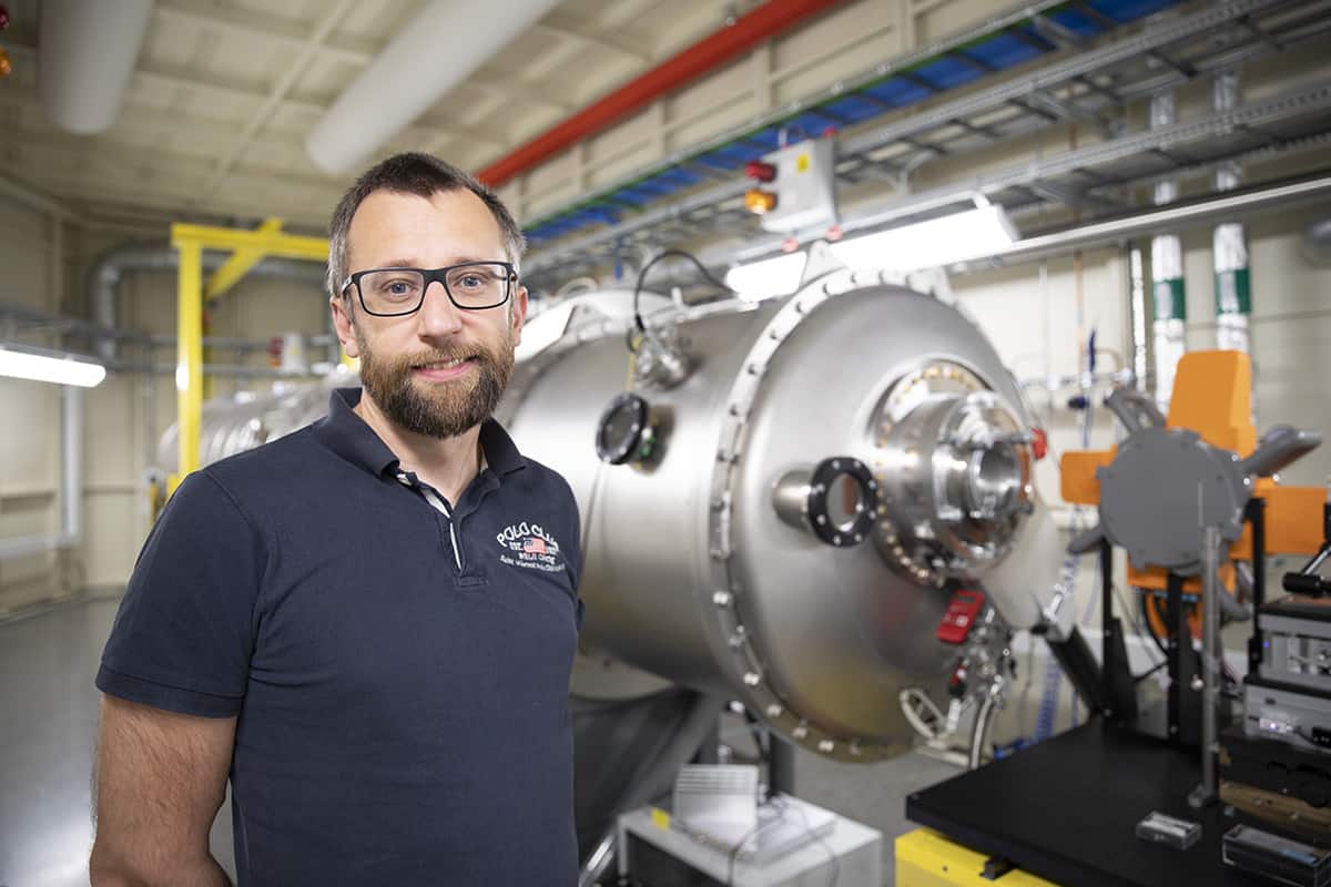
Daniel Söderberg “One of Treesearch’s core priorities is to help our stake-holders access advanced research infrastructures.” (Courtesy: Treesearch)
Collectively, there are more than 510 researchers (and 190 projects) represented within the Treesearch consortium, with the work broadly carved up across four thematic areas: wood and wood components (structure and modification); biorefinery for materials and chemical systems; fabrication of material systems; and advanced material concepts (design and functionality). “One of Treesearch’s core priorities is to help our academic and industry stakeholders to access advanced research infrastructures across Sweden,” explained Daniel Söderberg, Treesearch director and head of the department of fibre and polymer technology at the Royal Institute of Technology (KTH), Stockholm.
A flagship Treesearch partner in this regard is the MAX IV synchrotron radiation facility in Lund. Globally significant, MAX IV is one of an elite cadre of large-scale X-ray sources that is shedding light on the structure and behaviour of matter at the atomic and molecular level across a range of fundamental and applied disciplines – from clean-energy technologies to pharma and healthcare, from structural biology to quantum science and cultural heritage.
In terms of core building blocks, this fourth-generation light source – which was inaugurated in 2016 – consists of a linear electron accelerator plus 1.5 and 3 GeV electron storage rings (with the two rings optimized for the production of soft and hard X-rays, respectively). As well as delivering beam to a short-pulse facility, the linac serves as a full-energy injector to the two storage rings which, in turn, generate X-ray photons that are extracted for user experiments across 16 specialist beamlines.
The latest addition to MAX IV, the ForMAX beamline, opened for user experiments in November 2022 and is dedicated to research on sustainable wood-based materials from the forest (though it will also support X-ray studies on other complex materials such as food, textiles and bone). Collaboration is once again front-and-centre, with the 100 million SEK (£7.5 million) construction costs of ForMAX funded by the Knut and Alice Wallenberg Foundation (a Swedish philanthropic organization that supports scientific research), while the 80 million SEK operational budget over 10 years is covered by commercial partners (mainly companies in the pulp and paper industry). ForMAX beamtime is allocated accordingly, with 50% of the experiments to be conducted by Treesearch members and the remainder offered via open calls to the wider research community.
“Through ForMAX, Treesearch academia and industry partners have a dedicated access point to the MAX IV research environment,” explained Söderberg. “As such, the beamline underpins a broad, unique competence in synchrotron science and will, over time, enable the development of sustainable wood-based products to replace today’s plastic products.”
Into the woods
For the most part, ForMAX beam time will support fundamental and applied studies on wood-based materials, providing in-situ structural characterization from nanometre to millimetre length scales by combining full-field X-ray microtomographic imaging, small- and wide-angle X-ray scattering (SWAXS) and scanning SWAXS imaging in a single instrument. For context, the ForMAX microtomography system uses incident X-rays to generate planar cross-sections of a sample that can be used to recreate a virtual 3D model (at length scales from 1 mm down to 1 micron). The SWAXS set-up, on the other hand, relies on two discrete detectors to collect X-ray spectra scattered from a sample at different angles: WAXS yielding structural information down to the 1 nm scale, with SAXS used for soft-matter studies – of polymer, colloidal and biological assemblies, for example – up to several hundred nm in size.
Better together Kim Nygård, ForMAX beamline manager, heads up a specialist technical team providing experimental support to Treesearch members and other scientific users of the beamline. (Courtesy: Anna Sandahl, MAX IV)
That versatility will enable researchers to investigate wood’s structural hierarchy and composite nature – from the fibre network and cellular structure at the macroscopic scale, through ordered assemblies of the fibril matrix structure and cell walls at the nanoscopic scale, right down to the cellulose which forms the (partially) crystalline building blocks at the macromolecular level. “ForMAX will improve our understanding of the complex structure-function relationship in forest-based materials and food products across multiple length scales,” Kim Nygård, ForMAX beamline manager, told Treesearch Insight.
ForMAX is a flexible instrument that enables the study of materials in situ during processing and under realistic conditions
Kim Nygård, ForMAX beamline manager
A notable feature of ForMAX is the beamline’s multimodal imaging capability, combining full-field microtomography and SWAXS sequentially in the same experiment. “Fast, efficient switching between set-ups allows collection of imaging and scattering data on the same sample,” noted Nygård. In other words: full-field microtomography to provide the user with an overview of 3D structure and regions of interest, with localized SWAXS then used to investigate structure and orientation at the nanoscopic level (see also “Versatile by design: the ForMAX experimental station”, below).
“ForMAX is a flexible scientific instrument that also provides the temporal resolution to study materials in situ during processing and under realistic conditions such as applied temperature or pressure,” added Nygård. A unique feature is the beamline’s RheoSWAXS capability, integrating a state-of-the-art rheometer (supplied by Anton Paar, an Austrian metrology company) with polarized-light imaging and SWAXS to study orientational dynamics of wood-based samples across a range of length scales and under steady and oscillating shear conditions. Since shear-aligned cellulose nanocrystals exhibit structural colour, such studies could, for example, pave the way for the use of printed cellulose suspensions instead of traditional inks in future biobased packaging.
Getting ready to shine
Although ForMAX has only been up and running for little more than six months, early-adopting Treesearch partners are already showing the way with their initial experimental runs. A case in point is an industry-academia collaboration on fibre-based sustainable food packaging involving Swedish packaging giant Tetra Pak and researchers at Chalmers University of Technology inGothenburg.
Using ForMAX’s SWAXS imaging techniques, Linnéa Björn of Chalmers and Eskil Andreasson of Tetra Pak told Treesearch Insight attendees how the joint team – working closely with ForMAX staff scientists – is studying the nanostructure of fibre-based materials in an effort to optimize the composition and volume manufacture of paper straws.
If that seems like a narrow focus, the wider commercial imperative is clear: while there is growing market demand for more sustainable alternatives to plastic packaging, manufacturers like Tetra Pak must ensure that paper-based materials will remain food-safe, recyclable and durable against liquids and humidity. Put simply, the task for the Chalmers–Tetra Pak team is to understand the correlation between wetting of the paper straw with different liquids (water and orange juice, for example) as well as the impact of process treatments on the nanoscale structure.
“Our first experiment at ForMAX provided analysis into how paper-straw material responds to changes in the environment in real-time, as well as how the straw interacts with different types of liquids under stringent conditions,” explained Andreasson, a technology specialist in virtual modelling at Tetra Pak. “These insights will be applied to develop the paper straws of the future in our computer modelling tools, helping us to improve their functionality.” Further Tetra Pak collaborations are already in the works at ForMAX, including the use of real-time 4D X-ray microtomography to study water transport mechanisms in sustainable paper straws.
We can exploit synchrotron technology to optimize our manufacturing processes or enhance the fundamental understanding of our products and their performance
Christophe Barbier, senior research manager, Billerud
The application of synchrotron X-rays in product development was reinforced by Christophe Barbier, a senior research manager in paper physics with Billerud, the Swedish pulp and paper manufacturer which also specializes in fibre-based packaging materials for food, drink and medical applications. “We can exploit synchrotron technology in several ways,” he explained at Treesearch Insight. “To optimize our manufacturing processes, for example, or enhance the fundamental understanding of our products and their physical performance, as well as to drive product superiority and competitive differentiation.”
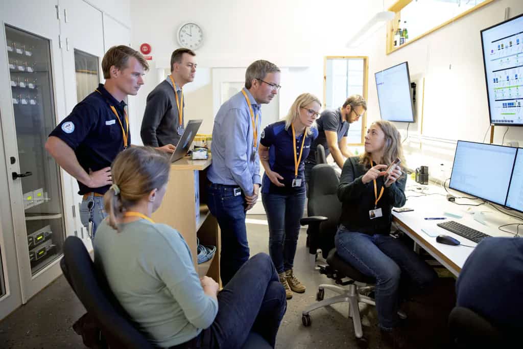
Collaborate and innovate Tetra Pak’s Eskil Andreasson (middle), Linnéa Björn (seated right) and colleagues get busy in the ForMAX control room. (Courtesy: Anna Sandahl, MAX IV)
Barbier and his colleagues are long-time converts to “big science” who have seen the benefits of synchrotron light sources up close. Previously, the team has booked beam time at DESY’s PETRA III facility in Hamburg, Germany, to study the fundamentals of mechano-sorptive creep (an effect that can see stacked cardboard boxes of fresh produce, for example, unexpectedly buckle due to tensile load when ambient warehouse temperature or humidity exceed certain limits).
“We set out to establish that synchrotron-based X-ray scattering techniques can detect the effects of mechano-sorptive creep on the ultrastructure of pulp fibres,” noted Barbier. “The results are sufficiently encouraging to warrant continued SWAXS investigations of the phenomenon and, we hope, to eventually develop appropriate countermeasures.”
Proximity being what it is, Billerud is also gearing up for further studies at MAX IV’s ForMAX beamline. Current lines of enquiry, in collaboration with the 4D Imaging Lab at Lund University, include the use of X-ray microtomography to characterize “multi-ply” packaging (comprising multilayer or composite materials) and correlate its microscale properties versus bulk performance under load in forming machines.
“Treesearch is our door-opener to large-scale research facilities like MAX IV,” Barbier concluded. “There is huge potential for such facilities to address knowledge gaps in many areas relating to forest materials and sustainable products.”
Versatile by design: the ForMAX experimental station
To maximize flexibility for visiting scientists, the ForMAX end-station is built around a single experimental table, with tomography microscope/cameras and SWAXS detectors added in a modular manner. This arrangement ensures efficient switching between different modes of operation, allowing combined full-field tomography and SWAXS experiments in a sequential manner.
In this way, the in-house design of the experimental station is tailored to the specific needs of ForMAX’s user community. Key building blocks include:
Two beam-conditioning units, including slits, beam diagnostics equipment and two sets of secondary optics to achieve a large range of beam spot size at the sample position (from up to approx. 5 mm field-of-view in tomographic imaging down to approx. 1 micron real-space resolution in scanning SWAXS experiments).
An experimental table with 200 mm vertical and horizontal (transverse) translation ranges and 200 kg load capacity.
A motorized detector gantry so that the user can move the WAXS detector and tomography microscope in and out of the X-ray beam path (i.e. allowing combined full-field microtomography and SWAXS experiments in sequential mode).
A 9 m long, evacuated SAXS flight tube with SAXS detector inside on a motorized XYZ trolley; sample-to-detector distance can be varied between approximately 1.2 and 7.5 m.
Our guest in this episode of the Physics World Weekly podcast is Emily Grubert, who is a civil engineer and environmental sociologist at the University of Notre Dame in the US. In a wide ranging interview, she chats about her research, which focuses on justice and deep decarbonization.
Much of Gubert’s work explores how we will make the transition from our current carbon-intensive economy to a low-carbon future – and she points out that exactly how this will be done is far from settled. She talks about carbon capture and storage, a controversial (and mostly hypothetical) way of removing carbon dioxide from flue gases or even from the atmosphere.
Grubert also talks about how we can build climate-change resilience into buildings and how society can respond to climate-change driven migration by preparing communities in cooler regions for an influx of people who have had to move because of the effects of climate change.
Brand new journal
Grubert is editor-in-chief of the journal Environmental Research: Energy, which has just opened for submissions. She talks about her plans for the journal and why there is an urgent need for an open access publication that brings together researchers across the many disciplines working on the transition to zero-carbon energy systems.
And if you are interested in a career in climate-change research, Grubert makes the argument for pursuing an interdisciplinary education.
The scientific community must act to prevent an exponential growth in the carbon footprint of computational science as the ubiquity of artificial intelligence, algorithms and data science increases. That is the warning from researchers at the University of Cambridge who say that those involved in computational science – from individuals to institutions – should take responsibility for reducing greenhouse-gas emissions.
There has been increased interest in recent years in the climate impacts of scientific research. Much of this has focused on conferences – particularly emissions from attendees’ flights – and the carbon footprint of laboratories.
But there is one aspect of research that is often overlooked: high performance and cloud computing. The researchers argue that while the environmental impact of laboratories and travelling can be easier to determine, the impact of running algorithms is less clear and often underestimated.
Indeed, information and communication technologies (ICT) can have a substantial environmental impact. In 2020, for example, the ICT sector produced between 1.8% and 2.8% of global greenhouse gas emissions — more than the aviation industry (1.9%).
There have been a small number of studies highlighting the environmental impacts of computing in science, particularly in astronomy and astrophysics. For instance, research in 2020 found that the average Australian astronomer produced around 15 tonnes of CO2 equivalents per year just from their supercomputer usage – which was almost four times their annual emissions from flights.
“Science has led to great benefits to society, but this has come with a significant – and not always well understood – impact on the environment” notes Loïc Lannelongue, a mathematician and physicist who works on biomedical data science at the University of Cambridge.
Energy concerns
To tackle this, Lannelongue and colleagues have come up with a set of principles for best practices in environmentally sustainable computational science. The first step, the team say, is for those involved in computational science to take responsibility for reducing greenhouse-gas emissions. This requires transparency and for the carbon footprint of computations to be estimated and monitored.
Such steps could be achieved, the researchers say, through training, more centralised data infrastructures and hardware procurement considering equipment’s lifetime emissions as well as funding bodies requiring estimates of carbon footprints.
Computational scientists have a real opportunity to lead the way in sustainability, but this is going to involve a change in our culture and the way we work
Loïc Lannelongue
According to the authors, reducing the carbon footprint of the electricity used is one of the quickest ways to reduce computational greenhouse-gas emissions. Relocating to a different setting or country is one way to do this. For example, electricity produced in Australia is three orders of magnitude more carbon intensive than electricity in Iceland.
The researchers caution, however, that scientists should not forget about the footprint of data storage, which is often exacerbated by duplication of datasets so multiple groups can have a copy.
The team also argue that education and research are needed to raise awareness and drive innovation. Recent research has shown that different programming languages and coding practices can make to the energy efficiency of algorithms, for instance, highlighting the need for well-trained software engineers.
“Computational scientists have a real opportunity to lead the way in sustainability, but this is going to involve a change in our culture and the way we work,” says Lannelongue.
Echocardiography – an ultrasound scan of the heart – is the most frequently used imaging modality for assessing cardiac function and diseases. The technique, however, requires specialized skills that are often in short supply. An alternative option could be to use chest X-rays, one of the most common and widely available medical exams, primarily employed for diagnosis and management of lung diseases. But while the heart is visible in chest radiographs, the relationship between chest radiographs and cardiac health is poorly understood.
Aiming to bridge this gap, a research team led by Daiju Ueda of Osaka Metropolitan University has developed a deep-learning model that uses artificial intelligence to detect valvular disease and classify cardiac function from chest radiographs with unprecedented accuracy. The researchers publish their results in The Lancet Digital Health.
Deep-learning models that are trained and tested on a single dataset can be prone to overfitting, in which the final model only works well for images in the training dataset. To prevent this, Ueda and colleagues developed their model using data from four different institutions, with a total of 22,551 chest radiographs plus associated echocardiograms collected from 16,946 patients.
The researchers used 17,293 radiographs from three institutions to train the deep-learning model, plus 1947 radiographs from the same sites as internal test datasets. For external testing, they employed 3311 radiographs from 2617 patients at a separate institution.
After labelling the chest X-rays using the echocardiography reports as ground truth, the researchers trained their model to learn features connecting the two datasets. They examined six types of valvular heart disease – mitral regurgitation, aortic stenosis, aortic regurgitation, mitral stenosis, tricuspid regurgitation and pulmonary regurgitation – classifying the severity of each disease as none, mild, moderate or severe. They also classified three measures of cardiac function: left ventricular ejection fraction, tricuspid regurgitant velocity and inferior vena cava dilation.
To evaluate the diagnostic performance of their deep-learning model, the researchers calculated the area under the receiver operating characteristic curve (AUC) for nine primary classifiers – a cutoff of none–mild versus moderate–severe for each of the six valvular heart diseases, plus cutoffs of 40% for left ventricular ejection fraction, 2.8 m/s for tricuspid regurgitant velocity, and 21 mm for interior vena cava dilation – for the internal and external test datasets.
The team found that the model could accurately classify cardiac functions and heart diseases, information typically obtained from echocardiography, using information from the chest radiographs. The overall mean AUCs for the primary classifiers were 0.89, 0.90 and 0.92 for the internal test datasets, and 0.87 for the external test dataset (values closer to 1 indicate better classification).
Focusing on the external test dataset, the model could precisely categorize the six types of valvular heart disease, with AUCs ranging from 0.83 to 0.92. The AUC for classifying left ventricular ejection fraction was 0.92, while the AUC for both tricuspid regurgitation velocity and interior vena cava dilation was 0.85.
“To the best of our knowledge, this study is the first to create and validate a deep learning-based classification model for cardiac functions and valvular heart disease using chest radiographs from multiple institutions,” the researchers write.
They point out that the model has several advantages over echocardiography-based evaluation of cardiac disease. Chest X-rays are easy and quick to record and the model can be applied rapidly with low computational requirements. After its initial implementation, the model could be used without any specialized skills and at any time. In addition, it should be possible to use existing chest radiographs to provide information on cardiac function when necessary, without needing additional tests.
“It took us a very long time to get to these results, but I believe this is significant research,” says Ueda in a press statement. “In addition to improving the efficiency of doctors’ diagnoses, the system might also be used in areas where there are no specialists, in night-time emergencies, and for patients who have difficulty undergoing echocardiography.”
“In the future we hope to evaluate the real-world applicability of our model in various clinical settings,” co-author Shannon Walston tells Physics World. “It is crucial for us to understand how our AI-based model can be integrated seamlessly into clinical workflows, and how it can contribute to improved patient care.”
A rapidly rotating white dwarf star that contains two opposing hemispheres – one covered by hydrogen and the other by helium – has astronomers scratching their heads over how it got that way. The star, nicknamed “Janus” after the two-faced Roman god of transition, was discovered by the Zwicky Transient Facility (ZTF) at Palomar Observatory in the US, and one possible explanation is that it is the result of a strong but lopsided magnetic field generated by the merger of two white dwarfs.
White dwarfs are the remains of Sun-like stars that have ceased nuclear reactions in their interiors, puffed off their outer layers, and experienced gravitational contraction of their remnant cores. The resulting objects are about the size of Earth, but with the mass of a star.
Though white dwarfs are born hot, they gradually cool as they age. This cooling affects their structure. At temperatures above 35,000 K, their surfaces are covered by a layer of hydrogen that envelops a sub-layer of helium. Once the surface temperature cools to 35,000–25,000 K (the exact temperature depends on the star’s mass), this helium layer begins to convect. If the upper hydrogen layer is thin enough, it can dissipate in the roiling helium.
About 40% of white dwarfs have made this transition from hydrogen dominance to helium dominance. However, since the transition normally occurs in a matter of seconds, no one has ever seen it happening – until, perhaps, now.
Stuck in transition?
Officially designated ZTF J203349.8+322901.1 (the numbers are its right ascension and declination co-ordinates on the sky) and located over 1300 light-years away, the Janus white dwarf attracted the attention of the California Institute of Technology astrophysicist Ilaria Caiazzo because of its rapid changes in brightness. Additional observations by Palomar and other facilities showed that the star completes one rotation every 15 minutes, during which its brightness varies from a maximum when its hydrogen-covered face is pointed towards Earth, to a minimum when we see the opposing hemisphere covered in helium.
The question is, why? “We might have finally caught a white dwarf in the act of transitioning,” Caiazzo tells Physics World. In fact, based on the findings of the team Caiazzo assembled to investigate the discovery, Janus seems to have got stuck in transition. On one of its hemispheres, helium convection seems to have consumed the hydrogen, but mysteriously the same does not appear to have occurred on the other. Writing in Nature, the team suggests that a sufficiently strong magnetic field offset from the white dwarf’s centre could be inhibiting helium convection on one hemisphere and not the other, but this explanation is tentative. Suffice to say, nobody has ever seen a white dwarf of two halves before.
“There’s no model that predicts this,” says team member Pier-Emmanuel Treblay, an astronomer at the University of Warwick, UK. “In astrophysics, when something is messed up and needs to be finely tuned, people often invoke magnetic fields, and this is a perfect example of that.”
About 20% of white dwarfs are magnetic, and some have field strengths of up to 1 billion Gauss. By comparison, Earth’s magnetic field is half a Gauss, while the magnetic field strength on the surface of the Sun is about one Gauss. For Janus, the team estimates the field must be 1000–1 million Gauss. Any stronger, and it would distort the star’s spectral lines.
“For Janus, we assume that there is a magnetic field because it would be very hard to explain the different composition on the two faces otherwise,” Caiazzo says. However, she adds, “We still don’t know why only some white dwarfs are magnetic and where this huge diversity in field strengths comes from.”
A white-dwarf merger?
Janus’ strong and lopsided magnetic field, its rapid rotation rate, its high mass (between 1.20 and 1.27 solar masses) and its two-faced composition all point to a quite remarkable white dwarf. For Tremblay, this indicates that other factors may be at play. “There must be something special about this white dwarf in addition to a magnetic field,” he says.
Tremblay speculates that Janus could have formed through the merger of two white dwarfs – an event that could have created internal magnetic dynamos. “The fast rotation, and the magnetic field generation and asymmetry, they all point to binary evolution and a merger,” he says.
Tremblay is also sceptical about the magnetic field being an offset dipole. The internal magnetic field structure of white dwarf stars is not yet well understood, and in his view, invoking an offset dipole could hide a higher-order magnetic field geometry.
“In my opinion, it means that the magnetic field might not be dipolar,” Tremblay says. “Instead it might be a quadrupole, with four poles, for example. It doesn’t necessarily mean that the field is offset from the centre.”
Implications for distance measurements
When white dwarfs explode as type Ia supernovae, their well-understood brightness allows astronomers to treat them as standard candles – a vital tool for measuring distances across the cosmos and the expansion rate of the universe. However, astronomers are still not sure how many type Ia supernovae occur when a single white dwarf accretes too much matter from a companion star and explodes, and how many occur due to the merger of two white dwarfs that, when combined, exceed the Chandrasekhar mass limit of 1.44 solar masses and explode.
If Janus is indeed the product of a merger of two smaller white dwarfs, finding more examples of half-transitioned white dwarfs will enable astronomers to constrain the numbers of such systems and how much they might contribute to the population of type Ia supernovae.
A laser wakefield accelerator (LWFA) that guides its laser beams along curved channels while accelerating electrons has been created by Jie Zhang and colleagues at Shanghai Jiao Tong University in China. The new technique could be a key step towards the development of compact, inexpensive alternatives to conventional particle accelerators.
In an LWFA, a dense plasma is created by focusing an intense laser pulse into a gas. As it moves through the gas, the pulse creates a region of alternating electric fields – a “wakefield” – that resembles a water wave that forms in the wake of a moving boat.
By riding these waves, electrons within the plasma can be accelerated to very high energies over very short distances. As a result, this technique shows great promise for developing accelerators that are much smaller than conventional systems. Such compact devices would be very useful for medical and research applications.
Reinjection woes
For electrons to reach relativistic speeds, the acceleration must happen multiple times, with electrons from one LWFA stage being injected into the next. This is not easy, as team member Min Chen explains, “since the wake is tens of microns size and its velocity is very close to the speed of light, the electron reinjection is extremely difficult”. While some recent studies have achieved reinjection using techniques such as plasma lenses, researchers have only managed to inject a small fraction of electrons into a second stage.
In 2018 Zhang and Chen’s team introduced a new approach as Chen describes, “In our scheme, the electrons always travel inside a straight plasma channel, where they can be focused by the laser wakefield. The second fresh laser is then guided by a curved plasma channel and merged into the straight channel, just like a highway ramp.”
By allowing the electrons to travel along one unbroken stage, instead of injecting them at the beginning of every new stage, this approach would enable the researchers to retain far more of the particles during acceleration.
Wobbling plasma
At first, the team’s goal might have appeared overambitious. If a beam was even slightly off-centre as it merged with the straight channel, it could cause the plasma wakefield to wobble – throwing the electrons off their straight paths, and diminishing their acceleration.
Zhang’s team addressed this challenge by varying the curvature of the channel, which created variations in the density of the plasma inside. With just the right curvature, the researchers found that they could stop the laser beam’s positioning from oscillating – so that when electrons were injected into the straight part of the channel, the resulting wakefield was stable enough to accelerate the particles to higher speeds.
Through their latest experiments, the researchers discovered a further advantage of their approach. “We found that in some cases, not only can the laser be guided, it can also generate a wakefield inside the curved channel and accelerate electrons,” Chen explains. “Usually these were only found in a straight plasma channel. It means both laser and high energy electrons can be guided in such curved plasma channel.”
The team believes that its early results are an important milestone. “Our experiment shows how relativistic electrons can be stably guided by a curved plasma channel, which is the critical step of our staged wakefield acceleration scheme,” Chen says. “In the future, such channels could be used for wakefield acceleration and electron guiding.”
If they can demonstrate higher numbers of acceleration stages using multiple curved channels, Zhang’s team hopes that teraelectronvolt energies may one day be within reach for LWFAs at just a fraction of the size and cost of modern particle accelerators. “For the moment, we can say our study solves a critical step for staged laser wakefield acceleration and shows the potential for a compact synchrotron radiation source,” Chen says.