In-beam PET provides the first glimpse of a proton FLASH beam

FLASH radiotherapy – an emerging treatment technique in which radiation is delivered extremely rapidly at ultrahigh dose rates – offers the potential to spare healthy tissues while effectively killing cancer cells. For proton therapy, FLASH treatments could also prove economically favourable by substantially increasing patient throughput, alongside improving treatment outcome and significantly decreasing radiation-related side effects.

With its prospects of one day revolutionizing radiation oncology, FLASH has received a tremendous amount of research attention. But before it can be routinely employed in the clinic, researchers need to understand the biomedical mechanisms underlying the FLASH effect, demonstrate that it does indeed reduce normal tissue toxicity, and characterize its impact on medical personnel and instrumentation.

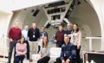
Towards this goal, a team headed up by Karol Lang at the University of Texas at Austin is investigating the use of positron emission tomography (PET) to help in the transition to clinical FLASH therapy. The researchers have now demonstrated the first ever recorded PET imaging and dosimetry of a proton FLASH beam.

“We have successfully realized tests which open a new PET modality with proton FLASH beams leading to improved monitoring of irradiations and image-guided FLASH proton therapy,” the researchers write in Physics in Medicine & Biology.

PET verification

In-beam PET scanning can verify proton range by mapping positron emitters (such as 15O, 13N or 11C) generated along the beam path as the protons interact with tissues in the patient. During FLASH delivery, the instantaneous positron intensity could be up to 1000 times higher than in conventional proton therapy. In addition, the sub-second FLASH spill means that the signal does not experience the washout seen during longer beam deliveries. This strong and fast signal could enable unique monitoring of proton therapy, but also poses challenges for the PET instrumentation, which must function within an intense radiation zone.

For their experiments, Lang and collaborators used the 75.8 MeV proton FLASH beam at the MD Anderson Proton Therapy Center. This beam delivers a dose rate of about 163.7 Gy/s and has an intensity of about 3.5 x 10¹⁰ protons during each 101.5 ms FLASH spill. They developed a PET “mini-scanner” using two PET modules assembled from LYSO:Ce scintillation crystals. Each module incorporates two 8 × 8 arrays of crystals, with each crystal coupled to a silicon photomultiplier.

The researchers initially characterized the proton FLASH beam using detectors known to operate well in high radiation environments. They first irradiated a PMMA block with the FLASH beam and measured the emitted radiation using a cadmium–zinc–telluride-based M400 gamma imager, which can measure annihilation gamma rays and prompt gammas at up to 140,000 counts/s.

The resulting spectra showed peaks due to positron annihilation gammas (511 keV) from induced isotopes and pair production interactions of prompt gammas, as well as prompt gammas from 12C emitted from the target. Overlaying a 2D image reconstructed from the annihilation gammas onto an optical image of the experiment provided visualization of the 2D size and range of the FLASH pencil beam in the phantom, during and immediately after irradiation.

Proof-of-principle

Following this initial assessment, the researchers assessed the PET mini-scanner’s potential for imaging and dosimetry of a FLASH beam irradiating a cylindrical PMMA phantom. They used the PET modules to measure the yield and time evolution of activated positron emitters in the phantom, which was centred on the beam axis. The system recorded results over 200 s, which included three 101.5 ms FLASH spills.

During this 200 s acquisition, the system detected 163,394 coincidences (events detected in two opposing crystal arrays within a 10 ns timeframe) in 3815 distinct pairs of readout channels. Plotting the number of observed coincidences over time revealed clear jumps in phantom activation by the three FLASH spills. However, due to the high rate of hits in the PET crystals, which the readout and data acquisition system were unable to process, the PET system readout dead-timed during each spill.

“The bottom line is that our system works well with after-spill events (due to activated positron-emitters) but requires a new approach to image in-spill prompt gammas due to excitations of a multitude of isotopes,” says Lang. “We have ideas how to do it and we are proposing it to the NIH [National Institutes of Health].”

PET images reconstructed from lines-of-response

The researchers determined the line-of-response (LOR) for each coincidence crystal pair and used these LORs to reconstruct images with a voxel size of 1.5 mm3. Despite a limited field-of-view (the setup geometry meant that only the lower crystal arrays were sensitive to back-to-back gammas emitted from the activated phantom), the images clearly showed the path of production of positron-emitting isotopes.

This ability to count events and LORs demonstrates the dosimetric capability of the in-beam PET mini-scanner. The researchers point out that that full analysis of the PET images and dosimetry is underway, but that this work offers a first glimpse of “such unprecedented possibility for a FLASH beam”.

“The main motivation for this report is to share with the public our recent results of studying FLASH beam data before more elaborate measurements can be conducted. Our results deliver encouraging news about PET imaging of FLASH beams,” they conclude.

Lang tells Physics World that the team recently submitted another publication reporting on beam exposures of six phantoms and analysis of data recorded both during the 101.5 ms spill and up to 20 min afterwards. “These unprecedented studies pave a way to a new important PET modality that will improve the overall outcome of proton therapy,” he says.

JWST finds ‘smoking gun’ evidence of early galaxies transforming the universe

Using the James Webb Space Telescope (JWST), an international team of astronomers has found compelling evidence that early galaxies were responsible for the reionization of the early universe. This is the process by which neutral hydrogen atoms are ionized, making the universe transparent to light at wavelengths that would have been absorbed by the atoms. The research was done by members of the EIGER collaboration, which is using the JWST’s Near Infrared Camera (NIRCam) to study light from quasars in the early universe.

The cosmological era of reionization occurred around a billion years after the Big Bang. Prior to reionization, neutral hydrogen gas between early galaxies absorbed light at certain wavelengths. Then something caused the cosmos to heat up, ionizing the gas to create a plasma. These regions of plasma were less efficient at absorbing light, creating “bubbles” of transparency in the universe.

These bubbles were much larger than the galaxies themselves, with diameters of about 4 million light–years across. Over the hundred million years or so, the bubbles grew and joined together, and eventually the entire universe became transparent. However, exactly what caused this reionization is an important cosmological mystery.

Default explanation

“It was presumably some source of ionizing radiation,” team leader Simon Lilly tell Physics World. “It’s fair to say the default explanation was always that it was ultraviolet light from the first stars and galaxies that formed early in the universe,” explains Lilly, who is based at ETH Zurich in Switzerland. “Some of those stars will be hot enough that they produce a lot of ultraviolet photons, and in principle, that could do it.”

Other sources of ionizing radiation have also been suggested. These include the generation of ultraviolet radiation and X-rays by the acceleration of material as it falls into black holes.

Now, Lilly and colleagues have studied reionization by using the JWST to observe light from an ancient quasar. This is a supermassive black hole that swallows large amounts of material, causing the emission of huge amounts of radiation. By studying how this light passed through ancient galaxies on its way to Earth, the team discovered a correlation between the locations of these galaxies and the locations of patches of reionized gas. This allowed the researchers to conclude that something within these galaxies, possibly young stars, ionized the surrounding space.

This is the smoking gun, that it was galaxies that did the reionization

Simon Lilly

Lilly together with Daichi Kashino at Japan’s Nagoya University and colleagues describe their findings in a paper in The Astrophysical Journal. This paper is one of three published in that journal related to this research. The second paper is by ETH Zurich scientist Jorryt Matthee and colleagues and looks at the properties of the galaxies, and the third paper looks at the quasar itself and is by Anna-Christina Eilers at the Massachusetts Institute of Technology.

The team will now investigate five other quasars and their lines of sight to Earth in order to further explore the connection between galaxies and reionization.

“We’re not the first to discover galaxies at these epochs, and indeed, the JWST is finding galaxies at significantly earlier epochs, but what the telescope really enabled was for us to produce very large, homogeneous samples of galaxies,” Lilly said. “I would have no hesitation in saying we couldn’t have done this without the JWST.”

For Lilly, who has been associated with the telescope since its early planning stages in the 1990s, this result is an important validation that the JWST is useful beyond taking incredible images of the universe; it has real experimental value too.

“We’re seeing the universe as it was a few 100 million years after the Big Bang, and we’re doing physics there,” Lilly concluded. “We are actually answering questions with the JWST, not just standing in awe of the majesty of the universe.”

Life in space impacts human brain structure

Spaceflight – both short-duration space shuttle missions and longer periods living on the International Space Station (ISS) – alters the human body, including widespread changes in the brain. By studying brain scans of astronauts before and after space travel, a multi-institutional research team has found that ventricles – fluid-filled cavities in the brain – expand significantly in longer spaceflight missions, and that inter-mission intervals of less than three years may not be long enough for them to fully recover.

Time spent in space induces displacement of intracranial fluid and an upward shift of the brain within the skull, causing cortical crowding and narrowing of the sulci, the grooves in the cerebral cortex, at the top of the brain. With future space exploration including more long-duration missions, it is imperative to understand the effects of space travel on the brain.

With this goal, Rachael Seidler and Heather McGregor of the University of Florida and co-researchers studied the brains of 30 astronauts, evaluating changes in grey matter (GM) volume, white matter (WM) microstructure, extra-cellular free water (FW) distribution and ventricular volume from before to after spaceflights. The group included both novices and experienced astronauts, on missions ranging from two weeks to 12 months.

In a prospective study conducted between 2014 and 2020, the researchers obtained pre- and post-flight data (T1-weighted and diffusion-weighted MRI scans) from 13 astronauts who spent six months and two who spent 12 months in space. They also examined retrospective MRI data from the NASA Lifetime Surveillance of Astronaut Health Repository. This included eight astronauts who completed a short-duration mission of up to two weeks, and seven who spent six to 12 months on the ISS.

The goal of the study was to understand whether longer missions or shorter recovery periods between missions have a particularly significant impact on the brain. The team examined whether and how spaceflight-induced brain changes are associated with individual differences in spaceflight experience, factoring in the duration of the mission, the experience level of the astronaut, the number of previous missions and the time elapsed since a previous mission.

The researchers compared pre- and post-flight diffusion-weighted MRI data for the astronauts, assessing group-level changes in GM volume, ventricular volume, FW fractional volume, and FW-corrected WM diffusion indices. They developed models that adjusted for individual differences in age at the time of launch, sex, current mission duration, and the number of days between landing and the post-flight MRI scan. For each astronaut, they calculated FW difference images reflecting changes in FW fractional volume and performed ventricular volume analysis.

Pre- to post-spaceflight changes in ventricle volume

These analyses demonstrated that ventricular volume increased during spaceflight, with longer missions resulting in greater ventricular enlargement that tapered off after six months in space. The study did not resolve the length of time for the ventricles to fully recover post-flight, or to what extent they recover. But based on the findings, described in Scientific Reports, the team suggests that at least three years or longer intervals between spaceflight missions are needed for intracranial fluid to return to normal levels and for the ventricles to fully recover.

The study also confirmed previous published findings that spaceflight induces grey matter shifts and FW redistribution. The researchers determined that longer missions induce greater fluid shifts. Among the experienced astronauts, the number of years elapsed since the previous mission was significantly associated with post-flight volume changes for all four ventricles.

Seidler and colleagues point out that the rate of ventricular expansion during spaceflight exceeds that seen with normal aging, suggesting that post-flight ventricular enlargement is not a consequence of brain atrophy with aging, but instead arises from cerebrospinal fluid changes. They suggest that this enlargement may be a compensatory response to accommodate fluid shifts toward the head that occur in microgravity.

The researchers note that variation in astronaut age did not account for observed individual differences in structural brain changes, and that there were no differences in spaceflight-induced structural brain changes between novice and experienced astronauts. Rather, brain changes differed according to the number of prior flights the experienced astronauts had completed. “This suggests that the brain is impacted by the cumulative effects across multiple flights and perhaps separate bouts of adaptation to microgravity and the spaceflight environment,” they write.

“Collectively, our work suggests that longer missions, multiple flights and shorter inter-mission recovery time induce greater intracranial fluid changes. Moreover, the findings suggest that neuroplasticity changes resulting from adaptation to microgravity may not be dependent on previous spaceflight experience,” they conclude. Seidler tells Physics World that the team is now conducting a new study with long-term post-flight follow up, up to five years after a mission.

Why it would be better if everyone thought like a scientist

In the 1950s many science-fiction movies and TV shows took it for granted that, by the 21st century, we’d be living in a society that understood and respected science. Science and engineering had made significant contributions to the Second World War including the development of radar and nuclear power. Over the years, our daily lives have been transformed, lengthened and enriched by science in ways that have exceeded the imaginations of science-fiction writers.

It is disturbing, then, that a significant portion of the public deny scientific evidence on a wide range of topics from climate change to the safety of vaccines and even the shape of the Earth. This science scepticism is highly selective. Few doubt or distrust science when it brings flatscreen TVs or smartphones, but some draw the line when it conflicts with their cherished preconceived notions. For many, their views on vaccines or climate change are fixed in stone – unwavering no matter how much evidence or logical arguments are presented to contradict their opinion.

Psychology and neuroscience have identified a phenomenon termed “belief perseverance” that accounts for why everyone resists new information that conflicts with our preconceived notions. More evidence or better arguments are not the answer. The more information that comes in that conflicts with a pre-existing worldview, the more our brains reject it – known as the “backfire effect”. The worldviews of many are tied to their political affiliations, and when new information such as the effectiveness of masks and vaccines during the COVID-19 pandemic becomes politicized, it can be a serious problem.

Scientists are in a unique position to counter the backfire effect. We have been trained to be open to new information, test it for validity, and most importantly, be willing to change our minds in the light of new information. For a scientist, there is a singular pleasure in discovery, in learning that something that one previously took as established fact is not in fact. We are always on the lookout for something new and unanticipated, while simultaneously guarding against errors and mistakes.

When I was a graduate student, another student told me: “When you see something remarkable in your research, don’t think Nobel prize, think ‘How did I screw this up?’ Assume it’s wrong and try to find your mistake. If there’s no error, great. But if there is an error, and you don’t find it, you can be sure that someone else will.”

To counter the defensive reactions to new information that challenges our preconceptions, everyone should learn to think like a scientist

There are two important points here. The first is that reality exists, and that data are data. If you are seeing a real effect, others will confirm it. And if not, they won’t. Second, be open to doubt and uncertainty. There are a host of observations and results for which we do not have a valid scientific explanation. Scientists do not see this as a flaw in the scientific method, but as an opportunity. When faced with something we don’t understand, scientists don’t throw up their hands, rather, they roll up their sleeves.

Scientists will often disagree about how to interpret a particular result, but we all agree on what constitutes a valid observation. For example, only after thorough checking for mistakes or statistical or experimental artefacts would over 97% of climate scientists reach agreement on a warming global temperature driven by anthropomorphic causes, indicating that one can trust its validity. After all, there is nothing a scientist loves more than proving that their colleagues are wrong – it’s almost as good as making a new discovery. Expertise matters and in cases such as this, you want to give weight to those who specialize in climate science, for they are intimately familiar with all the ways one can be fooled or led astray.

Guiding principles

To counter the defensive reactions to new information that challenges our preconceptions, everyone should learn to think like a scientist. Of course, there are many examples where a scientist has refused to accept data that contradict a favourite theory or model. It takes continued and sustained effort to learn to be your own harshest critic and to admit that something you took to be true is in fact not so. Rather than dogmatic certainty, one must embrace doubt and be open to changing one’s mind.

Science is, of course, not a set of answers, but rather a process, a way of asking questions of the world

The essence of thinking like a scientist, and escaping from the trap of the backfire effect, can be summarized in one single question: What if I’m wrong? Asking this is all it takes to think scientifically. Science is, of course, not a set of answers, but rather a process, a way of asking questions of the world. Science also consists of agreed-upon criteria for what constitutes a proper answer. Sometimes these facts are presented as “science”. But new studies can lead to new information, and in the best-case scenario, our understanding of the world and how it works improves.

New discoveries may lead to previously established results being shown to be not the full story. That does not mean that we can’t know anything – rather, it is the promise that tomorrow our understanding will continue to improve. The challenges we face, from pandemics to climate change to AI, are too great to let the backfire effect dictate the choices we make. We are constantly being bombarded by people who know that when we are reacting emotionally, we can be more easily manipulated.

In the 21st century, science should be recognized as universal. The principles that improve our lives should not be ignored or rejected when politically inconvenient.

Electron ‘kick’ removes single atoms from 2D material

A beam of electrons can “kick” single atoms out of a two-dimensional sheet of hexagonal boron nitride (hBN) in a controllable way, defying predictions that electron irradiation would be too damaging for this purpose. Even more remarkably, the physicists behind the discovery predict that a higher-energy version of the same technique could preferentially remove nitrogen atoms from the hBN lattice, which is unexpected since nitrogen is heavier than boron. The empty spaces, or vacancies, left behind by the “missing” nitrogen atoms could have applications in quantum computing, communication networks and sensors.

 Nitrogen vacancies in hBN have optical properties that make them ideal for use in emerging quantum and optoelectronic devices. The downside is that they can be difficult to isolate, but  researchers at the University of Vienna led by experimental physicist Toma Susi have now found a way to do it using a technique called aberration-corrected scanning transmission electron microscopy (TEM).

 “Transmission electron microscopy allows us to image the atomic structure of materials and it is particularly well suited to directly reveal any defects in the lattice of the sample,” explains Susi. “Aberration correction provides us with the resolution to observe single atoms – it’s like using eyeglasses to see more clearly – but it can also be used to remove these atoms.”

Previously, TEM measurements were usually conducted under relatively poor vacuum conditions. In these circumstances, the gas molecules that remained in the instrument could easily damage hBN samples by etching away atoms in the material’s crystalline lattice. The high-energy electron beam can also damage the sample via elastic collisions with the electrons in the beam or electronic excitations.

Lattice damage is greatly reduced

Susi and colleagues overcame these problems by operating the TEM in near ultrahigh vacuum conditions and testing different electron-beam energies between 50 and 90 keV. They found that the lack of residual gas molecules under the improved vacuum suppresses unwanted etching effects, which occur extremely fast and would otherwise prevent single atoms from being controllably removed.

What is more, the team found that the TEM could create single vacancies of either boron and nitrogen at intermediate energies. Although boron is twice as likely to be ejected at energies below 80 keV because of its lower mass, at higher energies, the team predict that nitrogen will become easier to eject, thus allowing this vacancy to be preferentially created. “To create these vacancies, nothing special is needed,” Susi tells Physics World. “The electrons used for imaging have enough energy to knock out atoms in the hBN lattice.”

The fact that the researchers performed measurements over many electron energies allowed them to collect robust statistics on how the missing atoms are generated, something that will be useful for developing a future theory for how vacancies can be created using a TEM.

“Now that we are able to predict how much we need to irradiate the material at each energy to kick out nitrogen or boron atoms, we can design experiments that optimize the desired distribution of vacancies,” Susi says. “We’ve also pioneered atomic-level manipulation by directing the electron beam at individual lattice sites.

“We previously thought hexagonal boron nitride would damage too quickly to be suitable for such treatment. We’ll have to reconsider that now.”

Susi says that the next step will be to generalize the results beyond hBN. “With better theoretical models, we could predict how the beam interacts not only with hBN but potentially other materials, such as graphene and bulk silicon,” he says.

The researchers detail their work in Small.

Impact flashes help protect satellites from space junk, novel uses for ultrasound

NASA tracks about 23,000 pieces of space debris bigger than a grapefruit orbiting Earth. This space junk is of human origin and includes defunct satellites, bits discarded by space missions, and pieces created by the fragmentation of the previous two categories. Furthermore, NASA estimates that there are millions of pieces in orbit that are too small for the space agency to keep track of.

Some of this junk is travelling as fast as 28,000 km/h and even a small piece can do serious damage if it collides with a satellite. As a result, the people who design spacecraft are keen to protect their missions from space junk.

Now,  Gary Simpson, KT Ramesh, and colleagues at Johns Hopkins University in the US have done a spectacular study that provides further insights into how tiny bits of high-speed space junk impact objects. The study is quite literally spectacular because of the videos that they have taken of the impacts.

Ultrafast jet

The team fired stainless steel spheres at an aluminium plate at a speed of 10,000 km/h. The resulting impact flashes were captured by a high-speed camera (see video) and the team studied the intensity and spectral nature of the light. The images show the emergence of an ultrafast jet of material that blasts out from the point of impact.

The team found that the composition of the target and the size of the jetted particles could be inferred from the flash – which could prove useful for studying the impact of space junk on satellites. The research is described in PNAS Nexus.

We do like a good story about ultrasound here at Physics World, and we have written about research covering everything from medical applications to ultrasound-based tractor beams. Today, I was pleased to discover two uses of ultrasound related to food and drink.

Frozen chops

If you eat meat, you might be tempted to defrost a frozen chop in the microwave if you don’t have the patience for defrosting it at room temperature. Now, researchers in China and Australia have investigated whether it’s a good idea to use ultrasound to defrost pork.

Zhongyuan Chen and colleagues found that ultrasonic assisted thawing decreased the thawing time by as much as 65% when compared to conventional water-immersion thawing. The pork also exhibited better water retention and tenderness. You can read more in Food Physics.

Meanwhile in Spain, researchers at the University of Castilla-La Mancha and the University of Murcia have developed a way to use ultrasound in a key step in the making of rosé wine called maceration. Maceration involves the leaching of tannins and other compounds from the skins of grapes. Some of these compounds give red and rosé wine its colour and flavour.

Colouring wine

High-powered ultrasound has already been used in the maceration of red wine, but it had not been clear if it could be used for making the much paler rosé. This involves shorter maceration times to avoid creating wines with an undesirable dark colour and flavours.

Now, Encarna Gomez Plaza and colleagues have shown that ultrasound-driven maceration (called sonication) created wines with “superior aroma”. Indeed, the researchers say, “Sonication gave rise to wines with intense red berry and flowery odours, with scores higher than those of wine from macerated grapes”.

They describe their work in the Journal of the Science of Food and Agriculture.


UK scientists dismayed over lack of progress on Horizon Europe membership

Scientists have expressed their disappointment at the lack of progress in negotiations over the UK re-joining the €95bn Horizon Europe research programme. Reports last week had suggested that UK and European Union negotiators had agreed a draft deal that could be signed this week. But a final deal has failed to materialize, leaving British membership of the programme, which it had previously played a strong part of, still uncertain.

The UK government has long maintained that it wishes to re-join Horizon Europe, which began in 2021. Britain had been a full and highly successful member of previous EU research programmes for decades. Its ongoing participation was agreed at the end of 2020 as part of the post-Brexit trade deal between the UK and EU. Membership stalled, however, and became a bargaining chip in disagreements over Northern Ireland, which have now been resolved.

There were expectations that UK prime minister Rishi Sunak would confirm the agreement during a meeting with European Commission president Ursula von der Leyen on the fringes of the NATO summit in Vilnius in Lithuania, which took place on 11–12 July. On the way to Vilnius, Sunak reiterated the government’s preference to associate to Horizon, but added that it must be “on terms that work for the UK and are in the UK’s best interests”.

Despite Sunak and von der Leyen meeting, no deal was announced, with von der Leyen taking to Twitter to note that the pair had discussed Ukraine as well as “broader geopolitical challenges, including China, and bilateral issues”. She did not, however, mention Horizon Europe. The EU did announce, though, that New Zealand has now joined Horizon Europe as an associate member.

No progress

As part of a statement released by European Movement – a cross-party group campaigning to reverse Brexit – Martin Rees, UK Astronomer Royal, outlined the danger of further delays. “We cannot waste any more time. New Zealand is now on board and we should be too,” says Rees. “Sunak may think he’s securing greater value via protracted negotiations, but prolonging the delay leads to further missed opportunities and will make it harder for UK science to restore its standing and its collaborations.”

That view is echoed by Carsten Welsch, an accelerator physicist from the University of Liverpool. “If we want to be serious about being a science superpower, we need a scheme like Horizon Europe – not just for the funding, but to support collaboration and knowledge exchange across borders,” he told Physics World. “It is disappointing to see that not more progress has already been made, given that the importance of full association of the UK with Horizon Europe”.

Welsch notes that non-association is damaging UK science, adding that he had been awarded a major grant “only to subsequently see it being transferred to another institution” – an experience he described as “heartbreaking”. “We have seen outstanding researchers leave the UK and transfer their prestigious [European Research Council] grants to other countries,” says Welsch. “This loss of talent will be very difficult to turn around.”

Ask me anything: Lilly Liu – ‘We need team work: it’s impossible for one individual or team to solve a problem’

Lilly Liu

What skills do you use every day in your job?

I teach undergraduates and head up a team of more than 10 people – so managing, teaching and communication skills are vital. This type of project management does not, however, just involve organizing people. It also requires an in-depth scientific knowledge to solve different technical questions and an ability to know how best to distribute resources to achieve our end goals. At the same time, when our group’s PhD students and postdocs run into setbacks and frustrations, I have to encourage them through these difficulties and help them develop their research ideas, interpret experimental results and write up papers and reports. I always try to learn about my colleagues’ long-term career plans so I can point them to the right training and help them build the right skills for the future.

What do you like best and least about your job?

Whenever we get new results, we need to know how to interpret and make sense of the data. So what I like best is working with our PhD students and postdocs to go over the data, try to figure out the underlying science, and establish if anything new is going on. In doing so, we can identify gaps in the research that have to be addressed to achieve the end goals and understand the materials better.

I also enjoy crafting research proposals. Many find this process tedious and difficult, but I like it because by addressing the questions demanded by the relevant funding agency, my ideas and hypotheses about a particular problem become clearer. Whenever I start drafting a proposal, I usually only have a general idea about how to solve a problem of interest to industry. But as I refine my proposal with my team, we can discuss the ideas in detail and in a systematic way.

In addition, I like working with others as a team or a wider consortium from academia and industry. These days it would be impossible, especially in nuclear or aerospace, for one individual or one team to solve every problem. We need team work, with everyone bringing their own skills and specialities.

On the flip side, while I enjoy basically everything I do, the high workload takes some of the fun away. Over the past few years, I’ve been trying to polish my time-management skills and optimize processes to help with this. For example, I’ve taken training on a Female Leadership Initiative from my university and gathered some tips from my cohort, which I’ve found extremely helpful.

What do you know today that you wish you knew when you were starting out in your career?

When I was starting out, I thought that having a permanent position in academia meant I’d be free to do whatever research I wanted. But when I became a group leader, I realized it’s not like that. You’re at a different stage of your career, with very different responsibilities from being a PhD student or research fellow. Had I known this, I’d have taken the chance to fully enjoy every stage of my career, first as a PhD student, then as a postdoc and an independent research fellow as well as now. The pandemic has also reminded me to cherish life even more and to enjoy every single day I live.

India launches Chandrayaan-3 mission to the lunar surface

India’s third Moon mission has been successfully launched today at 14:35 local time from the Satish Dhawan Space Centre in Sriharikota in the state of Andhra Pradesh. Costing $75m, Chandrayaan-3 consists of lunar lander and rover and was fired into space by the country’s Launch Vehicle Mark 3 rocket.

India’s first mission to the Moon – the Chandrayaan-1 orbiter – was launched in 2008 and featured 11 payloads, six of which were built by outside countries. The follow-up mission – dubbed Chandrayaan-2 – was launched in 2019 and consisted of an orbiter as well as lander and rover.

While the orbiter was successfully inserted into lunar orbit, the lander, dubbed Vikram, failed to touch down softly on the Moon due to a breaking issue as it neared the surface. Its planned target was between the craters Manzinus C and Simpelius N, close to the lunar south pole.

With Chandrayaan-3, India is now trying again to land on the Moon in a similar spot as the Chandrayaan-2 mission with a rebuilt Vikram lander that contains a six-wheeled rover named Pragyaan.

The mission is now making its way to the Moon and if all goes to plan then the landing is planned for 23 or 24 August. If that is successful, the lander will then release the rover to make a series of sorties over 14 Earth days with a range of about 500 m. This will allow it to study the lunar rocks and soils with its five instruments that include laser and X-ray spectrometers.

If successful, India will join China, Russia and the US in successfully landing a craft on the Moon. Yet India would be the first nation to land at the lunar south pole.

Fractional quantum Hall state appears in ultracold atoms

Physicists at Harvard University in the US have created a novel strongly interacting quantum liquid known as a Laughlin state in a gas of ultracold atoms for the first time. The state, which is an example of a fractional quantum Hall (FQH) state, had previously been seen in condensed-matter systems and in photons, but observations in atoms had been elusive due to stringent experimental requirements. Because atomic systems are simpler than their condensed-matter counterparts, the result could lead to fresh insights into fundamental physics.

“Some of the most intriguing phenomena in condensed-matter physics emerge when you confine electrons in two dimensions and apply a strong magnetic field,” explains Julian Léonard, a postdoctoral researcher in the Rubidium Lab at Harvard and the lead author of a paper in Nature on the new work. “For example, the particles can behave collectively as if they have a charge that is only a fraction of the elementary charge – something that does not occur anywhere else in nature and is even ruled out by the Standard Model for all fundamental particles.”

The way in which such fractional charges arise is still not fully understood because it is difficult to study solid-state systems at an atomic scale. This is why it is so desirable to study the behaviour of FQHs in synthetic quantum systems such as cold atoms, which act as quantum simulators for more complex condensed-matter phenomena.

In the latest study, for example, members of the Harvard team directly observed particles in their atomic system moving around each other in a circular pattern, rather like “dancers in a waltz”, says Léonard.  “This vortex motion is too small to see in a solid-state sample, but we are able to resolve it in our experiment,” he tells Physics World.

Making atoms behave more like electrons

To create the Laughlin state, Léonard and colleagues used overlapping laser beams to form a periodic lattice potential made from light. They then placed atoms into each lattice site and tuned the parameters of the beams so that the atoms were free to “hop” between sites. This setup mimics the periodic potential experienced by electrons in a crystalline solid, Léonard explains. “The only difference is that our artificial crystal is more than 1000 times larger, so we can observe and control each ‘electron’ with an optical microscope,” he says.

One major challenge for the Harvard team was to mimic the electrons’ response to magnetic fields. While negatively charged electrons experience a force (the Lorenz force) in a direction perpendicular to their motion when placed in a magnetic field, the atoms that play the role of electrons in the new platform are electrically neutral, meaning that this force is absent. The researchers therefore had to “trick” the atoms into behaving more like electrons in a magnetic field.

To do this, they relied on the fact that when electrons circumnavigate a magnetic field, their wavefunction acquires a phase. This is known as the Aharonov–Bohm effect, and Léonard explains that they were able to create an equivalent in cold atoms. “In our experiments, we made use of several laser beams that applied exactly this phase to the atoms’ wavefunctions,” he says.

Possibility of observing anyons

The team also faced challenges in creating the strong, precisely engineered magnetic field required to observe FQH states, which had previously remained out of reach for laboratory experiments, Léonard adds. “We have now shown for the first time that it is possible to study strongly correlated systems under a magnetic field in a quantum simulator,” he says. “It is therefore now possible to study such states on a microscopic level and glean new insights into them. We may even discover completely new phenomena that have so far remained inaccessible.”

While the number of atoms in the FQH Laughlin state observed by the researchers is small, at just two atoms across 16 lattice sites, the team believes the system size could be increased. “A larger system will allow us to gain an even better view of the physics that underlies the FQH effect and one aspect we are particularly excited to observe are the excitations in such systems,” says Léonard. “These are believed to be neither fermions nor bosons, but so-called anyons, which are a completely new type of particle that fall outside our usual classification of quantum statistics.”

Copyright © 2026 by IOP Publishing Ltd and individual contributors