Synthetic skin removes the need for human volunteers in mosquito bite trials

Mosquitoes are often considered the world’s most dangerous animal. While feeding on blood through the skin they transmit pathogens that cause deadly diseases such as malaria, dengue, Zika and yellow fever. According to the World Health Organization, mosquito-borne diseases are responsible for more than 700,000 deaths each year.

Studying mosquito feeding behaviour could help develop countermeasures to decrease disease transmission. Such studies, however, have historically used live animals or human volunteers as a food source. What’s really needed is a high-throughput automated assay that can screen potential new mosquito repellents without the need for live volunteers. Researchers at Rice University and Tulane University have now developed a biomaterial-based platform that could be a promising step towards that goal.

“It’s a huge game changer,” says mosquito expert Dawn Wesson from Tulane’s School of Public Health and Tropical Medicine in a press statement. “If we can study how mosquitoes feed, what they do in the process of feeding, we can better understand their potential for transmitting diseases and possibly do things to stop them from feeding.”

“We hope that this platform could rapidly identify promising candidates for more effective repellents to decrease the spread of disease in the future,” says first author Kevin Janson from Rice University.

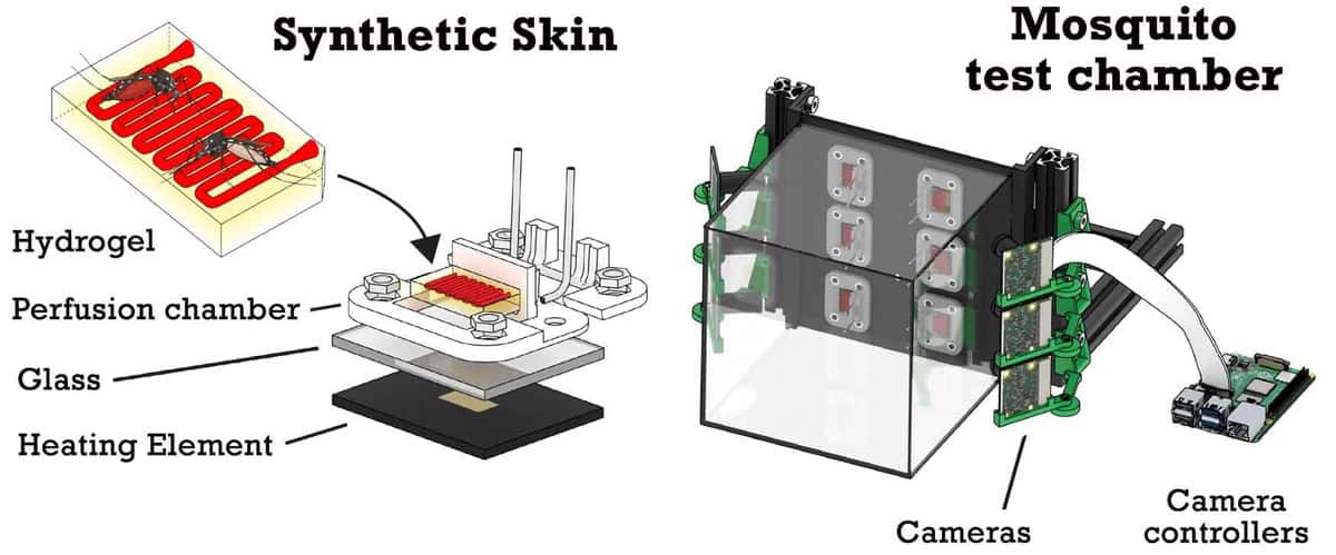
The mosquito feeding platform, described in Frontiers in Bioengineering and Biotechnology, uses biocompatible hydrogels to mimic skin. The researchers 3D printed rectangular patches of hydrogel containing a simple vascular network that can be filled with blood or other liquids. They note that the hydrogels can be printed on a large scale at a low cost and refrigerated until needed.
The mosquito feeding platform

The hydrogel patches are mounted on perfusion chambers to provide a continuous supply of blood, along with a heating element for temperature control. For feeding tests, up to six of these patches are housed within a glass cage to confine the mosquitos, with a Raspberry Pi camera aimed at each patch to recordthe insects’ landing locations and feeding patterns.

Over a period of six months, Janson and colleagues collected more than 180 recordings of mosquito feeding patterns. For these experiments they filled the hydrogels with animal blood, introduced 20–30 mosquitos into the cage, and recorded 30–45 min of feeding activity.

The researchers then used these videos to train a machine-learning model to detect mosquitoes in the camera’s field-of-view. They also trained the model to identify whether a mosquito had an engorged abdomen (from feeding) or not (non-feeding).

The trained model could identify mosquitoes with 98% precision, and was able to classify abdomens as engorged or not with 96% precision. Janson notes that the machine learning model can automate experimental analysis and provide results far more quickly and consistently than a human could.

To feed or not to feed

To determine whether mosquitoes feeding on the hydrogels were attracted to the blood itself, the researchers performed experiments using three cages containing hydrogels perfused with blood, red ink or phosphate-buffered saline (PBS) as a control, all heated to 37°C. They introduced about 20–30 mosquitoes into each cage and observed their feeding behaviour.

Blood-infused hydrogels

While the mosquitoes spent a substantial amount of time around all of the hydrogels, they only fed on hydrogels containing blood. This experiment confirmed that hydrogels do not inherently attract mosquitoes, and that a chemical component of blood, rather than its visual appearance, attracts the insects.

The researchers also used their mosquito feeding platform to evaluate the effectiveness of two repellents: 25% DEET and a plant-based repellent derived from the oil of lemon-eucalyptus plants (OLE). They again prepared three feeding cages, containing hydrogels coated with DEET, OLE or PBS. All hydrogels were perfused with blood heated to 37 °C.

The experiments successfully validated the repellent effects of DEET and OLE: none of the mosquitoes given repellent-coated hydrogels fed on the blood, while 13.8% of mosquitoes in the control cage did. The team notes that this relatively low feeding rate in the control is due to the small surface area of the hydrogels, which could be increased in future platforms.

The platform is currently optimized for laboratory mosquitoes, mostly Aedes aegypti in this study. It could, however, be adapted to analyse mosquitoes in the wild, which often exhibit different feeding tendencies. Deploying the platform in the field poses several challenges, but the researchers believe these could be addressed, for example, by using car batteries as a mobile power source and replacing blood with an artificial protein source.

Currently, the team is using the platform to investigate the viral transmission of dengue during mosquito feeding; future studies will involve malaria parasites. “Given the scale at which our hydrogels could be produced and the low cost of other components, we speculate that our platform could be adapted to support high-throughput testing in the future,” Jansen tells Physics World.


Controlling foam in bottom-up beer, why some icicles have a rippled shape

I live in southern England, where the perfect head of beer is absolutely no head at all. So it’s always a shock to travel elsewhere and be served a glass of beer with a tall, frothy head. A good head is apparently a sign of quality in beer, so people who make and serve the stuff (outside of England) are keen to get the head just right.

Now, physicists in South Korea and Germany have done an experimental and numerical study of beer foam that could help barkeepers to rapidly pour a glass from the bottom-up, while ensuring that the desired amount of foam is created.

If you are wondering what bottom-up pouring is, I can tell you because I just had the pleasure of seeing it in action in a Japanese restaurant in London. The beer is served in a cup with a hole in the bottom that is surrounded by a magnet. Beer is pumped into the hole and when the cup is full, a magnetic disk seals the hole. The result is a rapid fill and a beer with absolutely no head – or at least that’s how it was served in London.

Multiphase solver

The study is claimed to be the first investigation of beer froth using a multiphase solver, as team member Wenjing Lyu explains. “Simulation of a bottom-up pouring process using a multiphase solver is a complex task that involves modelling the physical and chemical interactions that occur during the process, such as fluid dynamics, heat and mass transfer, and chemical reactions.”

Lyu adds “By using a multiphase solver, it is possible to accurately predict the behaviour of the system and optimize the design of the nozzle outlets and the cup geometry to ensure the fastest possible bottom-up pouring under various conditions such as pressure, temperature, and carbonation.”

While the researchers were focused on filling speed, I hope that the work will allow users of bottom-up systems to achieve the perfect head of beer for their customers. While punters in London seemed very happy with a zero-foam pint, I don’t think that the German members of the team – some of whom are from Bavaria – would be happy to drink a foamless stein of beer during Oktoberfest.

The researchers describe their study in AIP Physics of Fluids.

Spiky structures

Sticking with the physics of fluids, have you ever wondered why icicles tend to have a rippled shape, with alternating ridges and valleys running down the length of the spiky structures? This mystery has puzzled physicists for centuries, and now the answer may have been found by Menno Demmenie and colleagues of the Institute of Physics and the Van ’t Hoff Institute for Molecular Sciences at the University of Amsterdam.

The team built an icicle machine in which water is trickled in sub-zero temperatures. The team then first determined the ideal flow rate of water required to create an icicle. If the water comes too fast, it just drips onto the floor, and if it comes too slowly, the icicle grows up rather than down.

By changing the salt content of the water and using colouring to monitor flow, the team came up with an explanation for why ripples form. When pure water was used, few ripples were seen and the icicles resembled candles. However, with salty water, the salt gets pushed out of the bulk ice and onto the surface of the icicle. This creates a film of salty water that flows along the surface of the icicle, forming the rippled structures.

The research is described in Physical Review Applied.

Ask me anything: Zahra Hussaini – ‘We’re working on autonomous driving technology that has the potential to transform lives’

Zahra Hussaini

What skills do you use every day in your job? 

Most of my job is collaborative problem solving. SREs are software engineers that focus on the overall reliability and performance of large software systems, and we work in close collaboration with the software engineering teams that built them. I model and reason about software in the same way that I was taught to think about the natural world in my physics classes. Sometimes, this involves reading code and predicting how a scenario will play out from first principles. Other times, it’s about designing and running experiments to measure how a system behaves under extreme conditions. Whatever the method, the goal is to find the edges of what the system can do and develop project ideas for how to expand its capabilities.

Technical communication is another job skill that I practised during my physics education. I’m rarely working on a problem alone, which means I need to be able to talk about complex problems and their potentially complex solutions simply and succinctly. When I’m writing project plans, I have to be clear about the assumptions and approximations I’m making; and to clearly describe the logical steps I took to reach a conclusion.

In addition to doing project work, SRE team members share the responsibility of dealing with software outages in real-time. My team runs exercises where we simulate outage scenarios and practise our responses. This part of the job combines diligently preparing for emergencies with quick, creative thinking.

What do you like best and least about your job?

What I like most about my job is the people that I work with. A healthy team culture is essential for every other part of the job. We do better work when people feel safe asking questions and sharing ideas. We build more reliable systems when we view mistakes and failures as an opportunity to learn rather than to blame. It’s important to me (and to Waymo) that we build a culture that makes it easy for people to do their best work because our mission is big. We’re working on autonomous driving technology that has the potential to transform lives.

My least favourite thing is that I don’t get to feel like an expert as much as I’d like. SREs have a lot of breadth because we’re a cross-functional team that works with many different software engineering teams. It’s common to get projects that require getting up to speed in a new area quickly. It’s easy to slip into feeling like I am incompetent and always playing catch up since I’m never done learning. I have to remind myself that this is a normal part of the job, and I have to balance my desire to understand everything with the need to get things done. 

What do you know today, that you wish you knew when you were starting out in your career?

I wish I knew that it was okay to be lost. When I was younger, I felt like I needed certainty and a clear direction if I wanted to achieve anything worthwhile in my career. I wrote and rewrote 10-year plans, trying to find my future. I didn’t have exposure to physics career options other than academia, so getting a PhD was an unwavering fixture of my many plans. For years, I suppressed signs telling me that a research career wasn’t for me, hoping that I could find a way to stick to the plan. When I did finally decide to leave for a career in the tech industry, I had a profound feeling that I had failed. This wasn’t true of course, but it took me a while to realize that and to let go of my rigid planning mentality.

I wish I knew that it was okay to explore, that there are many different paths to happiness, and that you can be good at things without them being the right thing for you. These days, my career plan has three words: follow your curiosity.

Join Physics World’s team of student science communicators

Back in 2017 Physics World set up its student contributor networks – a programme for early-career scientists to work alongside our award-winning journalism team to write and publish news stories for the global scientific community. These contributor networks, made up of PhD students with a passion for science communication, offer a fantastic opportunity for students to improve and refine their scientific writing skills.

We currently have three networks, for PhD students working in quantum science and technology, materials/nanoscience, and medical physics/bioengineering. With training and mentorship from members of the Physics World editorial team, the students in these networks have written articles covering the most exciting new research in their fields. All have benefited from the chance to connect with other members of the network and publish their work on a site read by hundreds of thousands of professional scientists all over the world. Students even get their own page on Physics World, such as these pages of current contributors Rojin Jafari, Maria Violaris and Hardepinder Singh.

Many of our original contributors have now completed their PhDs. Some are now pursuing careers in science communication, writing for us (and other media outlets) as paid freelancers. Others have remained in academia or entered industry jobs, where they can apply their communication skills to write better grant applications, research papers and reports.

What this means is that we are now looking to sign up the next generation of PhD students to join our three contributor networks.

So are you a PhD student with a talent for writing and a passion for communicating the latest research to a broader scientific audience? We’ll provide initial training on how to craft compelling science news stories, and we’ll also ensure that you get regular feedback from other student contributors as well as our professional journalists, providing ongoing support to hone your scientific writing skills.

We expect each contributor to write one or two articles (of about 500 words long) per quarter summarizing the results and significance of some recent research in their field – either a newly published paper or a recent conference presentation. As part of the team, we would also like contributors to provide pre-publication feedback on articles written by other students.

The goal is that after one or two years in the network, every contributor will have a body of well-written, published work that they can include on their CVs – either as evidence of strong communication skills when applying for jobs in academia or industry, or as a springboard to a career in science communication. There’s more information available here.

Join the team

If you‘d like to get involved in the project, and are currently a PhD student in one of the three areas mentioned above, please send the following information to pwld@ioppublishing.org by 31 March 2023:

  • A short (300 words or less) explanation of why you’d like to join the network and what you think you’d bring to it. Previous experience in science communication is not required, but if you have some, please say so.
  • A short (500 words or less) description of what your PhD is about, written so that a physicist in a completely different field (e.g. astronomy) can understand what you’re doing and appreciate why it’s interesting and important.

We look forward to hearing from you.

Advances in in silico trials of medical products: evidence, methods and tools

Want to learn more on this subject?

In silico trials (ISTs) for medical drugs and devices have gained increased popularity as cost-effective alternatives to their clinical counterparts. ISTs promise dramatic reductions in the resources needed for assessing novel technologies and for generating evidence in support of regulatory evaluation for safety and effectiveness. Some have suggested significant cost reductions comparing an all in silico approach versus an equivalent clinical trial with humans. Others have argued for, and reported on, incremental implementation of the in silico methodologies that complement or refine the design of clinical trials based on predictions from the in silico trial outcomes.

This webinar has been launched in collaboration with a special issue in Progress in Biomedical Engineering, a new high-impact Reviews journal from IOP Publishing. The special issue is still welcoming article proposals from the community, and participants are encouraged to discuss contributions with the guest editors.

Want to learn more on this subject?

Alejandro Frangi, University of Leeds, UK and KU Leuven, Belgium. Alex is Diamond Jubilee chair in computational medicine and Royal Academy of Engineering chair in emerging technologies at the University of Leeds, Leeds, UK, with joint appointments at the School of Computing and the School of Medicine. He directs the CISTIB Center for Computational Imaging and Simulation Technologies in Biomedicine. He is Turing fellow of the Alan Turing Institute. Alex is the scientific director of the Leeds Centre for HealthTech Innovation and director of Research and Innovation of the Leeds Institute for Data Analytics.

Aldo Badano, US FDA, USA. Aldo holds a senior biomedical researcher service appointment at FDA and currently serves as director of the Division of Imaging, Diagnostics, and Software Reliability, OSEL/CDRH. His interests are in the characterization and modeling of medical imaging acquisition and visualization systems. Aldo is a fellow of SPIE, AAPM, and AIMBE.


Panelists

Marc Horner, ANSYS, USA
Marc Horner is a senior principal engineer, leading technical initiatives for the healthcare industry at Ansys. Marc joined Ansys after earning his PhD in chemical engineering from Northwestern University in 2001.

Abhinav Jha, Washington University in St Louis, USA
Abhinav K Jha is an assistant professor of biomedical engineering, radiology, and electrical and systems engineering at Washington University. His research interests are in developing task-specific computational medical imaging solutions.

Rebecca Shipley, UCL, UK
Becky Shipley is professor of healthcare engineering at UCL, and director of the UCL Institute of Healthcare Engineering, which brings together engineers, medical and clinical scientists to develop medical and digital technologies. Her research interests lie in mathematical and computational modelling in medicine and biology with an emphasis on multidisciplinary approaches which integrate data from biological experiments, medical imaging and patients.

Ed Margerrison, US FDA, USA
Ed is the director for the Office of Science and Engineering Laboratories at the Center for Devices and Radiological Health, US FDA. The Office is responsible for providing technical expertise and analyses in support of the regulatory processes within CDRH. In addition, the c300 scientists and engineers engage in representing the Agency on International Standards organizations, provide scientific guidance for policy, and “futureproof” the Center for technologies making their way into novel medical devices.

Puja Myles, MHRA, UK
Puja is director of Clinical Practice Research Datalink (CPRD), a real-world data research service provided by the Medicines and Healthcare products Regulatory Agency (MHRA). Since 2018 she has been working on the development of high-fidelity synthetic data, used to support the regulation of machine learning algorithms in healthcare.

Ehsan Abadi, Duke University, USA
Ehsan Abadi is an imaging scientist at Duke University. His research focus is in quantitative imaging and optimization, CT imaging, lung diseases, computational human modelling, and medical imaging simulation.

Ilse Jonkers, KU Leuven, Belgium
Ilse Jonkers leads research on the quantification of joint loading, using multi-scale modelling-based analysis to understand the effect of pathological movement on cartilage degeneration. She is also the director of iSi Health, the KU Leuven Institute of Physics-based modelling for in silico health.

Blanca Rodriguez, Oxford, UK
Blanca Rodriguez is professor of computational medicine, Wellcome Trust senior research fellow and head of the Computational Biology and Health Informatics Theme at the University of Oxford. Her team develops human-based interdisciplinary methodologies to accelerate medical therapy development, through industry collaborations.

About this journal

Progress in Biomedical Engineering is a new interdisciplinary journal publishing high-quality authoritative reviews and opinion pieces in the most significant and exciting areas of biomedical engineering research.

Editor-in-chief: Metin Sitti, Max Planck Institute for Intelligent Systems, Stuttgart, Germany


Attosecond electron pulses are claimed as shortest ever

The shortest electron pulses ever made in the lab have been claimed by researchers in Germany. By firing ultrashort laser pulses at a tungsten nanotip, the team created electron pulses just 53 attoseconds (53×10–18 s) long and then characterized the pulses using a new technique.

When a short, intense laser pulse is fired at a material it can cause the emission of an extremely short pulse of electrons. These electrons are then driven back into the material before re-emerging – a process that can provide important information about the material’s properties. The challenge in doing such experiments is knowing how long the electron pulses are and characterizing the electrons that are emitted.

Now, Eleftherios Goulielmakis and colleagues at the University of Rostock have developed a new technique to create and study ultrashort electron pulses. In their experiment, an intense laser pulse is fired at a tungsten nanotip that is just 70 nm in diameter at its apex. This causes the emission of a pulse of electrons from the tip. Crucially, the laser pulse is so short that it comprises less than one cycle of the light (about 2 fs). This ensures that the emitted electron pulse is not buffeted around by an oscillating electric field, but instead the electric field gives the electrons a precisely controlled kick in the right direction.

Time-varying shift

After being ejected, the electrons are immediately drawn back to the nanotip, and then backscatter off its surface. This imprints the electron pulse with information about the structure and dynamics of the nanotip. In the next the step of the experiment, the re-emerging electrons are hit with a second laser pulse that is much weaker than the first. This introduces a time-varying shift, or “chirp” in the energy of the pulse, which allows the team to better characterize the pulse using an electron spectrometer.

At the highest peak intensity used for the driving laser pulse, about 1000 electrons were released from the nanotip. The team also found that the electron pulses endured for just 53 as, making them the shortest electron pulses to be made and characterized in the lab. The experiment also marks the first time that the time-varying profile of an attosecond electron pulse has been determined accurately.

The new technique could have exciting implications for science – particularly in electron microscopy. With the ability to probe matter at both picometre spatial scales and attosecond timescales, the technique could open a new window into the nanoworld.

Beyond its research applications, Goulielmakis and colleagues hope their discoveries could pave the way for ultrafast microcircuits in which electron pulses travel directly through a vacuum rather than in conducting wires. This could lead to new types of electronic devices that operate thousands of times faster than conventional electronics.

The research is described in Nature.

The chatbot revolution: how physicists are using large language models in academia

In this episode of the Physics World Weekly podcast, we explore the use of large language models (chatbots) in physics. Our guest is the theoretical physicist Matt Hodgson, who uses chatbots both as a teaching tool and as an aid in writing computer code.

Based at the UK’s University of York, Hodgson points out that the physics community has been late to the game when it comes to chatbots. While he believes that the artificial-intelligence systems are having a mostly positive effect on academia, he says that we should be aware of potential downsides of chatbot use – particularly in the early stages of undergraduate education.

New technique produces colour X-ray images quickly and efficiently

A new technique produces X-ray images in colour quickly and efficiently using a specially-structured device called a Fresnel zone plate (FZP). The technique could have applications in nuclear medicine and radiology, as well in non-destructive industrial testing and materials analysis.

X-rays are frequently used to determine the chemical composition of materials thanks to the characteristic “fingerprint” of fluorescence that different substances emit when exposed to X-ray light. At present, however, this imaging technique requires focusing the X-rays and scanning the whole sample. Given the difficulty of focusing an X-ray beam down to small areas, especially with typical laboratory X-ray sources, this is a challenging task, making images time-consuming and expensive to produce.

Single exposure and no need for focusing and scanning

The new method, developed by Jakob Soltau and colleagues at the Institute for X-ray Physics at the University of Göttingen, Germany, allows an image from a large sample area to be obtained with just a single exposure, while doing away with the need for focusing and scanning.

Their approach uses an X-ray colour camera and a gold-plated FZP placed between the object being imaged and the detector. FZPs have a structure of opaque and transparent zones that are often used to focus X-rays, but in this experiment, the researchers were interested in something else: the shadow the FZP casts on the detector when the sample is illuminated.

By measuring the intensity pattern that reaches the detector after passing through the FZP, the researchers gleaned information on the distribution of atoms in the sample that fluoresce at two different wavelengths. They then decoded this distribution using a computer algorithm.

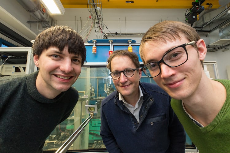
Jakob Soltau, Tim Salditt and Paul Meyer in the laboratory where they carried out this research

“We know the set of algorithms that can favourably be used for this very well from phase-retrieval in coherent X-ray imaging,” Soltau explains. “We apply this to X-ray fluorescence imaging using the X-ray colour camera in our experiment to distinguish between the different energies of the detected X-ray photons.”

Thanks to this full-field approach, the researchers say that just one image acquisition is enough to determine the chemical composition of a sample. While the acquisition time is currently on the order of several hours, they hope to reduce this in the future.

Potential for imaging biological tissues

The team says the new technique has many potential applications. These include nuclear medicine and radiology; non-destructive industrial testing; materials analysis; determining the compositions of chemicals in paintings and cultural artefacts to verify their authenticity; analysis of soil samples or plants; and testing the quality and purity of semiconductor components and computer chips. In principle, the technique could also be used to image incoherent radiation sources such as inelastic X-ray (Compton) and neutron scattering or gamma radiation, which would be useful for nuclear medicine applications.

“As a research group, we are very interested in the three-dimensional imaging of biological tissues,” Soltau tells Physics World. “Combining tomographic imaging, for example, with a detector recording the transmitted X-ray beam to obtain a map of the electron density (a technique known as phase contrast propagation imaging) with our novel full-field fluorescence imaging approach would allow us to image structures and (local) chemical compositions of the sample in one scan.”

In this first demonstration of the new technique, which is detailed in Optica, the Göttingen team achieved a spatial resolution of about 35 microns and a field of view of around 1 mm2. While the number of resolution elements imaged in parallel remains relatively low, this could be increased by using a FZP with smaller zone widths or by increasing the sample area being illuminated towards larger fields of view. Another challenge will be to reduce acquisition times without increasing unwanted background noise from elastically scattered radiation.

The researchers would now like to try their technique with synchrotron radiation, which is much more intense than the X-ray light available in most laboratories. A further advantage is that synchrotron radiation consists of high-energy beams of charged particles generated using electric and magnetic fields, giving it a narrow bandwidth that should allow for higher spatial resolution and shorter acquisition times. The team has booked time on DESY’s PETRA III synchrotron beamline in June for this purpose.

Wearable ultrasound sensor provides continuous cardiac imaging

Cardiac ultrasound imager

A wearable cardiac ultrasound imaging system that can even function during a workout has been developed by researchers from the University of California San Diego. The team hopes that the postage-stamp-sized sensor, which can assess both the heart’s function and structure, will make long-term cardiac scanning accessible to a large population.

According to the British Heart Foundation, in the UK alone, 7.6 million live with a heart or circulatory disease and some 460 people die each day from the same. While cardiac diseases are the leading cause of death among senior citizens, they are also becoming increasingly common among the young.

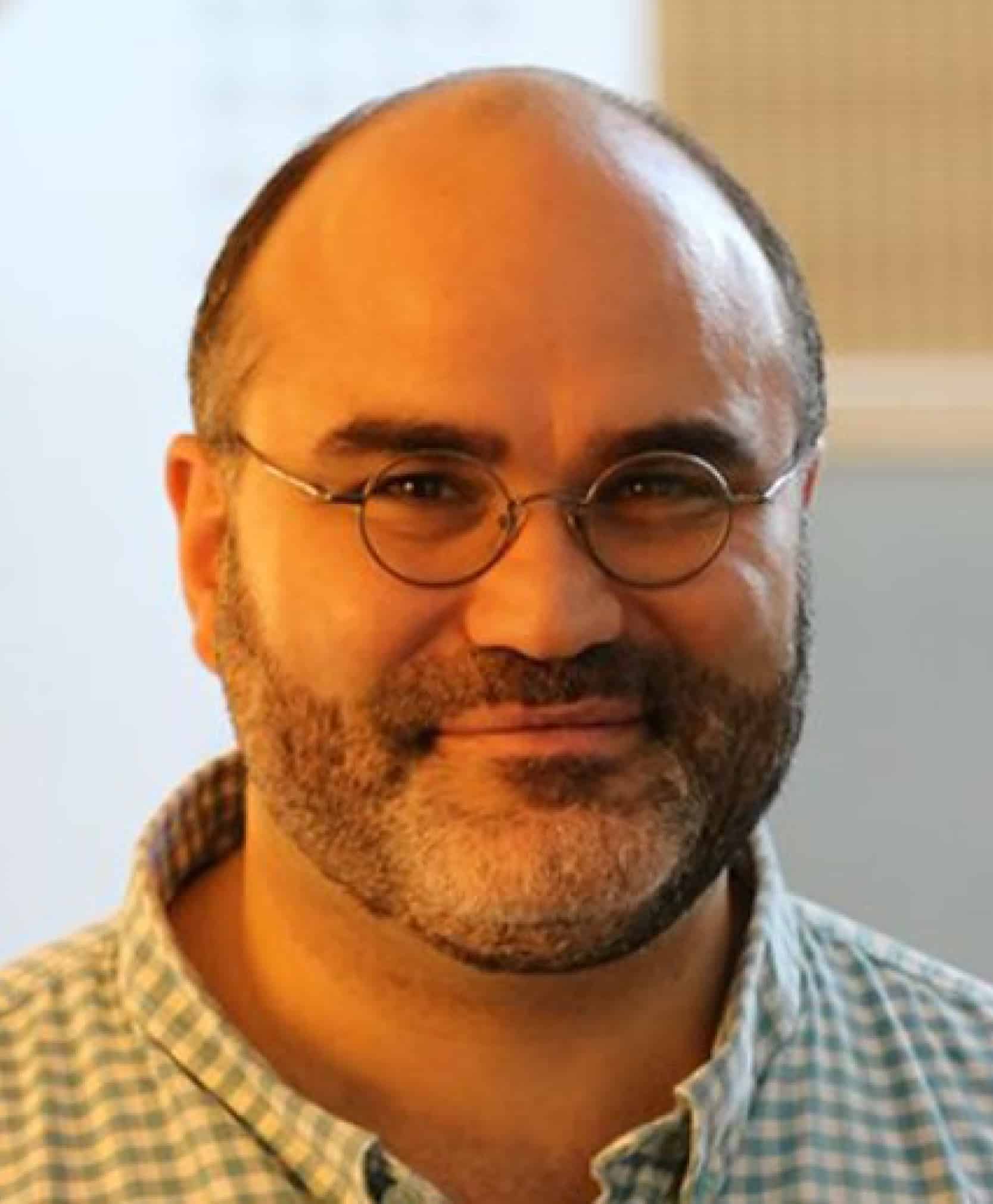
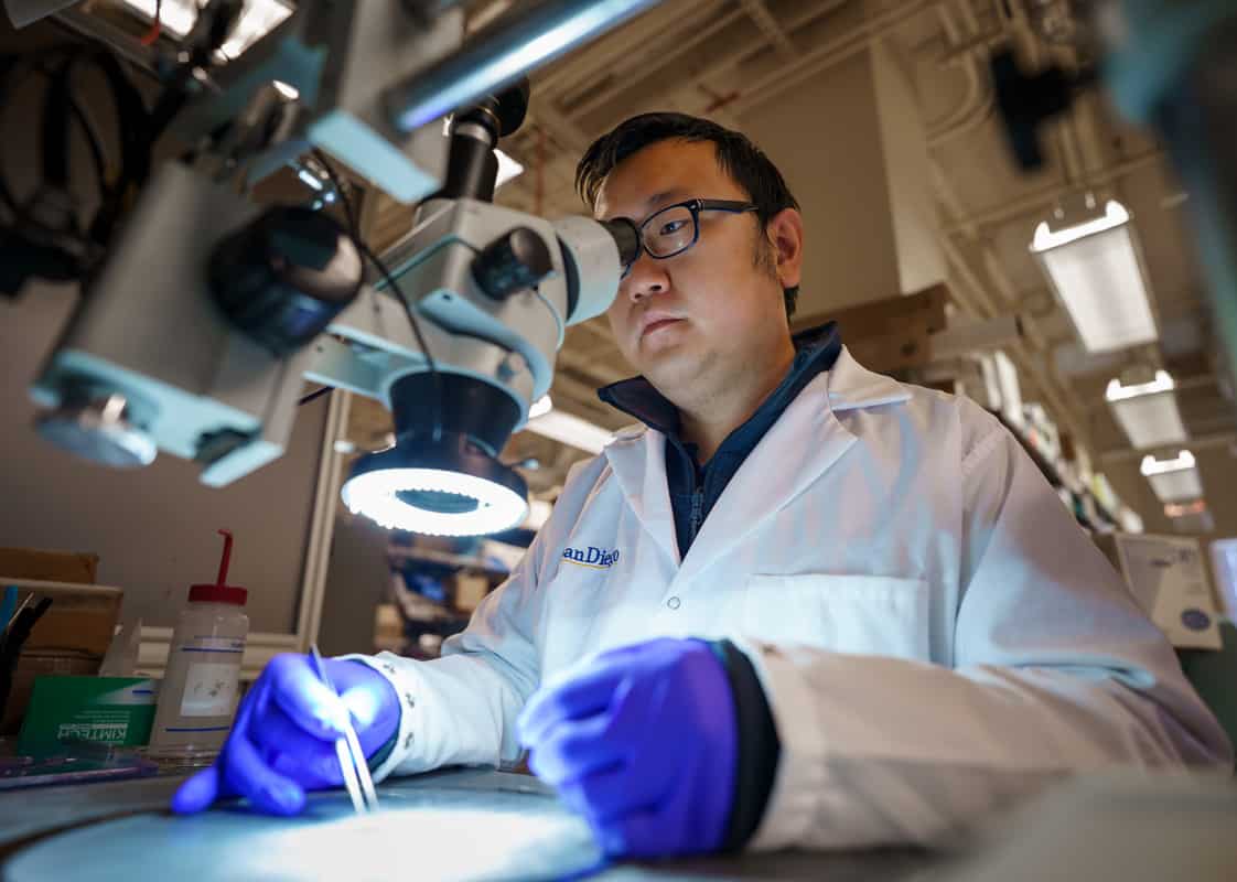
Hongjie Hu

“The heart undergoes all kinds of different pathologies,” explains co-first author and materials scientist Hongjie Hu. “Whether it be that a strong but normal contraction of heart chambers leads to the fluctuations of volumes, or that a cardiac morphological problem has occurred as an emergency, real-time image monitoring on the heart tells the whole picture in vivid detail.”

The problem with cardiac imaging is that echocardiograms typically require highly trained technicians and bulky scanning machinery, whereas CT and PET scans can be uncomfortable for some and come with the added factor of exposing patients to radiation.

Furthermore, many issues with heart function are intermittent, or only become apparent when the body is in motion, the researchers note. Large, fixed pieces of equipment are ill-suited for long-term monitoring, and certainly cannot image moving patients.

In contrast, says project lead and nanoengineer Sheng Xu, the new sensor can be worn for 24 hours in one go, enabling “anybody to use ultrasound imaging on the go”.

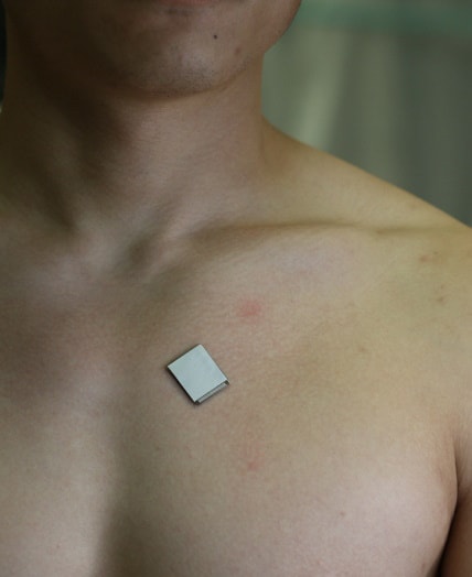
Central to Xu and colleague’s system is a wearable, stretchy and adherent patch – 1.9 × 2.2 cm in size, just under a millimetre in thickness and as soft as skin – that emits and receives ultrasound waves. The patch images the structure of the heart in real time and with high spatial and temporal resolution.

Furthermore, artificial intelligence algorithms built into the device allow it to determine how much blood the heart is actually pumping – a vital measurement, as failure to pump enough blood is often at the root of many cardiovascular diseases.

Wearable ultrasound sensor

The design of the patch makes it ideal for use on bodies in motion. As co-author and nanoengineer Xiaoxiang Gao notes, it can be attached to the chest with minimal constraint to the subject’s movement, even providing a continuous reading of cardiac activities before, during and after exercise. At present, the patch needs to be connected to a computer via cables, but the researchers say they have developed a wireless circuit to remove this limitation.

“The increasing risk of heart diseases calls for more advanced and inclusive monitoring procedures,” says Xu. “By providing patients and doctors with more thorough details, continuous and real-time cardiac image monitoring is poised to fundamentally optimize and reshape the paradigm of cardiac diagnoses.”

Cardiovascular imaging expert Alastair Moss of the University of Leicester, who was not involved in the present study, says that the system has the ability to “transform” the way doctors monitor and treat heart disease among high-risk patients. “High-quality imaging can help save lives for people with heart disease,” he says. “It is astonishing to think that we will be able to take echocardiography outside of traditional healthcare settings and place it directly in the hands of patients.”

Steffen Petersen – a cardiologist at Queen Mary University of London, who was also not involved in this study – agrees, lauding in particular the patch’s ability to provide continuous data feeds during daily activities. He adds: “The potential for such technology in cardiology is huge and assessing cardiac structure and function in such a way will lead to further inventions and clinical indications.”

With their initial study complete, Xu and colleagues are looking to improve the sensor design, miniaturize its power system, and generalize the deep-learning model so it can be used by a larger population of patients. They will also be moving to commercialize the technology – via their start-up Softsonics – over the coming years, with the expectation that a single unit will likely cost around $80 (£66).

The study is described in Nature.

Innovation underpins novel biophysics research

The 67th Annual Meeting of the Biophysical Society aims to provide a dynamic forum for researchers who are working at the interface between the life, physical and computational sciences. Running from 18 to 22 February in San Diego, US, the meeting offers a valuable opportunity for attendees to share their latest research findings while also learning about the emerging techniques and applications being developed by scientists from all over the world.

“The last few years represent an epochal moment for science in human history,” comment programme chairs Baron Chanda and Janice Robertson, both from Washington University in St Louis. “Basic science discoveries were translated into treatments for human diseases at an astonishing pace, and advances in biophysics at various levels has been central to these developments.”

The conference, which is expected to attract upwards of 5000 delegates, combines technical symposia and interactive daily poster presentations with programmes focusing on career development, education and science policy. “This meeting will showcase incredible breakthroughs in new drug developments and protein structure prediction,” continue Chanda and Robertson. “Other symposia will highlight the emerging complexity of cellular membranes, RNA and genomic organization.”

Scientific workshops during the event will feature developments in computational drug discovery, and spectroscopic and microscopy approaches for high-throughput research. The Saturday Subgroup symposia enable attendees to meet within their scientific communities, while social activities throughout the event allow more informal networking with friends and colleagues.

Meanwhile, the technical exhibit offers an opportunity for delegates to explore the latest innovations in equipment and techniques. Some of the vendors are also hosting presentations to explain how their solutions can be used for biophysical studies – read on to find out more.

Precision instruments enable biophysical research

For 25 years Mad City Labs has provided precision instrumentation for biophysicists, including nanopositioning systems, micropositioners, atomic force microscopes (AFMs) and RM21 single-molecule microscopes. For a start, the company’s piezo nanopositioners feature PicoQ sensors, which combine ultralow noise with high stability to yield sub-nanometre precision. Their stability makes them ideal for biophysics and microscopy applications, and they also provide the ideal building blocks for nanoscopy when paired with the company’s micropositioners.

Mad City Labs

AFMs from Mad City Labs achieve atomic-step resolution while also being affordable and fully customizable with automated software and calibration, and for full flexibility all of the company’s nanopositioners and micropositioners are compatible with its AFM instruments. Meanwhile, the RM21 single-molecule microscope offers high stability, precision alignment and direct access to the optical pathway, plus they can be used with nanopositioners to enable advanced nanoscopy methods.

Also available is the MicroMirror total internal reflection fluorescence (TIRF) microscope, a multi-colour instrument that combines an excellent signal-to-noise ratio with efficient data collection. An array of options are available to enable the microscope to be used for different single-molecule techniques. A further addition to the company’s product range is the Mad-Deck XYZ stage, an automated stage platform that is ideal for Interferometric scattering microscopy.

During the Biophysical Society meeting Mad City Labs will host a presentation to showcase research that has been enabled by the company’s microscopy solutions. “Open & Flexible Microscopy Systems for Combined Single Molecule Methods” will run on Sunday 19th February, 1:30 – 3:00 pm, and will feature three talks by leading academics:

  • “Quantification of Lipid Diffusion Dynamics on Live Cell Membranes through Interferometric Scattering Microscopy” by Francesco Reina of the Leibniz Institute for Photonic Technologies and the Friedrich Schiller University Jena, Germany
  • “A Single-Molecule View on Dynamic Chromatin Access of Epigenetic Regulatory Factors” by Beat Fierz at the École Polytechnique Fédérale de Lausanne, Switzerland
  • “Magnetic Tweezers Investigations of the Type 1A Topoisomerase of Mycobacterium Smegmatis” by Maria Mills at the University of Missouri, US

More details and full abstracts are available online.

  • Find out more by visiting Mad City Labs at booth 508

Confocal microscope simplifies time-resolved imaging

Quantitative time-resolved fluorescence techniques such as fluorescence-lifetime imaging microscopy (FLIM) are increasingly being used in cell biology to monitor processes such as phase separation, conformational changes, and the interactions between proteins. So far, however, expert knowledge has been needed to obtain accurate and reproducible results from these tools, which has slowed down their widespread adoption.

The Luminosa confocal microscope

PicoQuant’s new Luminosa confocal microscope overcomes that challenge by combining state-of-the-art hardware with cutting-edge software to deliver high-quality data while also simplifying daily operation. The software includes context-based workflows to improve the reproducibility of experiments, while features such as sample-free auto-alignment and calibration of the excitation laser power make experiments more efficient. At the same time, every optomechanical component is fully accessible to enable the development of new methods.

Application specialists Marcelle König and Evangelos Sisamakis will present two use cases during the meeting. First, they will show how the instrument can make it easier for researchers to use single-molecule fluorescence resonance energy transfer (smFRET) in their experimental studies. For example, the FRET efficiency and stoichiometry can be calculated during the experiment, corrected according to standard protocols, and displayed in real time.

Second, they will describe how Luminosa can be used to streamline FLIM experiments. The instrument’s rapidFLIM module can record images at several frames per second with high photon-count rates, which the software handles with a novel dynamic binning format to enable high-speed automated analysis of FLIM images. Join their talk on Sunday 19 February, 12:30 – 2:00 pm, in Room 10.

  • For a live demonstration of the Luminosa confocal microscope, and to find out about other innovations from PicoQuant, visit booth 309.


Copyright © 2026 by IOP Publishing Ltd and individual contributors