Particle accelerators play important roles across a wide range of science, medicine and engineering. They also tend to be very large and expensive facilities, which means that beam time on accelerators – be it for developing new materials or treating cancer – is precious and in short supply.
Lasers offer a way of accelerating particles in much smaller and cheaper facilities. In this episode of the Physics World Weekly podcast we meet a physicist who has founded a company called Tau Systems, which aims to build a laser-driven accelerator facility. Based at the University of Texas at Austin, Manuel Hegelich explains how the technology works and how it could be used to create free-electron lasers that would benefit scientists working on a wide range of topics.
Also in this episode, we chat about physicists who have won Nobel prizes in fields other than physics.
The James Webb Space Telescope (JWST) and the Hubble Space Telescope have captured the moment the $330m Double Asteroid Redirection Test (DART) craft hit a small asteroid.
At 7:14 p.m. EDT on Monday, DART impacted the asteroid Dimorphos, which together with another larger body – Didymos – forms a binary, near-Earth asteroid system. The aim of the DART mission is to be the first to demonstrate “kinetic impact” by putting Dimorphos in a slightly different orbit around Didymos.
The JWST observed the impact over five hours, capturing 10 images, while Hubble took more than 40 pictures before and after the impact. The image of the impact shows a tight, compact core with plumes of material appearing as wisps streaming away from the centre of where the impact took place.
In the Hubble images, astronomers estimate that the brightness of Dimorphos increased three times after impact and they also found, surprisingly, that the brightness held steady for some eight hours after impact.
Hubble and the JWST will now continue to observe the system over the coming weeks. Such observations by space-based telescopes as well as ground-based instruments will allow scientists to study the surface of Dimorphos, how much material was ejected by the collision, how fast it was ejected and reveal the distribution of particle sizes in the expanding dust cloud.
This information can then be used to understand how effectively a kinetic impact can modify an asteroid’s orbit.
Early breakthrough: Allan Cormack with his original computer axial tomography (CAT) scanner. (Courtesy: AIP Emilio Segrè Visual Archives, Physics Today Collection)
Physicists have always had an interest in biological and medical physics, with Francis Crick and Maurice Wilkins famously sharing the 1962 Nobel Prize for Physiology or Medicine for elucidating the structure of DNA (along with the biologist James Watson).
But two other huge breakthroughs in medical physics – the introduction of X-ray computed tomography (CT) and magnetic resonance imaging (MRI) – also won their inventors a physiology or medicine Nobel prize.
Tackling tomography theory
Even before Wilhelm Röntgen won the first-ever Nobel Prize for Physics in 1901 for discovering X-rays, we’ve known that they can be used to image the interior of the body. This rapidly led to the introduction of a range of medical applications; but it was the development of CT scanning – in which X-rays are sent through the body at different angles to create cross-sectional and 3D images – that vastly expanded the potential of medical X-ray imaging.
That work was recognized in 1979 when the physicist Allan Cormack was awarded the Nobel Prize for Physiology or Medicine “for the development of computer-assisted tomography”, an honour he shared with engineer Godfrey Hounsfield.
Born in Johannesburg, South Africa, Cormack had been intrigued by astronomy at an early age. He then went on to study electrical engineering at the University of Cape Town, but after a couple of years abandoned engineering and turned to physics. After completing a BSc in physics and an MSc in crystallography he moved to the UK to work as a doctoral student at the University of Cambridge’s Cavendish Laboratory. Cormack returned to Cape Town as a lecturer and, following a sabbatical at Harvard University, in 1957 became assistant professor of physics at Tufts University in the USA. Unusually for a Nobel laureate, Cormack never actually earned a PhD.
At Tufts, Cormack’s main pursuits were nuclear and particle physics. But when he had time, he pursued his other interest – the “CT scanning problem”. He was the first, from a theoretical point of view, to analyse the conditions for demonstrating a correct radiographic cross-section in a biological system.
Having developed the theoretical underpinnings of tomographic image reconstruction, he published his results in 1963 and 1964. Cormack noted that at that time, “there was practically no response” to these papers, so he continued his normal course of research and teaching. In 1971, however, Hounsfield and colleagues built the first CT scanner and interest in CT scanning escalated.
What’s interesting is that Cormack and Hounsfield built a very similar type of device without collaboration, in different parts of the world. Thanks to their independent efforts, CT scans are now ubiquitous in modern medicine, employed for applications such as disease diagnosis and monitoring, as well as guiding tests such as biopsies or treatments such as radiation therapy.
The emergence of MRI
The next Nobel Prize for Physiology or Medicine to go to a physicist was in 2003 when Peter Mansfield was recognized (along with the US chemist Paul Lauterbur), for “discoveries concerning magnetic resonance imaging”, which paved the way to modern MRI. The technique provides clear and detailed visualization of internal body structures and is now routinely used for medical diagnosis, treatment and follow-up. Crucially, unlike X-ray-based scans, MRI does not expose the subject to ionizing radiation.
MRI pioneer: Peter Mansfield’s research helped pave the way to modern MRI. (Courtesy: University of Nottingham)
Mansfield originally studied physics at Queen Mary College in London, where his graduate research focused on building a pulsed nuclear magnetic resonance (NMR) spectrometer to study solid polymer systems. After receiving his PhD in 1962, he undertook further NMR research at the University of Illinois at Urbana–Champaign, before returning to the UK to take up a lectureship at the University of Nottingham (where he worked until his retirement in 1994).
Mansfield’s PhD and postdoc led him to the idea of using NMR for human imaging (a technique originally called nuclear magnetic resonance imaging, but soon rebadged as just MRI to avoid alarming patients). And it was during his time at Nottingham that Mansfield made some of the key breakthroughs leading to his Nobel prize.
In the mid-1970s, Mansfield produced the first MR images of a live human subject: the finger of one of his research students. His team went on to develop a whole-body MRI prototype, which he volunteered to be the first to test. Despite fellow scientists cautioning that it may be potentially dangerous, Mansfield was “fairly convinced there would not be a problem”.
As for Lauterbur, he discovered that introducing gradients in the magnetic field made it possible to create two-dimensional images of structures that could not be visualized by other techniques. Mansfield further developed the use of gradients, showing how the detected signals could be mathematically analysed and transformed into useful images. He is also credited with discovering how to drastically reduce MRI scanning times, using the echo-planar imaging technique.
These days, tens of millions of MRI exams are performed each year around the world, and in 1993 Mansfield was knighted for his services to medical science. There’s even a beer (the 4.2% ABV Sir Peter Mansfield ale) named in his honour.
Physics World‘s Nobel prize coverage is supported by Oxford Instruments Nanoscience, a leading supplier of research tools for the development of quantum technologies, advanced materials and nanoscale devices. Visit nanoscience.oxinst.com to find out more.
Whorls of FLASH: (a) Optimized scan patterns; (b) comparison of standard (non-optimized) line-by-line and scan-pattern optimized (SPO) patterns. The resulting pencil-beam scanning dose rate (PBS-DR) distributions are shown as coloured overlays. (Courtesy: Int. J. Radiat. Oncol. Biol. Phys. 10.1016/j.ijrobp.2022.08.053)
The ultrahigh dose rates used in FLASH radiotherapy may increase the therapeutic window by protecting normal tissues against radiation damage. In addition, some researchers believe that FLASH proton beams might be available with commercially available cyclotron-accelerated proton beams. But when FLASH is combined with the most advanced type of proton therapy, lateral pencil-beam scanning (PBS), the very PBS proton deliveries used to treat complex cancers with unparalleled precision also impact the local dose rates critical to achieving the FLASH effect.
“We were trying to optimize FLASH through optimization of dose rate, without compromising plan quality in terms of radiation dose,” says lead author Rodrigo José Santo. “We were trying to set up a pipeline that would consistently optimize FLASH coverage for different tumour shapes and sizes, without re-optimizing the treatment plan and considering FLASH as a local effect dependent on the pencil-beam delivery pattern.”
The result: optimizing FLASH proton therapy treatment plans without compromising dose rate.
PBS as a travelling salesperson
The travelling salesman problem poses the following question: “Given a list of cities and the distances between each pair of cities, what is the shortest possible route that visits each city exactly once and returns to the origin city?”
This problem, long studied by combinational optimization researchers, is a barometer for genetic algorithms used in computer science and operations research. José Santo, who is currently a doctoral student at UMC Utrecht but was a master’s student when the work was performed, realized that genetic algorithms could be used to solve his own problem – optimizing the order in which proton pencil beams are irradiated to maximize FLASH coverage.
The researchers’ resulting approach uses a voxel-based metric defined by fixed-dose thresholds to determine when irradiation of that voxel starts and ends. The algorithm evaluates dose rate for each pencil beam separately and assumes that FLASH is a local effect and that total irradiation time is a critical FLASH parameter.
The algorithm is run on different solutions in parallel, though it occasionally shares information between them. The average distance between pencil beams is included as a cost function to minimize the total distance travelled in the plane transverse to the beam direction. The algorithm is applied sequentially after pencil-beam positions and weights are optimized and without compromising the plan quality in terms of (nominal) absorbed dose.
The researchers tested their algorithm on treatment plans using transmission proton pencil beams for 20 patients with early-stage lung cancer and lung metastases. (Lung lesions are ideal sites for FLASH, the researchers say – current FLASH proton treatments involve high-energy beams that pass through the patient rather than the Bragg-peak beams harnessed for conventional proton therapy.)
Median FLASH coverage improved from 6.9% for standard line-by-line scan patterns to 29% with PBS optimization. The researchers observed that PBS-optimized plans have a whorl-like appearance. The FLASH window changed only slightly for marginally different beam currents.
Since other research groups are primarily working to optimize FLASH at the treatment planning level, the researchers say that it’s challenging to compare their own PBS-optimized results to other FLASH proton therapy studies – to their knowledge, this study is the first to perform pencil-beam delivery pattern optimization for FLASH proton therapy. They are now focusing on optimizing PBS delivery for larger targets and integrating dose-rate optimization into their existing dose optimization pipeline.
“Radiation therapy is still [being] continuously improved, and the FLASH effect is a promising path to better treatment outcomes for patients. Proton therapy, combined with optimization algorithms such as the one we have developed, is an important step towards achieving exactly that,” José Santo says. “Our manuscript underlines that there is a lot of room for further optimization of FLASH proton therapy as a treatment modality, even with current beam hardware.”
“I have dealt with many different transformations…but the quickest that I have met was my own transformation in one moment from a physicist to a chemist.”
Rutherford, who at the time of his nomination was based at McGill University in Canada, had been awarded the chemistry prize “for his investigations into the disintegration of the elements, and the chemistry of radioactive substances”.
First as a postgraduate student at the University of Cambridge in the UK and then at McGill, Rutherford had shown that atoms are not stable but can radioactively disintegrate into other elements. In particular, by finding that thorium gives off a radioactive “emanation”, he discovered radon gas (we now know that thorium decays via radium into radon).
But the irony is that Rutherford had never formally studied chemistry. As a teenager at Nelson College in New Zealand, his best subject was mathematics and he had also taken courses in mechanics, sound and light. According to his biographer John Campbell, however, he “avoided chemistry, as he knew that the teacher was only one step ahead of the class”.
Later, as a master’s student at Canterbury College in Christchurch, Rutherford even turned down a suggestion for an experimental project that would involve looking for the molecular building blocks of life by studying electrical discharges in gases, as he felt he did not know enough chemistry.
Quite why Rutherford, the eminent physicist, should then have received a chemistry Nobel prize has been examined in detail by the theorist Cecilia Jarlskog from Lund University in Sweden, who dug into the Nobel archives.
She discovered that between 1907 and 1908, Rutherford actually received 12 nominations for a physics Nobel prize – and only four for chemistry.
In 1907 the Nobel Committee for Physics rejected Rutherford on the grounds that “his observation of the decay of a chemical element (radium) should be awarded with the chemistry prize rather than the physics Nobel prize.” As Jarlskog puts it, the physicists basically felt that “radium is a chemical element and that’s chemistry”.
The following year the chemists once again seemed keener on making Rutherford one of their own, with the chemistry committee going so far as to write an extensive, 15-page report on him. And they, of course, won the day.
Then, as now, the message is that the dividing line between physics and chemistry is a human construct that nature does not respect. Of course, Rutherford could – and perhaps should – also have won a Nobel Prize for Physics for later discovering the atomic nucleus and for being able to transform one element (nitrogen) into another (oxygen). Indeed, he received 11 further nominations for the physics prize from 1912 onwards (and seven for chemistry).
But he didn’t and perhaps that serves him right. After all, Rutherford is famous for his cutting remark that: “All science is either physics or stamp collecting.”
We’ll see if any physicists win the 2022 Nobel Prize for Chemistry this year. If they do, they’ll have joined an eminent band that includes not just Rutherford but also other people who did physics or studied the subject at university, such as:
• Marie Curie (1911 for discovering radium and polonium);
• Alan Heeger (2000 for discovering and developing conducting polymers);
• John Goodenough (2019, for developing lithium-ion batteries).
Physics World‘s Nobel prize coverage is supported by Oxford Instruments Nanoscience, a leading supplier of research tools for the development of quantum technologies, advanced materials and nanoscale devices. Visit nanoscience.oxinst.com to find out more.
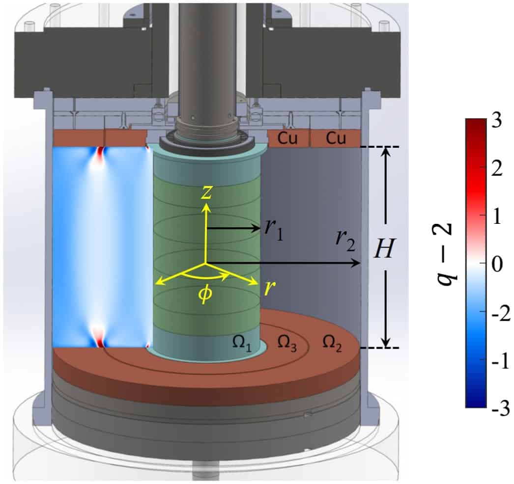
Lab top accretion disc: Cutaway diagram of the experimental setup used by Wang and colleagues. The data shown in the left portion of the gap between cylinders illustrates the simulated shear profile of the fluid. (Courtesy: Y Wang et al/Physical Review Letters)
Researchers in the US have designed an experiment that attempts to simulate the complex dynamics of astrophysical accretion discs more closely than ever before. Yin Wang and colleagues at Princeton University did this by adapting previous experimental techniques to avoid unwanted flows in their simulated disc, while more closely representing the magneto-rotational instability that is believed to emerge in real accretion discs.
Accretion discs are swirling vortices of matter that form as massive objects such as black holes and newly forming stars gather gas and dust from their interstellar surroundings. The influx of this material leads to planet formation and produces the intense radiation that is emitted from the vicinity of some black holes.
For gas and dust to move nearer to the massive object, it must transfer angular momentum to the outer edge of the disc – and an explanation of how this happens has eluded astronomers. One leading theory is that this transfer is driven by turbulent flows in the disc. To explore this idea, previous studies have used a Taylor Couette setup in which a fluid fills the gap between two concentric cylinders that can be rotated independently.
Astrophysics in the lab
By rotating the outer cylinder more slowly than the inner cylinder, and carefully controlling their respective motions, researchers can closely recreate the motions of evolving accretion discs as closely as possible. Their aim here is to determine whether turbulent flows could really be responsible for their angular momentum transfer.
However, beyond the clear limitation that these motions are not driven by gravity, the fluid must also be contained vertically by upper and lower caps. This introduces secondary flows to the fluid, with no analogue in real accretion discs. One recent study done in Paris reduced the influence of these unwanted flows by applying a vertical magnetic field to a liquid metal disc – more closely recreating the electrical conductivity of real accretion discs. However, the Parisian team did not fully recreate the desired turbulent flows.
One possible driver for turbulence in accretion discs is magneto-rotational instability (MRI): which could better explain how a differentially-rotating, electrically conducting fluid can be destabilized by a magnetic field. This concept has been widely studied theoretically, but still hasn’t been confirmed in Taylor Couette experiments because of difficulties in setting the appropriate parameters.
Conductive liquid
Wang’s team have addressed this challenge by using a fluid called galinstan, which is a liquid alloy of gallium, indium and tin that is about twice as viscous as water, and some 100 million times more conductive of electricity. To eliminate secondary flows, they also implemented a pair of electrically conducting caps, which rotated independently at speeds intermediate to the inner and outer cylinders.
As they applied a vertical magnetic field along the cylinders’ rotation axis, the researchers measured the fluid’s magnetic Reynolds number, which characterizes how a magnetic field interacts with a conducting fluid. Crucially, they observed this value passing a certain threshold: beyond which the strength of the magnetic field passing through the inner cylinder began to increase nonlinearly – indicating that MRI had been triggered.
Simulations have also been able to reproduce this behaviour, so the team’s observations are an important step forward in researchers’ ability to reproduce accretion disc dynamics in real experiments; and ultimately, in answering the long-standing mystery surrounding the transfer of angular momentum in accretion discs.
Learning to enhance the collaborative process. (Courtesy: Shutterstock/Lightspring)
Collaboration is a defining feature of 21st century research, with more and more people routinely traversing formal disciplinary boundaries in the quest for fundamental and applied scientific discovery, even when it takes them beyond their core domain knowledge and expertise. During this webinar we want to take a practical approach to support researchers in their collaborative practice. It will help researchers to overcome common barriers to research collaboration, preparing them to form high-quality collaborations within a supportive organizational culture. Suitable for researchers at all levels, serving a global audience.
Annette Bramley is the N8 research partnership director and chief collaboration officer. She is a public speaker highly regarded for her expertise in research culture and collaboration. She is an ambassador for excellent collaborative research that has a genuine impact on the world. Her particular passion is multidisciplinary partnership, team science and collaboration. With a first-class degree and DPhil in materials science from the University of Oxford, she brings real-world understanding from the laboratory combined with experience of more than 20 years guiding researchers in a range of disciplines at the Engineering and Physical Sciences Research Council (now part of UK Research and Innovation). Her LinkedIn recommendations give a sense of how her input has helped in the success of a great number of people and projects.
Sharing scientific information is as old as science itself. Early scientific pioneers agreed that it was important to discuss ideas, show experiments to others and read what other scientists were doing. Today’s scientists continue this tradition when they discover something new and interesting about the world, publishing their work in journals and discussing it at conferences. Doing so allows findings to be disseminated and helps others with their own research. But for this vital step to take place, knowledge must be transferred – in other words, science must be communicated.
Centuries ago, those interested in such pursuits were few and far between. Today, however, the results of scientific research are spread far and wide – and sometimes even beyond the confines of science. Some scientists, for example, want to communicate their research in the hope that policy makers make more informed decisions. This interaction between scientists, the public and policy makers can even raise the profile of “citizen science” initiatives by attracting attention to their aims.
In the past few decades, however, a disconnect has emerged between scientists who generate knowledge and the journalists, bloggers and science communicators who disseminate it to the public. This has reinforced the view held by some scientists that these popularizers distort the findings of their research to generate a better headline and more readers. But it is not just the popularizers’ fault; researchers often lack the skills to effectively communicate their research to journalists and the public.
Indeed, I have witnessed this culture at first hand. During my postgraduate studies, I came across few PhD supervisors who supported, or harder still, encouraged their students to get involved in science communication. The opportunity to participate in outreach events was often viewed as a “tick-box exercise” to demonstrate transferable skills. Such activities, it was felt, got in the way of the “real work” of pure scientific research.
As a consequence, scientists who engage with the public are often less well regarded by their peers – there seems to be a false dichotomy that you can be a good scientist or a popularizer, but not both. This picture is slowly changing, in part due to the COVID-19 pandemic, which has forced scientists to explain their findings and offer their opinions to the public. The last few years have shown that talking, explaining, listening and learning are important skills in the collective effort to control the pandemic. So how can we carry on this trend?
Communication as a skill
Science communication used to be viewed as a simple process, with a clear progression of information from scientist to journalist to the wider public. This broadly describes the outdated, and slightly patronizing, “deficit model” of science communication, where the public was only required to pay attention. But science is becoming increasingly interdisciplinary, with more scientists from different fields collaborating with each other, while the Internet is radically changing how the public access and share information. These developments have blurred the boundaries with the traditional flow of scientific information.
Perhaps we should rather consider science communication as a continuum. The communication skills scientists need to explain their findings to collaborators from different scientific backgrounds are not all that different from the skills needed to communicate with journalists or to non-scientists. Moreover, scientists who have an active social media profile can enter direct discussions with the public on their research. With this in mind, I think we should place more emphasis on teaching the next generation of scientists that effective communication is an indispensable research skill.
Doing so would not only raise the profile of science communication but also build direct links between scientists and the public who fund them. Adopting this approach would also create accessible scientific role models. Young people are much more likely to go into science if they can see someone they identify with who inspires them. Making research accessible and engaging to a wide audience can inspire future generations to continue with research.
Yet we must not fall into the trap of thinking that role models alone determine what career we pursue. As a young person who is passionate about physics myself, the lack of professional physicists with non-degenerative physical disabilities did not stop me from going into science. So, as well as presenting young people with positive role models, it is also important to give them the confidence to blaze their own trail through life.
In this information-hungry age, it will always be essential to have people dedicated to disseminating scientific information to the public across all forms of media. Yet if we are to achieve the highest quality scientific communication, current researchers must up their game and not just view the activity as something reserved for those outside academia.
Women and men have such different citation patterns that it is possible to accurately predict a scientist’s gender from such data alone. That’s the finding of a new study that investigates how men and women cite – and are cited by – their communities (Proc. Natl. Acad. Sci119 e2206070119).
Led by network scientist Kristina Lerman from the University of Southern California, the authors studied 766 members of the US National Academy of Sciences (NAS), which included 120 women. They matched the scholars to their profiles on Microsoft Academic Graph, which contains metadata on over 150 million academic publications.
After identifying the scientists’ genders by checking pronouns on the individual’s biographies, the researchers created an “ego citation network” for each scientist. This contained “directional links”, indicating which other scientists – represented by nodes – the individual had cited, and which scientists had cited them.
It is well known that female scientists receive fewer citations than their male counterparts, but the new study reveals that women reciprocate a significantly higher fraction of citations than men do. A woman’s network also has more “connectedness”, suggesting that women tend to work in more closely knit research communities.
The study found as well that women have fewer peers – though these tend to be highly productive colleagues – and that women have a greater proportion of female scientists in their networks.
Rich get richer
The researchers then trained a machine learning algorithm on 75% of the data that was randomly selected. Using the other 25% to test the system, they found that the algorithm can accurately predict a scientist’s gender based on the citation networks – correctly doing so about 80% of the time.
The citation networks showed few significant differences based on the prestige of an author’s affiliated institution, although NAS membership is highly skewed towards more prestigious institutes. The researchers also found that women are under-represented across all seven fields they looked at. Just 8% of NAS physicists were women – the lowest proportion of all the fields studied.
Lerman thinks the gender differences in citation networks could be down to two aspects. “There is a preference by both genders to cite men, and preferential attachment — or the ‘rich get richer’ effect — is the well-known mechanism of rewards in science, where the already better-known researchers get more credit,” she says. “We are now working on a manuscript that shows how a large gender disparity can emerge from these components.”
In this webinar, Thierry Gevaert, coordinator at the UZ Brussel, will share his experience of the THALES 3D MR SCANNER for the acceptance, commissioning, and QA for both MRIdian and Halcyon linacs. Thierry will highlight the benefits of using the THALES system for their two machines. During the webinar, an overview of future released features will also be presented.
Find out how you can use the THALES 3D SCANNER as a tool for fast and efficient commissioning procedures of bore-type linacs. Learn how to minimize machine occupancy and benefit from cost-effectiveness.
Thierry Gevaert graduated as a medical physicist at the Vrije Universiteit Brussel (VUB) and defended his PhD, “Clinical implementation of frameless radiosurgery”, in 2012. In 2016, he was nominated professor at the VUB and became the chief of medical physics at the UZ Brussel (UZB). His appointment in the department of radiotherapy involves research, teaching and clinical practice. Thierry is a board member, certified in therapeutic physics and is engaged in mentoring medical physics for graduate students, residents and medical engineers. His research and clinical interests include stereotactic radiosurgery (SRS), stereotactic body radiotherapy (SBRT), motion management, the development of high-tech radiation techniques and image-guided radiotherapy (IGRT), all with an emphasis on clinical implementation of translational research. Thierry has made numerous international and national presentations and published more than 50 peer-reviewed papers. Since 2015, he has been an auditor for the Novalis Certification programme.
Thierry Mertens
Thierry Mertens has a PhD in physics and nearly 15 years experience in medical physics and radiotherapy, with the major commitment to develop innovative quality assurance solutions to support the medical end-users with their clinical tasks. As business development manager for LAP since 2016, Thierry has been instrumental in the development of the THALES 3D MR SCANNER and has had the opportunity to work closely with pioneer users of the MRIdian system. He represents the customer voice within the LAP R&D team, ensuring that the end-user needs are fulfilled.
Tim Everaert
Tim Everaert graduated as a medical physicist at Ghent University in 2019. In 2020, he started his position as a medical physicist at UZ Brussel. His main focus is based on research and clinical implementation of new techniques in radiotherapy with an emphasis on stereotactic body irradiation (SBRT), motion management, and image-guided radiotherapy (IGRT). He is one of the core team members of online adaptive with MR linac.






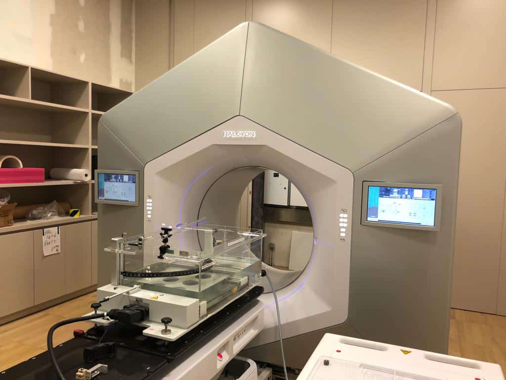 In this webinar, Thierry Gevaert, coordinator at the UZ Brussel, will share his experience of the THALES 3D MR SCANNER for the acceptance, commissioning, and QA for both MRIdian and Halcyon linacs. Thierry will highlight the benefits of using the THALES system for their two machines. During the webinar, an overview of future released features will also be presented.

