A sensing technique that can precisely detect the release and diffusion of dopamine from individual neuron release sites has been developed by researchers in Germany. Led by Sebastian Kruss at Ruhr University Bochum, the team created their sensor using specially-modified carbon nanotubes that fluoresce at specific near-infrared wavelengths on contact with dopamine.
Dopamine is a signalling molecule exchanged by neurons in the brain. It controls a wide range of processes in the brain including our sense of reward and motivation. Scientists know that issues with dopamine exchange are tied to multiple brain disorders, including Parkinson’s disease, schizophrenia and addiction. Yet so far, a clear understanding of how these conditions arise has been limited by the low spatial and temporal resolutions of existing techniques for imaging dopamine concentrations in biological tissues.
To tackle the challenge, Kruss’ team has fabricated a new type of nanosensor that uses modified, single-walled carbon nanotubes. When illuminated with visible light, these structures undergo fluorescence by emitting photons with near-infrared (NIR) wavelengths. This makes them ideal for imaging, because NIR light can propagate relativity long distances in biological tissues – providing sharper images than visible light.
Nucleic acid molecules
The researchers modified the nanotubes by binding them to a specific set of nucleic acid molecules, which themselves bind to dopamine. This caused the nanotubes to fluoresce at specific NIR wavelengths when they interacted with dopamine, with a light intensity that is proportional to dopamine concentration.
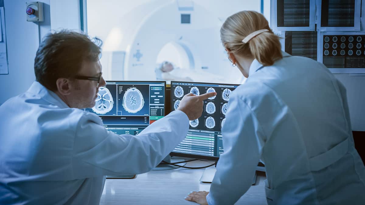
To create a 2D nanosensor, Kruss and colleagues painted a concentrated solution of the nanotubes onto a glass coverslip. This thin layer was then used to coat a culture of neuron cells. When these neurons were stimulated to produce dopamine, the nanotube paint immediately fluoresced at the distinctive NIR wavelengths.
The spatial and temporal resolution of the sensor is good enough to detect both discrete release events from specific sites, and the diffusion of dopamine between cells. By applying a machine-learning-based analysis tool, the researchers could visualize up to 100 release sites simultaneously, and discriminate between sites by the amount of dopamine they released.
Kruss’ team hope that their technique will enable neurologists to better understand the molecular- and cellular-scale mechanisms that drive the release of dopamine; and subsequently, to identify the factors which inhibit this signalling. This could eventually lead to new progress towards treatments for conditions including Parkinson’s disease and drug addiction. In addition, by binding different sets of molecules to carbon nanotube sensors, the technique could be extended to detect other types of signalling molecule.
Physicists in the UK have created a camera that can image the complex tangles of vortices that form inside a helium-3 superfluid. Developed by Theo Noble and colleagues at Lancaster University, the approach could help researchers to better understand the behaviour of quantum fluids.
When cooled to temperatures just above absolute zero, liquid helium-3 becomes a superfluid, which below a certain critical velocity, can flow without any loss of kinetic energy. The effect arises because at very low temperatures atoms of helium-3 – which are fermions – can form Cooper pairs. These pairs are bosons, which means that helium-3 can become a superfluid.
Physicists are fascinated by the dynamics of superfluid helium-3 at high flow velocities. Here, thermal fluctuations break Cooper pairs to create quasiparticles that propagate through the superfluid. These structures cannot exist within a certain energy range, which can prevent them from entering certain regions of a superfluid. As quasiparticles approach these regions, they will trap a partner to form a Cooper pair, leaving behind a quasiparticle called a hole, which propagates in the opposite direction – a process called “Andreev reflection”.
Tangled vortices
This process can be triggered by the quantized vortices that form around obstacles to the flow of a superfluid. In liquid helium-3, these vortices can exist as a disorderly tangle of strings just tens of nanometres thick and can shift the forbidden range of quasiparticles in the fluid by a certain amount – which varies with distance from the vortex.
A variety of techniques have been used to probe these structures: including measuring the magnetic fields surrounding helium-3 nuclei and passing sound waves through the fluid. Yet so far, physicists have struggled to image these tangles directly without the use of invasive techniques, such as artificial tracer particles.
The Lancaster team used a partially closed box within their superfluid to create quasiparticles using a vibrating curved wire. Some of the quasiparticles could move into the rest of the superfluid via a small hole in the box – thus creating a beam of quasiparticles. Upon leaving the box, the beam encounters another vibrating wire that creates a “turbulent tangle” of vortices. Quasiparticles that pass through the tangle are then detected using a 5×5 array of quartz tuning fork resonators.
New discoveries
This allowed the team to produce a series of pixelated images revealing the shadows of vortices, where the quasiparticle beam had been blocked by Andreev reflection. Using this method, the team has already made new discoveries about the properties of superfluid helium-3. For example, they observed many more vortices appeared on the inner edge of the curved wire than its outer edge, despite flow velocities being roughly the same on each side.
The team intends to study these effects in more detail through further improvements to the set-up: including larger pixel arrays, and higher operation speeds to enable video recordings. If achieved, these improvements could allow researchers to mimic a wide variety of complex, large-scale flow patterns in quantum fluids: including sudden accelerations in the rotations of neutron stars; and the break-up of Cooper pairs by incoming cosmic rays, or even by as-yet undiscovered dark matter particles.
Stereotactic radioablation is a novel, non-invasive treatment option for cardiac arrhythmias. The heart is dose sensitive and its motion contributes significantly to dose delivery uncertainties.
To increase dose conformality and minimize toxicity, we explore the parallel application of respiratory MLC-tracking and cardiac-gated radiation delivery for cardiorespiratory motion mitigation for the first time, using the Modus QA QUASAR MRI4D. Cardiac and respiratory motion management decreases dose uncertainties significantly and might therefore be highly beneficial for treating cardiac arrhythmia patients in the future.
Osman Akdag is a PhD candidate at the University Medical Center Utrecht (UMCU) in the Netherlands under supervision of Dr Martin Fast. He obtained his master’s degree in medical engineering at the University of Technology Eindhoven in 2019. During his master’s, Osman worked on diffusion-weighted cardiac MRI in a four-months internship at the King’s College Cardiac MRI group. He has written his master’s thesis on abdominal 4D-MRI at the UMCU. In his current research, Osman is focused on prototyping MRI-guided cardiac radioablation workflows on a hybrid MRI and radiotherapy treatment device. His research is published in renowned, peer-reviewed international journals. He also presented cardiac MRI and dosimetry-focused work at recent AAPM, ISMRM and ESTRO meetings.
For a few early-career scientists, the future is a preordained pathway written in the stars; for others, it seems, the future is just as likely to be found on the back of a grocery-store receipt. Take Christine Tremblay who, in the early 1980s, had just completed the first year of an undergraduate degree in engineering physics at Université Laval, Quebec City, and was all set for a summer job at the Canadian Post Office. It was then that she bumped into one of her lecturers in the local supermarket.
Upon learning of her plans, Tremblay’s would-be careers counsellor scribbled down an alternative option on the back of his till receipt, providing contact details for two professors likely to be in need of a research assistant over the summer break. Curiosity piqued, Tremblay went to the physics department, knocked on some doors and landed a three-month placement within Laval’s Centre for Optics, Photonics and Lasers (COPL).
While there, she worked on a prototype laser-based fingerprint detection set-up and rebuilt a rather sorry-looking – in fact, totally dismantled – CO2 laser system. “I loved that summer – the atmosphere in the department was amazing,” Tremblay recalls. “I was very much the new kid in town, working alongside all these talented graduate students, but I have a can-do mindset and was eager to learn from all of them.”
Tremblay never looked back. Her engineering physics degree was followed by a Master’s degree in integrated optics and a PhD in optoelectronics, after which she spent 14 years accumulating domain knowledge and applied know-how at Canada’s leading fibre-optic technology companies. These included INO (a private research centre that focuses on optics and photonics innovation for industry partners); Nortel (at the time, one of the telecoms industry’s biggest network equipment makers); EXFO (a specialist provider of fibre-optic test and measurement gear); and Roctest (which develops fibre-optic sensors and measurement kit for geotechnical applications).
Building a hierarchy of knowledge
That granular understanding rooted in the building blocks of the fibre-optic communications network – specifically, the laser transmitters, optical amplifiers, switches, receivers and fibres that underpin high-bandwidth data transmission – has informed and enriched Tremblay’s subsequent academic research career at École de technologie supérieure (ÉTS) in Montreal over the past 18 years. “It helps you to design real-world solutions for the network when you know what the optical components can deliver and what their limitations are in terms of performance and optimization,” she explains.
For Tremblay, one big attraction of the optical communications sector is the opportunity it gives her to work with people across a wide range of disciplines, such as electrical engineers, physicists, mathematicians, component integrators, computer scientists and more recently, machine-learning specialists. There’s also lots of collaboration between academia and industry, where the focus is very much on working with equipment makers’ R&D teams (as well as marketing and business development) to translate and commercialize research breakthroughs into network-ready technologies and applications.
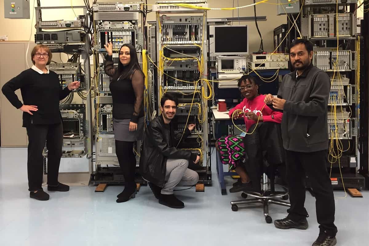
Multidisciplinary by nature Christine Tremblay (left) and members of the ÉTS Network Technology Lab. (Courtesy: ÉTS)
“My R&D pathway has allowed me to work across a broad and still-evolving physics and engineering canvas,” Tremblay says. “It helps, of course, that I’m inherently curious – someone who likes to push out in new directions to pursue fresh lines of enquiry. All sorts of opportunities follow when you’re open-minded and willing to connect and collaborate with partners working on interesting problems of their own.”
That R&D focus and diversity of approach is mirrored in Tremblay’s ÉTS research group, which she says is a “multidisciplinary melting pot” comprising two postdocs, three PhD students and another 10 team members (mainly MSc/MEng students and research assistants). Significantly, given the long-term gender imbalance in telecoms engineering disciplines, Tremblay’s group has included more women than men over the last five years – and she confesses to being “very proud to have such a talented mix of applied scientists and engineers working in my team”.
Training the next generation
Beyond her broad-scope research interests, Tremblay is also passionate about professional development and education. In particular, she has done much to train the next generation of communications engineers to support the installation, testing and maintenance of high-speed optical networks – chiefly through a long-standing association with the Optical Society’s flagship Optical Fiber Communication (OFC) annual conference.
Working alongside industry colleagues, Tremblay was academic co-instructor for two well-received continuing education courses at OFC: one on optical-fibre characterization and testing in long-haul and metro-area networks; the other on polarization-related measurements in fibre networks. “These were hands-on, practical workshops targeting a mixed audience of research students, early-career engineers and senior telecoms and photonics people from all over the world,” she explains.
Crucially, all of the optical kit used by the attendees was loaned free of charge by various test and measurement companies, with Tremblay having to “wheel and deal” in advance to call in favours from the likes of her former employer EXFO. Given the interactive nature of the training, Tremblay reckons she learnt as much from the delegates as they likely took from the instructors, making some long-lasting contacts too. “I was very glad and proud to contribute to the OFC professional development programme,” she notes. “Although a non-trivial overhead on top of my ÉTS research activities, it was great fun and incredibly rewarding in equal measure.”
These days, Tremblay is a full professor in the ÉTS electrical engineering department as well as the founding researcher and head of the institute’s Network Technology Lab. This advanced fibre-optic layer testbed, developed with the telecoms equipment maker Ciena over the past decade and more, comprises 2500 km of various fibre types linking an array of high-speed optical transmission systems. Alongside Ciena, her current R&D partners are Chalmers University of Technology in Sweden, the French engineering school Télécom SudParis and Canadian telecoms service provider TELUS.
As such, Tremblay’s research programme spans several broad areas of interest. It covers, for example, “smart optical networks”, in which machine learning is used to forecast the quality of transmission in optical fibre systems, as well as the performance monitoring of installed networks (including predictive methods for degradation and failure across multiple fibre plant deployments to inform engineering upgrades). Tremblay’s group also works on “filterless” optical network architectures (based on low-cost passive optical-routing technologies) as well as photonic device modelling and polarization measurements for sensing applications in telecoms networks.
Another emerging opportunity is quantum communications. “Quantum is largely aspirational for us just now, though we are already talking to relevant experts in Canada,” says Tremblay. If funding is forthcoming, however, her team plans to optimize the performance of secure, long-distance quantum key distribution across a classical optical network using engineered quantum-entangled photonic technologies.
Research pathways aside, Tremblay’s key message for today’s early-career physicists is the same as it was 40 years ago, back in that Quebec City grocery store. “Follow your instinct and be curious,” she concludes. “It’s equally important to be alert so that when new opportunities arise, you can take advantage of them.”
• AI autocontouring of organs in preclinical radiation studies for cancer
Frank Verhaegen
An overview will be given of the role of artificial intelligence (AI) in automatic delineation (contouring) of organs in preclinical cancer research models. It will be shown how AI can increase efficiency in preclinical research.
Speaker: Frank Verhaegen is head of radiotherapy physics research at Maastro Clinic, and also professor at the University of Maastricht, both located in the Netherlands. He is also a co-founder of the company SmART Scientific Solutions BV, which develops research software for preclinical cancer research. His interests are radiotherapy physics, imaging, preclinical research and Monte Carlo simulations.
• Deep learning prostate segmentation in three-dimensional ultrasound
Nathan Orlando
This presentation will explore the development and validation of a generalisable deep-learning-based automatic prostate segmentation algorithm for three-dimensional ultrasound images. Practical considerations for implementing deep-learning segmentation tools will be explored including the effect of dataset size, image quality, and image type on segmentation performance.
Speaker: Nathan Orlando is a fifth-year PhD candidate in the Department of Medical Biophysics at Western University and Robarts Research Institute in London, Ontario, Canada, supervised by Dr Aaron Fenster and Dr Douglas Hoover. His research has focused on improving ultrasound-guided prostate brachytherapy through both software- and hardware-based solutions. Prior to starting at Western University, Nathan completed a BSc (Hons) in physics at the University of Alberta in Edmonton, Alberta, Canada.
• Prospectively-validated deep learning model for segmenting swallowing and chewing structures in CT
Aditi Iyer
In this presentation, we present a deep learning-based method to automatically delineate the swallowing and chewing structures in CT. Its potential for use in radiotherapy treatment planning to improve efficiency is demonstrated through prospective validation.
Speaker: Aditi Iyer, is a senior scientific application developer in the Department of Medical Physics at Memorial Sloan Kettering Cancer Center, where she has served as a key contributor to the open-source Computational Environment for Radiological Research (CERR) software for six years. Her research interests include the application of machine learning and radiomics for image analysis and predictive modeling. Prior to joining MSKCC, she received her master’s degree from Purdue University, Indiana, where she worked on the estimation of multi-subject functional connectivity maps from fMRI data.
Speakers relationship with IOP Publishing
They are accepted authors of the focus collection.
Webinar chairs Georgios Papanastasiou and Guang Yang, guest editors of the joint Physics in Medicine & Biology and Machine Learning: Science and Technology focus issue, Focus on Machine Learning Models in Medical Imaging.
In recent years, deep learning gained a lot of attention and made impressive achievements in various applications. Incorporating deep learning in X-ray CT has become a non-reversible trend.
In this webinar, we’ll give a brief overview of deep learning technology. On this basis, focusing on the key issues in CT imaging, including denoising, artefact suppression, image reconstruction, we will discuss the methodology of incorporating deep learning into different data-processing missions by addressing the deep-learning framework, neural network design, loss functions, multiple domain learning, as well as some of our preliminary research results. Some of the key issues in the current field and technological-development challenges will also be discussed.
Yuxiang Xing
Yuxiang Xing received her PhD from the State University of New York at Stony Brook in 2003 and then joined Tsinghua University as a faculty member. She is currently a professor of the department of engineering physics at Tsinghua University, China. Since 2003, she has been devoted to research on the theories and technologies for the development and application of X-ray imaging systems. She has authored or co-authored more than 150 research publications and more than 50 patents. Her current interests include X-ray imaging physics, reconstruction methods for CT, radiation image processing and performance evaluation, especially cutting-edge deep-learning methods for CT reconstruction and artefacts reduction.
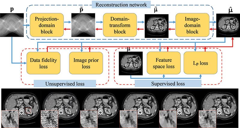
Conventional model-based image reconstruction (MBIR) for X-ray CT is often formulated as an optimization problem, the solution of which is the unknown image to be reconstructed.
Research in the past few years has shifted to replace components of these conventional MBIR methods by deep neural network models. Such integration can provide both improved image quality and certain interpretability of the deep learning architecture.
Jingyan Xu will present some existing approaches combining deep learning and MBIR, and discuss their strengths, weaknesses, and possible future extensions.
Jingyan Xu
Jingyan Xu obtained her PhD in electrical engineering from Stanford University. She is currently an assistant professor in the Department of Radiology at Johns Hopkins University. Her area of expertise lies in developing model-based image reconstruction methods and task-based image-quality evaluation for X-ray CT. More recently, she has been working on synergistic integration of deep learning and model-based reconstruction for CT image generation.
Recent innovations of computer calculation and artificial intelligence in radiotherapy resulted in rapid advancements in various aspects in cancer treatment such as treatment planning, patient-specific quality assurance, radiation dosimetry, and education for patients and radiation staff. Such computer applications not only improve the quality of radiation treatment, but also enhance the safety in the radiotherapy chain.
In this webinar, we shall draw attention to some significant developments in computer calculation and machine learning in radiotherapy. There will be a focus on Monte Carlo simulations in nanoparticle-enhanced radiotherapy, and the applications of machine learning in treatment plan evaluation.
Showcased will be an AI-chatbot created for education and training. In addition, Monte Carlo simulation is a mathematical method based on random sampling and is well known to be the benchmark in predicting radiation dose in treatment planning.
We will go through some Monte Carlo simulation results for dose enhancement in gold nanoparticle-enhanced radiotherapy. Machine learning is recently implemented in treatment planning and patient/radiation staff education in the radiotherapy chain. Also explored is how machine learning can help in treatment-plan evaluation and creating an AI-chatbot for the education of patients and radiation staff.
James Chow
Dr James Chow is a medical physicist in the Princess Margaret Cancer Centre, University Health Network and an associate professor in the Department of Radiation Oncology at University of Toronto. He is also an affiliated scientist of the TECHNA Institute for the Advancement of Technology for Health, University Health Network, and a member of the Temerty Centre for Artificial Intelligence Research and Education in Medicine, University of Toronto. He is a senior member of the Institute of Electrical and Electronics Engineers in the USA, and fellows of the Institute of Physics, UK, and Canadian College of Physicists in Medicine, Canada.
Deep learning is revolutionizing many areas of science and technology, particularly in natural language processing, speech recognition and computer vision.
In this talk, we will provide an overview into the latest developments of machine learning and AI methods and application to the problem of drug discovery and development at Isayev’s Lab at CMU. We identify several areas where existing methods have the potential to accelerate pharmaceutical research and disrupt more traditional approaches.
Olexandr Isayev
Olexandr Isayev is an assistant professor at the Department of Chemistry at Carnegie Mellon University. In 2008, Olexandr received his PhD in computational chemistry. He was postdoctoral research fellow at the Case Western Reserve University and a scientist at the government research lab. During 2016–2019 he was a faculty at UNC Eshelman School of Pharmacy, the University of North Carolina at Chapel Hill. Olexandr received the “Emerging Technology Award” from the American Chemical Society (ACS) and the GPU computing award from NVIDIA. The research in his lab focuses on connecting artificial intelligence with chemical sciences.
Researchers in China have synthesized a new type of high-temperature superconductor, clathrate calcium hydride (CaH6). The material, which is superconducting at temperatures of 215 K and pressures of 172 GPa (1.72 Mbar), is one of best high-temperature superhydrides made to date and the only clathrate hydride outside the family of rare-earth and actinide hydrides.
Superconductivity is the ability of a material to conduct electricity without any resistance. It is observed in many materials when they are cooled to below their superconducting transition temperature (Tc). In the Bardeen–Cooper–Schrieffer (BCS) theory of (“conventional”) superconductivity, this occurs when electrons overcome their mutual electrical repulsion and form so-called Cooper pairs that then travel unhindered through the material as a supercurrent.
Superconductivity was first observed in 1911 in solid mercury below a Tc of 4.2 K and the search for room-temperature superconductors has been on ever since. Finding a material that superconducts at such high temperatures would considerably improve the efficiency of electrical generators and transmission lines, while also making common applications of superconductivity, such as superconducting magnets in particle accelerators, simpler and cheaper.
Compressed hydrides
Physicists came a step closer to this “holy grail” of condensed-matter physics thanks to the copper oxide (cuprate) superconductors, which were discovered in the 1980s and 1990s and which include materials with Tc above 77 K, the temperature at which nitrogen becomes a liquid. Then, in 2015, the role of pressure came to the fore as researchers discovered that hydrogen sulphide has a Tc of 203 K when compressed to pressures of 150 GPa. This result spurred a flurry of interest in the compressed hydrides containing rare-earth or actinide elements.
Using quantum-mechanics-based calculations, two independent teams, led by Russell Hemley at George Washington University in the US and Yanming Ma at Jilin University in China, predicted in 2017 that lanthanum hydride (LaH10) could be superconducting. Hemley’s team went on to synthesize the material, and in May 2018 reported direct measurements of its conductivity indicating a Tc of 260 K at 180–200 GPa,posting a paper on the arXiv in August 2018 that was then published in Physical Review Letters. The team then reported additional measurements showing a Tc of up to 280 K in some samples in August 2018 at the Boston ACS meeting. A separate team led by Mikhail Eremets at the Max Planck Institute for Chemistry in Germany reported a Tc of 250 K for lanthanum hydride synthesized at pressures of around 170 GPa in work posted on the arXiv in December 2018 and subsequently published in Nature.
A class of currently unexplored superconductors
Now, Yanming Ma and colleagues at the State Key Laboratory of Superhard Materials, College of Physics, Jilin University, China have succeeded in synthesizing a new type of superhydride altogether, one that contains an alkaline-earth metal instead of a rare-earth metal or an actinide. The discovery “opens the door to a class of superconductors that is currently unexplored,” the researchers claim.
Although CaH6 was first predicted to be a superconductor a decade ago, it proved difficult to synthesize because calcium and hydrogen are highly reactive. When the two elements are brought together at low pressures, the result is often a hydride with an undesirably low hydrogen content.
In the new work, Ma and colleagues overcame this problem by using ammonia borane (BH3NH3) as a hydrogen source. This allowed them to synthesize CaH6 via a direct reaction between Ca and H2 at high temperatures and pressures.
“We used a special sample loading method to synthesize the material at close to 200 GPa and temperatures of 2000 K in a diamond anvil cell with microelectrodes that we carefully mounted on the tips of the anvil for subsequent electrical conductivity measurements,” team member Hongbo Wang explains.
“The dramatic loss of resistance is very similar to the other superconductivity transitions that we have previously studied under pressure,” he tells Physics World, “and we have reproduced the result many times.”
According to the researchers, their new work will help to advance our understanding of superconductivity and could lead to new classes of ternary calcium-based superhydrides. The team is now busy exploring a broader range of compositions based on their own and other group’s calculations. “We believe this system is just one of many superhydrides, with likely higher Tc values,” says Wang.