In this episode of the Physics World Weekly podcast, we talk about the pros and cons of injecting large amounts of aerosols into the atmosphere to offset global warming. Wake Smith of Yale University and Harvard University in the US explains how it could be done and emphasises that this solar geoengineering is not a solution to climate change but rather a “fire extinguisher” that could be used in an emergency.
Also on hand are Nicole Xu and Jason Geder of the US Naval Research Laboratory who explain how microscopic features found on the skin of sharks are inspiring the development of new hull materials for unpiloted underwater vehicles. They also chat about those controversial shark’s skin-inspired swimsuits that have been banned from competitions.
What are you? Who are you? Where are you going? These are three deceptively simple-sounding questions. But, from the Ship of Theseus to the “hard problem of consciousness”, thousands of years of thinking have shown that they have anything but simple answers. And perhaps nothing could have complicated matters more than the past century of scientific discoveries. It is the relatively recent insights into these three profound questions that are the subject of Youniverse: a Short Guide to Modern Science by novelist and “lifelong learner” Elsie Burch Donald.
Physicists might first think of the shock revelations of quantum mechanics (that matter, including what we are made of, appears to be fundamentally probabilistic, not deterministic) and of general relativity (that the fabric of space–time can be distorted). These topics are covered in the first, material-focused, section of the book, in answer to the question “What are you?”.
But, as Donald shows, physics is not the only discipline that has been thoroughly shaken up in the last 100 years. In the second section, which asks “Who are you?”, the book discusses our current understanding of genetics, neuroscience and psychology, while placing it in the context of longer-held knowledge about Darwinian evolution. Often taking a storytelling approach, the writing is engaging, and occasionally accompanied by useful illustrations, such as anatomical diagrams of the brain and a nerve cell.
The third question “Where are you going?” is addressed in the final section, where Donald follows the recent trajectory of progress to speculate about humanity’s possible next steps. She opines that “the future is set to become a contest between two scientific disciplines: genetic engineering and computer technology in the form of artificial intelligence”.
The book’s content is accessible throughout, and would suit any interested person in the early stages of their scientific education, whether at school or learning independently. But, from CRISPR to quantum computers, the breadth of topics covered means that most readers would probably learn something new from it, while enjoying a comprehensive summary of today’s science.
Artificial intelligence has potential to improve the operation of many essential tasks in various fields of medicine and biomedicine – from dealing with the massive amount of data generated by medical imaging, to understanding the evolution of cancer in the body, to helping design and optimize patient treatments. At last week’s APS March Meeting, a dedicated focus session examined some of the latest medical applications of artificial intelligence and machine learning.
In-depth image analysis
Opening the session, Alison Deatsch from the University of Wisconsin, Madison, discussed the use of deep learning for diagnosing and monitoring brain disease. “Brain disorders and neurodegenerative disease are some of the most costly diseases, both in terms of human suffering and economic costs,” she explained.
The reason is that most of these conditions – which include Alzheimer’s and Parkinson’s disease, autism spectrum disorder and mild cognitive impairment (MCI), among others – lack reliable tools for diagnosis and progression monitoring and, as such, are often misdiagnosed. And for monitoring the neurological effects of cancer or chemotherapy, there are no standardized diagnostic tools at all, Deatsch noted.
Neuroimaging, using modalities such as MRI, functional MRI, PET and SPECT, could fill this gap. “However, when it comes to analysing these images, they are rarely brought to their full potential in the clinic,” said Deatsch. “This is due in part to the time it takes to manually curate or quantify data and some inherent uncertainty.”
To address this obstacle, the field is moving from visual-based image analysis towards a more quantitative approach, exploiting computational techniques to maximize information output from neurological images. This includes deep learning methods such as convolutional neural networks (CNNs), which are the most prevalent in medical imaging, as well as recurrent neural networks (RNNs) that use time series data. This shift could both advance our understanding of neurodegenerative disease and enhance clinical decision making.
“Deep learning has seen a significant increase in its use for neuroimaging in the last five years,” Deatsch said, presenting some recent clinical examples. CNNs, for instance, have been used with MRI data to identify Alzheimer’s disease, predict the progression of MCI to Alzheimer’s and assess Huntington’s disease severity, with reported accuracies of between 70 and 90%. Deep learning has also been employed to analyse PET and SPECT data for Alzheimer’s or Parkinson’s disease diagnosis, with similar high performance. Deatsch also highlighted some multimodality studies using CNNs to analyse combined MRI and PET data, with accuracies of more than 80%.
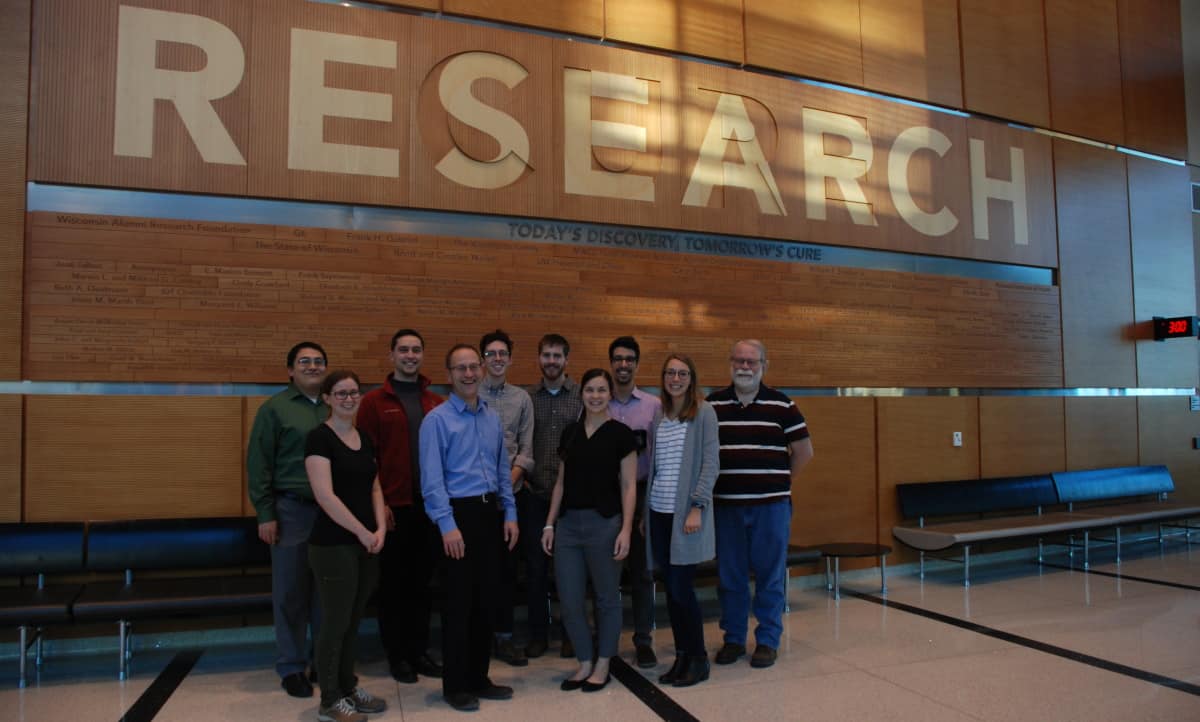
Deep learning team: Alison Deatsch (front row, third from left) and colleagues at the University of Wisconsin, Madison. (Courtesy: Alison Roth)
But despite the success achieved to date, challenges remain, which Deatsch and colleagues hoped to address in a recent project. They developed a novel deep learning model that can distinguish brain scans of patients with Alzheimer’s disease from normal controls, and investigated how various factors affected the model’s performance.
The team trained a CNN, with or without a cascaded RNN, to analyse 18F-FDG-PET and T1-weighted MRI scans. The CNN learns the spatial features and outputs a prediction of normal or Alzheimer’s disease. For patients with two or more scans, the RNN then learns temporal features and also outputs a classification for each patient. After training the model on several hundred PET and MRI scans, it achieved a maximum area under the ROC curve (AUC) of 0.93 and an accuracy of 81%.
Next, the researchers examined whether the imaging modality influences the model’s performance. They saw significantly better performance using PET data in both model types (with and without the RNN), possibly due to the larger variation between MR images. They also assessed whether adding longitudinal data has an impact and found that incorporating these data significantly improved performance for PET scans, but not for MRI.
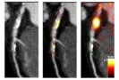
To validate the CNN’s generalizability, they tested it on an external data set, where it performed equally well on the new unseen data. Finally, they checked the interpretability of their model by generating attention heatmaps showing the brain regions responsible for the model’s decision. They note that such maps provide a step towards identifying a quantitative imaging biomarker for Alzheimer’s disease.
“There is a tonne of promise for deep learning with neuroimaging for neurological diseases,” Deatsch concluded. “There are still a few limitations to address, but I hope I’ve shown the significant potential contained within this field and that many future studies will continue.”
Radiotherapy safety check
Artificial intelligence can play many roles in medical imaging – not just for diagnostics, but also in tasks such as image registration and segmentation, to help in radiotherapy treatment planning, for example. This naturally leads to the incorporation of machine learning-based methods in other applications, such as ensuring the safety of radiation therapy.
Keeping radiotherapy safe: Qiongge Li.
Qiongge Li from Johns Hopkins University School of Medicine presented such an application: an innovative anomaly detection algorithm designed to increase patient safety. The idea is to use the new tool to ensure appropriate radiation dose schemes are delivered to every patient. “It is important to address the detection of prescription errors in radiotherapy, even if this is a rare event,” she explained.
Quality assurance checks of radiotherapy plans are usually performed via a peer-review chart round in which physicians reach a consensus on each patient’s dosage. This is a manual and time consuming process, however, and doesn’t catch every error. Li described one study in which simulated plan anomalies were inserted into the weekly peer-review chart round and only 67% of the prescription errors were detected.
Li and colleagues have developed an anomaly detection tool that uses historical data to identify atypical radiotherapy prescriptions. At the heart of the tool is a distance model that compares patient data to historical databases and determines two dissimilarity metrics: the distance between the new patient’s prescription features (number of fractions and dose per fraction) and historical prescriptions; and the distance between other features (patient age, radiotherapy technique and energy, and clinical intent) and those of historical patients with similar prescriptions.
The model automatically flags any detected anomalies, such as a prescription that is very different to any seen before, or a mismatch between the prescription and other features. Thresholds for flagging were defined using mean feature distances between all patient pairs in the historical database.
Anomaly detection tool: The model compares patient data to historical databases and automatically flags any outliers. (Courtesy: Qiongge Li)
The researchers trained their machine learning algorithm using historical data from 11062 thoracic cancer treatment plans. To validate the tool, they tested it on a set of unseen normal plans and plans created with simulated anomalies. These included, for instance, changing the number of fractions and dose per fraction to a non-standard combination, or changing the patient’s age from 90 to 10 and the treatment intent from cure to palliative – creating a mismatch between prescription and features.
The model demonstrated F1 scores (a combination of precision and recall) of 0.941, 0.727 and 0.875, for 3D conformal radiotherapy, intensity-modulated radiotherapy and stereotactic body radiotherapy plans, respectively. Three expert thoracic consultants also classified each case; the model and outperformed all three doctors in terms of recall, precision, F1 and accuracy.
Another benefit of the machine learning model is that it only takes around 1 s to run, compared with between 15 and 30 min required by the doctors. Li pointed out that training the model takes several days, but it only needs to be performed once. She also noted that a consensus between the three doctors performed slightly better than the model.
“Developing a fully automatic and data-driven tool for assisting peer-review chart rounds and providing that extra safety to our patients is in great need,” said Li.
The efficiency of some radiation detectors can be boosted by adding nanostructure arrays to scintillator materials. Charles Roques-Carmes and colleagues at the Massachusetts Institute of Technology have shown how the modifications increase the amount of light emitted by the scintillators by a factor of ten when the materials are exposed to either X-rays or high-energy electrons.
Scintillation occurs in a range of materials – solid, liquid and gas – when they are exposed to ionizing radiation. The radiation is absorbed by a scintillator’s atoms or molecules and some of the energy is re-emitted as light. The light can then be detected, allowing scintillators to be used as radiation detectors with applications including medical imaging, industrial quality control and particle physics experiments.
Researchers are always trying to develop better scintillators that produce more light when irradiated or have a shorter delay between the absorption of radiation and emission of light. Most studies so far have focused on developing new materials with brighter, faster, and more controlled scintillation – but this can be costly and time-consuming.
In their study, Roques-Carmes’ team took a simpler approach based on nanophotonics. They calculated that scintillation in materials can be enhanced by incorporating nanoscale features onto the surfaces of scintillating materials. The sizes of these features should be comparable to the wavelengths of the light that is emitted by the scintillators.
Unified theory
To explore this idea, the researchers first developed a unified theory of nanophotonic scintillators – which could predict from first principles how ionizing radiation interacts with the nanostructured surface any arbitrary material. Afterwards, they designed a method for integrating nanophotonic structures into existing scintillators. This could be done either by etching patterns onto the scintillator directly, or attaching a layer on top of the material, etched with an array of holes.
Roques-Carmes and colleagues then did a series of experiments that confirmed their calculations. They etched regular grids of circular holes onto the surfaces of two different types of scintillator – one that is used to detect X-rays and the other used to detect electrons. These holes were tens of nanometres deeps and had radii of about 200 nm.
In both scintillators the team measured a tenfold increase in light production in regions containing the nanostructures. Through further improvements, they hope that their generalized approach could lead to a new class of brighter, faster, and higher-resolution scintillators, with a hundredfold improvement on existing materials.
If achieved, this could lead to promising advances in a diverse array of applications: including higher-quality medical X-ray images that are produced using lower X-ray doses. This could significantly improve the safety of X-ray imaging, particularly for younger patients and those requiring routine screening. Elsewhere, the technique could lead to higher resolutions in particle detectors and electron microscopes, as well as faster and higher-quality inspections of manufactured parts.
Comprehensive, it seems, just got more comprehensive. That’s the key take-away following the acquisition of VALO Innovations, a German start-up specializing in ultrashort-pulse fibre lasers, by HÜBNER Photonics, one of the optics industry’s leading lights and home to an already broad portfolio of compact single-frequency CW lasers, diode-laser modules and nanosecond pulsed lasers across the full UV, visible and near-IR spectrum.
For its part, Hannover-based VALO Innovations represents an intriguing diversification opportunity, with proprietary fibre-laser technology and “market-unique short-pulse performance” (pulse lengths of less than 50 fs, up to peak powers above 2 MW) opening up – indeed underpinning – new ultrafast applications in bioimaging, spectroscopy and optogenetics. Here, Oliver Prochnow, CEO of VALO Innovations, tells Physics World why his team’s customer-centric approach to product development and a complementary offering in the ultrashort-pulse regime represent the perfect addition to the HÜBNER Photonics portfolio.
What are the anticipated upsides following the acquisition of VALO Innovations?
During our exploratory conversations last summer, both parties quickly realized that the acquisition was a win-win. From the HÜBNER Photonics side, VALO Innovations represents a compelling technology fit, complementing its established portfolio of high-performance laser products for advanced imaging and analysis applications, including the Cobolt lines of single-frequency CW nanosecond pulsed lasers, the C-FLEX laser combiners and the widely tunable, single-frequency CW laser C-WAVE. Put simply: ultrafast lasers represent a natural complement to the HÜBNER Photonics portfolio and VALO Innovations provides the ideal vehicle to fast-track that ambition.
Oliver Prochnow: “We’re already benefiting from the open, collaborative and supportive environment here.” (Courtesy: HÜBNER Photonics)
For the VALO Innovations team, meanwhile, we are now positioned to develop and scale our femtosecond fibre-laser offering within an established innovation and manufacturing ecosystem. That means full integration with the in-house R&D and product development functions at the parent company, plus the ability to benefit from economies of scale when it comes to component/subcomponent purchasing.
It’s worth adding that VALO Innovations and HÜBNER Photonics are also an excellent fit on a personal level, with a commonality of approach when it comes to our customer orientation, product development and commercial strategy.
How has your role evolved within HÜBNER Photonics?
As CEO of VALO Innovations, which is now a wholly owned subsidiary of HÜBNER Photonics, I am responsible for the parent group’s ultrafast fibre-laser market segment. That means owning and articulating the technology roadmap for the femtosecond product line while delivering our commercial growth targets in research and industry applications. Operationally, we are in the process of extending the office and laboratory space of the VALO Innovations division here in Hannover. We are also building up the team, recruiting talented scientists, engineers and product managers with specialist capabilities in ultrafast fibre-laser technologies and applications.
What differentiates VALO Series fibre lasers from the competition?
Our fibre-laser technology is based on the interaction between linear and nonlinear optical effects. In broad terms, we have found a way to control all phase contributions that affect pulse quality, a capability that enables us to generate very clean and extremely short pulse durations without any additional nonlinear broadening after the laser. As such, VALO Series lasers are compact and stable turnkey systems – including integrated group-velocity dispersion pre-compensation control – which enable us to scale the energy of ultrashort laser pulses while yielding the same clean pulses in the sub-50 fs regime.
How are commercial applications shaping up for the VALO Series?
We’re making inroads with a range of ultrafast imaging applications, including multiphoton microscopy (to study complex and dynamic biological processes deep within living tissue), optogenetics (which controls the activity of neurons or other cell types using light) and with amplifier seeding (in which femtosecond pulses increase the energy of selected laser pulses by several orders of magnitude).
Near-term focus: the VALO Innovations team is currently transferring best-practice production models from the wider HÜBNER Photonics laser portfolio. (Courtesy: HÜBNER Photonics)
It’s very much a mixed customer base, comprising research laboratories as well as industry OEMs who integrate our sources into their multiphoton microscopy/spectroscopy systems. For the latter, the benefits of ultrashort pulses are clear: a 40 fs pulse, for example, yields a x5 higher peak power versus a 200 fs pulse. That, in turn, means greater signal efficiency, enhanced penetration depth in tissue, better contrast and reduced photothermal damage.
What’s coming next on the development roadmap?
In the near term, we want to “industrialize” the femtosecond product line by transferring best-practice production models from the wider HÜBNER Photonics laser portfolio. Right now, the emphasis is on continuous improvement and internal benchmarking along a number of key coordinates, including manufacturability, device testing and quality control, long-term reliability, packaging and after-sales service. Looking ahead, we also want the VALO Series to be a scalable technology platform – both in terms of power levels and wavelengths – to broaden our access to emerging applications in life sciences and microscopy.
How important is customer engagement in shaping product innovation for the VALO Series?
We have an ongoing dialogue with our early-stage customers and R&D partners, figuring out what needs are not being met and how our lasers can help end-users to do their jobs better. Up to now, it’s been mainly me initiating these conversations, though in future much of the responsibility for requirements-gathering and prioritization will switch to our product managers.
It’s still early days, but what are the main operational benefits that you’re seeing since the acquisition?
Joining forces with HÜBNER Photonics feels like a once-in-a-lifetime opportunity to embed the VALO Innovations start-up model within an established laser manufacturing operation. We’re already benefiting from the open, collaborative and supportive environment here, with very close engagement between HÜBNER Photonics teams in Germany, Sweden and the US when it comes to product development and innovation. Another benefit is the uptick in lead generation given that VALO Innovations is now integrated with an experienced in-house sales team and global distributor network, while we also benefit significantly from our direct access to the group’s other core functions such as finance, legal and HR.
When water moves through nano-sized channels made of carbon, its flow rate is much higher than current theories of fluid dynamics predict. New work by researchers at the École Normale Supérieure (ENS) in Paris, France and the Flatiron Institute in New York, US suggests that a quantum version of friction plays a crucial role in resolving this conundrum. The team’s findings could have important implications for applications such as filtering salt from seawater or generating energy.
Experimentalists have known for more than a decade that, paradoxically, fluids such as water pass more easily through narrower carbon nanotubes – rolled-up sheets of carbon as little as one atom thick – than through wider ones. The effect is strongest for nanotubes constructed from more than one layer of carbon sheets. A further puzzle is that while fluids flow with hardly any friction through all sizes of channel thanks to the extreme smoothness of the nanotube walls, the friction that is present is hard to explain.
In 2016, Lydéric Bocquet and colleagues at the ENS revealed that the amount of friction increases for larger-diameter nanotubes. In principle, this could explain why water flows more easily through narrower tubes. However, the result was confusing because the walls of larger tubes are just as smooth as smaller ones.
A wall isn’t just a wall
In the latest work, Lydéric and Marie-Laure Bocquet of the ENS, together with Nikita Kavokine at the Flatiron Institute’s Center for Computational Quantum Physics, studied carbon nanotubes with diameters ranging from 20 to 100 nanometres. The researchers took a new approach, unusual in the field of fluid dynamics, by focusing on the nanotube walls. “In hydrodynamics, the wall is just a wall, and you don’t care what the wall is made of,” Kavokine explains. “We realized that at the nanoscale, it actually becomes very important.”
The researchers found that quantum effects at the wall’s carbon-water interface produce friction by allowing the flowing liquid to dissipate energy by scattering electrons in the carbon. These electrons interact with the water molecules electromagnetically because the latter are polar, with one end of the molecules being slightly positively charged and the other slightly negatively charged.
Kavokine and colleagues say that while the electrons in the carbon move along with the flowing water molecules, they tend to slightly lag behind. This effect, known as electronic or quantum friction, had only been considered in interactions between two solids or a single particle and a solid until now.
Increasing the quantum friction force
Kavokine goes on to explain that the electrons tend to vibrate collectively at a particular frequency. This behaviour is called a plasmon mode, and if water molecules can vibrate at the same frequency, they do so in unison with the electrons, increasing the quantum friction force. This finding explains why frictional effects are strongest for nanotubes with multiple well-aligned layers, since the interlayer motion of the electrons is synchronized with that of the water molecules.
Asked why this effect hadn’t been seen before, Kavokine notes that it is very slight even in carbon nanotubes and would be negligible for materials with rougher surfaces. He adds that the effect is also hard to mimic using molecular dynamics simulations, which fail to capture this type of friction because they rely on the so-called Born-Oppenheimer approximation. This approximation assumes that electrons adapt instantly to the motion of nearby atoms, which isn’t the case. “I think this work opens the door to many new quantum interfacial phenomena that we are only beginning to understand,” Kavokine concludes.
Reporting their work in Nature, the researchers now plan to return to their experiments and explore the practical consequences of their quantum friction theory. “We would like to systematically investigate the effect of electronic properties on friction,” Bocquet tells Physics World. “Our investigations will also continue on the theoretical side.”
The construction and operation of space missions, telescopes and other facilities produces more greenhouse-gas emissions than any other aspect of doing astronomy and cosmology research. That is according to a new study, which shows that if the field is to become more sustainable in future then the pace of building new observatories and space missions will need to be slower than it has been during the past few decades.
There has been an increased interest recently on the climate impact of scientific research. Much of the attention has focused on the effect of academic travel and other research activities such as the use of supercomputers. There has been less attention, however, on research infrastructure such as greenhouse-gas emissions from the construction and operation of space observatories, planetary probes and ground-based observatories.
To address this gap, astrophysicist Luigi Tibaldo from the Institut de Recherche en Astrophysique et Planétologie in France and colleagues estimated the greenhouse-gas emissions for nearly 50 space-based missions and 40 ground-based telescopes.
For ground-based facilities, the calculations assume that greenhouse-gas emissions are proportional to construction and operating costs, while for space and satellite missions, it is linked to the full mission cost and payload launch mass.
The team estimates that active astronomy research infrastructures worldwide will emit a combined total of more than 20 million tonnes of carbon dioxide equivalent (CO2e) over their lifetimes – similar to the annual emissions of countries such as Estonia, Croatia and Bulgaria.
The strong reduction in emissions that is required in the next decade will not be achieved if we continue building new infrastructures at the pace that is occurring right now
Luigi Tibaldo
Annual emissions from research facilities are estimated to be around 1200 kilotonnes of CO2e with around 520 ktCO2e attributed to space missions and 760 ktCO2e from ground-based observatories.
According to the researchers, this translates to 36 ktCO2e per year per astronomer and makes research infrastructures the single largest contributor to the carbon footprint of an astronomer, being around five times larger than the environmental cost of travel.
The researchers say that if astronomers want to cut their carbon footprint, they should focus on reducing emissions from research infrastructures. “Solutions are clearly available, the first step that is needed is that the existing infrastructures are decarbonised, for example by switching to renewable energy power sources,” says Tibaldo. “One important step we recommend is that all the facilities carrying out astronomical research carry out more detailed analysis of their greenhouse gas emissions… and make their results public.”
The team acknowledges that its emission estimates for individual facilities have an uncertainty of about 80% but says that other fields that are dependent on large research infrastructure will likely have a similar breakdown of emissions to astronomy.
Tibaldo warns that decisions that are made now on future research infrastructures will lock-in emissions from astrophysics research for decades to come. “We think that the strong reduction in emissions that is required in the next decade will not be achieved if we continue building new infrastructures at the pace that is occurring right now,” he adds.
A quantum gravity gradient sensor developed by researchers in the UK has been used outdoors to locate a small underground structure. Using a pair of vertically oriented atom interferometers that are probed by the same laser system, Michael Holynski at the University of Birmingham and colleagues were able to suppress noise in their sensor, thereby overcoming limitations of previous designs.
Variations in Earth’s gravitational field can reveal useful information about what lies underground including the locations of large features such as aquifers and fossil fuels. Gravity measurements could also be used to find smaller metre-scale features such as tunnels, but current measurement technologies are susceptible to vibrational noise – which must be averaged out by taking unfeasibly long measurements.
In the gravity sensor designed by Holyinski’s team, two clouds of ultracold rubidium atoms are held in separate magneto-optical traps that are in an “hourglass” configuration – with one cloud located 1 m above the other cloud in a long, cylindrical vacuum chamber. Each cloud is cooled to millikelvin temperatures and then released simultaneously so the clouds freefall in the vacuum chamber. A sequence of counterpropagating laser pulses is fired at the clouds. This makes each cloud operate as an atomic interferometer that can measure the local acceleration due to gravity. Finally, the difference in local gravity between the two clouds is extracted from the experiment.
As a result, the system operates as a gravity gradient sensor. This is a device that is sensitive to anomalies in the Earth’s gravitational field that are caused by structures such as buildings or underground voids.
Robust against noise
An important feature of the instrument’s design is that the same lasers are used to control and interrogate both atomic clouds. This and the fact that the difference between the outputs of the two atom interferometers is measured – rather than their absolute values – means that the system is robust against many kinds of noise. This includes fluctuations in the lasers, local vibrations and the tilt of the instrument. The compact nature of the instrument also means that it can be effectively shielded against stray magnetic fields.
Holynski and colleagues tested their gravity gradient sensor by doing an outdoor survey that was able to map out an underground tunnel at a spatial resolution of 0.5 m. Located on the University of Birmingham campus, the tunnel has a square profile that is 2 m on edge and the roof of the tunnel is about 0.5 m under the surface of a road. The measurement process was very speedy and the team reckon that a similar tunnel could be detected by making 10 measurements over about 15 min.
The researchers hope that their technique could soon be used to map out the world beneath our feet in unprecedented detail. Through further improvements, the sensor could be used in a diverse array of applications: such as charting complex cave systems, measuring the time-varying flow of groundwater, performing non-invasive surveys of archaeological sites, and reducing the risks of unforeseen ground conditions ahead of construction and infrastructure projects.
Portable electronic devices can be carried in a myriad of ways — sometimes, you may even find yourself waking up from a nap with your smartphone or headphones on your chest.
But that proximity to your chest may be a problem if you’re one of the millions of people worldwide who rely on a cardiovascular implantable device (CID), such as a pacemaker or defibrillator.
Results from a new study appearing in Circulation: Arrhythmia and Electrophysiology suggest that the magnetic fields surrounding several Apple and Microsoft devices may be strong enough to interfere with such CIDs, which contain sensors that may respond to magnets when in close contact.
“This study shows that magnetic interactions with pacemakers and implantable cardiac devices are not limited to smartphones, but can occur with many other electronic portable devices,” says Corentin Féry, a research associate at the University of Applied Sciences and Arts Northwestern Switzerland.
Measuring magnetic fields
Féry, who is lead author on the study, says that he and his colleagues focused on this research in collaboration with University Hospital Basel after a 2021 paper reported interactions between the magnetic field from the iPhone 12 Pro Max and CIDs. The researchers in those studies observed clinically identifiable magnetic interference in some individuals with CIDs. Féry decided to replicate that work and expand it to include other portable electronic devices.
CIDs are designed with a magnet mode that’s used when a patient is undergoing certain medical procedures, such as an MRI scan. According to the International Organization for Standardization, a CID should not switch into magnet mode below 10 gauss (10 G), a standard recognized by the US Food & Drug Administration (FDA). The 10 G isogauss line identifies the volume in which the magnetic field of a device exceeds 10 G and into which a cardiovascular implantable device should not penetrate (for reference, 10 G is approximately one fifth the gauss rating of a refrigerator magnet).
First author Corentin Féry.
Féry and his colleagues measured the magnetic fields around the iPhone 12 Pro Max, Apple AirPods Pro with charging case opened and closed, Apple Pencil 2nd Generation and the Microsoft Surface Pen. The magnetic mapper they use to delineate the 10 G isogauss line measures the magnetic fields around portable electronic devices using numerous magnetic sensors on three axes. The mapper, the researchers say, accomplishes the same task as a magnetic probe but faster and more accurately.
The researchers’ results suggest that individuals with CIDs should keep the portable electronic devices tested in this study at least one inch away — 2 cm for Apple devices and 2.9 cm for the Microsoft Surface Pen, to be more precise — from their CIDs to avoid activating magnet mode. This is well within Apple’s own recommendation of six inches (more information on product safety is reported in the “Important Safety Information” sections of the user guides for Apple products).
The American Heart Association also suggests that patients avoid keeping cell phones and similar portable electronic devices in a front chest pocket and to use these devices on the side of the body opposite an implanted device.
Raising awareness
So, what’s new? Though modern smartphones already contained magnets and emitted signals that patients with implanted devices needed to be aware of prior to the iPhone 12 Pro Max, teardowns show that this phone and ones like it have more magnets in them than in earlier versions. Apple’s wireless charging technology, MagSafe, also uses permanent magnets placed around the iPhone 12’s internal charging coil to quickly charge devices.
And, while patients with CIDs are already recommended to keep portable electronic devices that may create magnetic interference at least six inches away from implanted medical devices, the researchers say that these magnet-containing portable electronic devices are now ubiquitous. Some devices may be nonchalantly placed in a shirt pocket or on the chest, switching a patient’s CID to magnet mode.
“The six-inch limit [recommended by the FDA] is high and reaching 10 G at this distance with portable electronic device magnets is very unlikely. On the other hand, implant wearers can without realizing a potential risk, go below six inches with portable electronic devices including magnets; for example, the Microsoft Surface Pen can be put in a shirt pocket,” says Féry.
One major limitation of the study was that the team measured the magnetic fields and their influence on cardiovascular implantable devices ex vivo.
“The skin and the depth of the implant in the patient have an influence on a patient-specific minimum safety distance between the skin and the portable electronic device,” Féry says. “The distances we provide represent the worst case that one can have, because it simulates an implant that would be directly at the same level as the epidermis.”
Currently, the researchers are testing other portable electronic devices, such as smart watches and electronic cigarettes. They also hope to raise awareness: historically, magnets strong enough to trigger the magnet mode of a cardiac implantable device were easy to steer clear of. Now, small rare-earth magnets are used inside everyday technologies.
“We hope to raise the awareness of doctors and patients with cardiovascular implantable devices of the risk of disabling medical device therapy by portable electronic devices,” Féry says. “They should know that the risk is more widespread than previously thought and that it involves different categories of objects.”
The US’s divisive China Initiative has been expanded to focus not only on individual scientists with links to China but also on other foreign governments that may be attempting to acquire US technology through espionage and theft. The original programme, led by the US Department of Justice (DOJ), had drawn criticism that it was used to target academic researchers, particularly those of Chinese heritage. The new initiative will now be expanded to other foreign governments.
The China Initiative was introduced in 2018 to tackle efforts by China’s government to acquire US technology illegally. While the DOJ has successfully prosecuted cases of espionage and theft of technology and intellectual property, it has also used the initiative more than 20 times to prosecute academic scientists of Chinese descent who have research connections with Chinese universities. Most of those cases have failed to deliver guilty verdicts and instead led Asian-American scientists to fear that the DOJ was targeting them based on their ethnicity.
In October almost 2000 prominent Asian-Americans wrote to US president Joe Biden expressing “grave concern” over efforts to clamp down on scientists who have scientific collaborations with Chinese colleagues. In November, Matthew Olsen, an assistant attorney-general who heads the DOJ’s National Security Division, responded by reviewing the China Initiative.
His effort, which included conversations with critics of the initiative, led to the changes in the programme’s name and direction. “Safeguarding the integrity and transparency of research institutions is a matter of national security,” he says. “But so is ensuring that we continue to attract the best and the brightest researchers and scholars to our country from all around the world.”
As to the expansion of the programme’s reach beyond China, Olsen notes that Russia, Iran and North Korea “are becoming more aggressive and more capable in their nefarious activity than ever before”. The DOJ also announced that its prosecutors will consider more carefully than in the past whether they should pursue scientists accused of grant fraud, under the initiative, in civil rather than criminal courts.
Groups representing individuals of Chinese heritage cautiously welcomed the changes. “The end of the China Initiative is one step in the work we expect from the government to address serious concerns of racial profiling and misconduct in its surveillance and national security operations, and the over criminalization of issues related to research integrity,” says John Yang, president and executive director of the civil rights group Asian Americans Advancing Justice.