LGBTQA scientists (defined in these studies as lesbian, gay, bisexual, transgender, asexual or otherwise queer) who hide their identities while at work have been found to produce fewer peer-reviewed publications than those who are more open about their sexuality and gender identity. That is according to an analysis of two US-based surveys, the findings of which highlight the productivity cost of workplaces that are unwelcoming to sexual and gender minorities (PLOS ONE17 e0263728).
The surveys were conducted by Jeremy Yoder, a biologist at California State University Northridge, and colleagues. In 2013, the team carried out an online survey of more than 600 LGBTQA science, technology, engineering and maths (STEM) professionals. Participants were asked about their publication count, sexual and gender identity, openness about their LGBTQA identity at work, and career position.
The researchers found that those who did not disclose their LGBTQA identities in professional settings had written significantly fewer peer-reviewed publications. But they also found that those in more senior positions had a higher publication rate and were more likely to be open about their sexual and gender identities.
To tease out if respondents who were not open about their identities were mainly early-career scientists with naturally lower publication rates, or not, the team conducted another survey in 2016 of almost 1800 STEM professionals. This time they included non-LGBTQA scientists and, to control for career stage, asked respondents how long it had been since their first publication. They found that openly gay and bisexual men had a comparable number of publications to straight men, while women and LGBTQA scientists who did not discuss their identities at work all reported fewer publications.
The researchers’ modelling predicts that over a 20-year publication career, non-LGBTQA men will write 27 papers, on average, while non-LGBTQA female scientists will produce 17 papers. Over the same period, gay, bisexual, asexual or otherwise queer male and non-binary academics who are open about their identity will publish around 24 papers whereas disclosing female lesbian, gay, bisexual, asexual or otherwise queer female scientists will produce 20 papers.
Among those who do not disclose their identities at work, male academics were found to publish just 15 papers while female and non-binary scientists have the lowest productivity at 13 published papers.
“It makes a good deal of sense that working without being able to express a key aspect of your identity is often stressful and would reduce your sense of belonging in a particular workplace and overall career path,” Yoder told Physics World.
He adds that LGBTQA scientists are more likely to be “out” when they consider their workplaces to be safe and welcoming. “What we’ve seen in our work is that LGBTQA scientists are keenly aware of what acceptance they can expect from their colleagues – and when they find that acceptance, they’re better able to contribute to science,” Yoder adds.
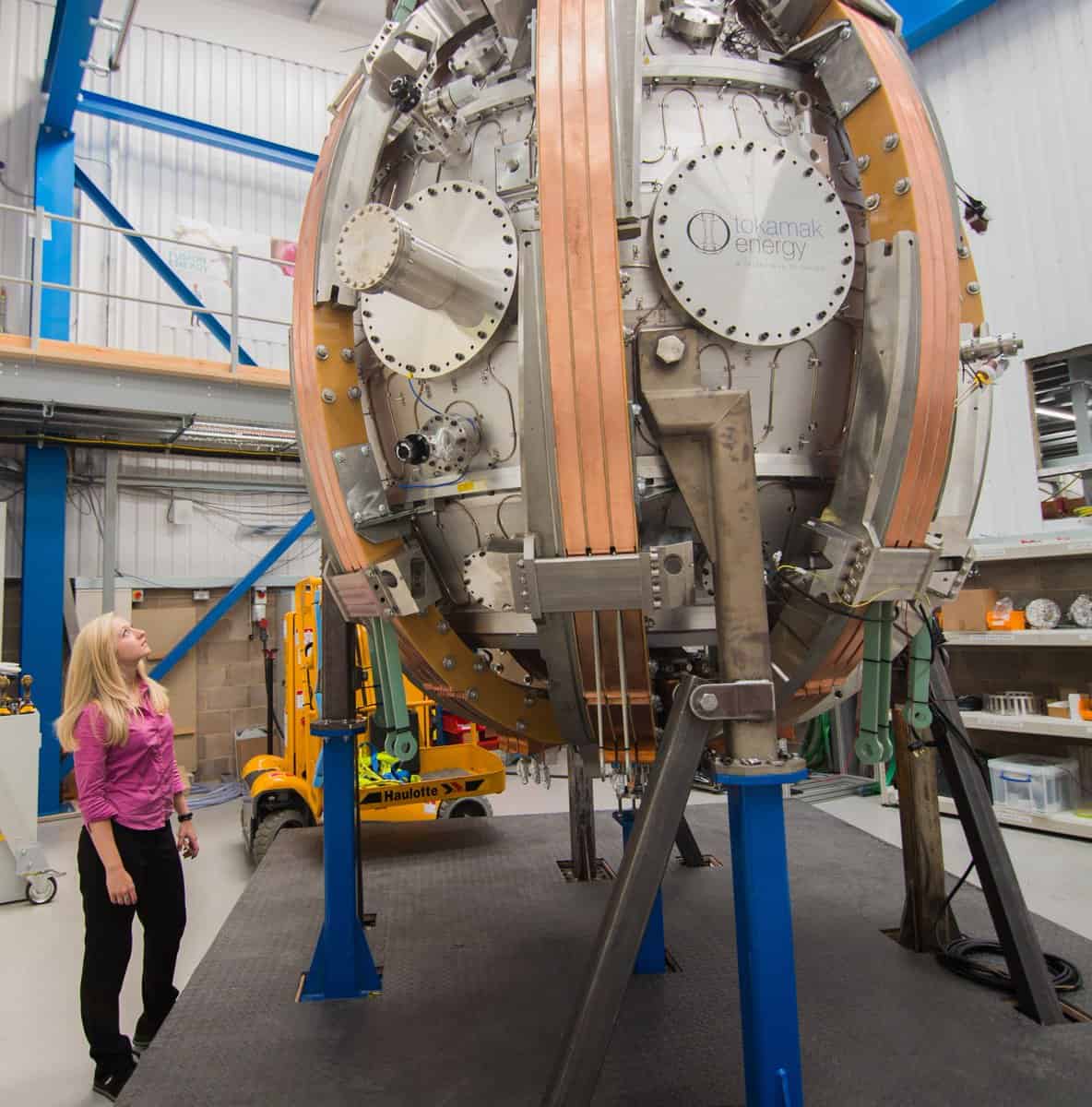
Private practices As one of a growing number of firms, Tokamak Energy aims to develop commercial fusion energy with its ST40 compact spherical tokamak. (Courtesy: Tokamak Energy)
Fusion is simple. Two light nuclei merge to form a bigger nucleus, releasing energy. It’s what powers the stars, but building a fusion reactor that can deliver power in a controllable way isn’t easy. Fusion needs high temperatures, high pressures and a decent confinement time. Those demands have, however, triggered some amazing approaches to make fusion power a reality.
Trouble is, no-one really knows which will work best. What’s more, once you achieve fusion, you need to generate more energy than you put in, so that the ratio Q > 1. But a “break even” result has so far never been achieved by a fusion reactor here on Earth. In fact, what you really want is a Q between 5 and 10 so that your reactor produces a useful amount of power.
Fusion has an increasing commercial angle too with 35 fusion firms around the world.
The approach being taken at the ITER fusion reactor, which is currently being built by a huge international consortium in southern France, is to confine a hot plasma with large superconducting magnets in a doughnut-shaped tokamak. Set to come online in 2025, ITER will point the way to a commercial fusion reactor called DEMO, which will be built by 2060. I visited in 2019 and ITER is truly incredible to behold.
But it’s not the only game in town. Last year, China’s Experimental Advanced Superconducting Tokamak achieved a temperature of 120 million kelvin for 101 seconds. That beat the previous record of 100 million kelvin held for 20 seconds by South Korea’s KSTAR reactor in 2020. There’s also the Joint European Torus (JET) in Oxfordshire, UK – the forerunner to ITER – which still holds the record for the highest Q ever (it got to Q = 0.67 in 1997). JET has just run tests that produced more than 59 MJ of energy over five seconds, more than doubling the output achieved in 1997.
The report found that most private fusion companies expect fusion power to be supplying electricity to the grid in the 2030s. If their efforts succeed, that would put them well ahead of ITER, which largely froze its design in about 2001 and hasn’t been able to exploit recent huge advances in high-temperature superconducting (HTS) magnets. Indeed, since the report was released, investment in private fusion firms has skyrocketed.
CFS – which was spun out from the Massachusetts Institute of Technology in 2018 – last year successfully demonstrated a 20 T HTS magnet. Simulations suggest that this magnet could be powerful enough to let the firm’s SPARC Tokamak reactor achieve net energy from fusion. Since then, CFS has raised $1.8bn to build the reactor, which will pave the way for ARC – the first commercially viable fusion power plant. Development could begin in 2025.
As for Tokamak Energy, this British firm’s ST40 spherical tokamak reactor with HTS magnets reached a stunning 15 million kelvin in 2018. The firm, which received its last funding of £67m in January 2020, is now targeting a 100 million kelvin plasma from its upgraded ST40 reactor. I wonder if 2022 could also be a big breakthrough year for the company?
Meanwhile, last year the UK government announced a short list for sites for a prototype fusion plant known as Spherical Tokamak for Energy Production, or STEP. Based on technology pioneered by the UK Atomic Energy Authority’s Culham Centre for Fusion Energy (CCFE), STEP could be up and running by 2040. The final location is due to be decided this year.
CCFE, where JET is located, has also been chosen by General Fusion as the site for its fusion demonstrator plant. It uses a spinning liquid jacket to hold a plasma, which is compressed rapidly into a sphere using powerful pistons. The fuel fuses and the resulting heat is absorbed by the liquid metal and used to turn a generator. Having last November announced a further $130m investment, the firm hopes to start work this year on the reactor, which could be ready by 2025.
Another player in the market is First Light Fusion, which raised $25m in 2020 and last May installed a “hyper-velocity gas gun” on its “Machine 3”. It fires a projectile at a fuel target, with the resulting shock waves squeezing the fuel so much that it gets hot enough to fuse. The average net cost of generating electricity over the plant’s lifetime could be as little as $25/MWh – roughly half that of an onshore wind plant.
Several approaches are looking more and more credible with each technical milestone achieved.
Then there’s Helion, a US firm that last year announced the largest single fundraise in private-fusion history. It secured a $2.2bn funding package to build their seventh-generation fusion reactor called Polaris using deuterium and helium-3 fuel to directly produce electricity. Helion’s reactors are expected to be about the size of a shipping container and could deliver about 50 MWe, with the plants in operation by 2024.
The race is on
Making sense of all these achievements – and knowing who will win the race – is not easy as each reactor is different and faces its own technical difficulties. One common challenge, however, is the “cycle-time” between each scale-up step as this will ultimately determine the speed at which the power plant hits the market. It’s clear to me, though, that several approaches are looking more and more credible with each technical milestone achieved.
No-one is quite sure how big the fusion market will be as the timing, cost and power output of potential reactors are all so different. But fusion has many advantages over fission, including a great safety record, no long-lived waste, and the potential for cheap fuel. If fusion reactors can gain regulatory approval and show that they have a competitive price tag, we could see a commercial plant in as little as five to 10 years.
I use multiple skills every day. Some of them are people skills, but others are about understanding the physics, so you get an interesting synergy there. You need to understand the subject matter of what you’re doing, but to develop a business you also need to be able to translate that into solving real-world problems and figure out how it applies to everybody’s lives. As a chief executive, you are responsible for getting your team together, ensuring good dynamics and, of course, business growth, so that’s something else I do every day.
What do you like best and least about your job?
What I like most is the scope and the fact that the quantum-technology sector is very new and there’s a lot to be discovered, not just in terms of science but also in terms of business and how the company can grow. That’s something that drives you and can get you through any difficulty. In academic research you tend to have a particular direction, but when you run a business you release yourself from those boundaries. You’re free to do whatever works best. It can be a curse and a blessing, but I focus on the latter.
What I like least is the quantity of tasks that need to be done, and the challenge of balancing the management work with the actual thinking and creative work. There is always a lot happening at the same time and I find that multitasking can be quite taxing.
What do you know today, that you wish you knew when you were starting out in your career?
This is the most difficult question, because there is a lot, but that’s probably a good sign because it means you have developed. It’s important to ask yourself why you are doing what you’re doing, and to really understand and be clear with yourself about the reasons. That allows you to make better decisions about what to pursue and how to build your career more efficiently. The more incoming opportunities you have, the more critical this question becomes. You should also ensure you are careful with your time.
Ask yourself why you are doing what you’re doing, and be clear with yourself about the reasons. This allows you to make better decisions about what to pursue and how to build your career
For people who have just graduated, I would advise them to explore topics outside physics too. Degrees are typically very technical and scientific, but they largely neglect the aspects of why you do physics and how it is applicable in the context of society, because there are not many people to teach that. It’s not easy to put that in a curriculum. It’s nearly common sense, but you need a professor who has worked in a lot of different settings to teach this, and there are very few out there. So it’s really important for students to start thinking about these questions. A lot of people tend to be quite narrowly focused and neglect other possibilities, but the opportunities are there if you can think outside the purely technical skillset. If you focus completely on physics and think that whatever else is happening is probably not as great, then you’re going to miss out.
The quality of China’s scientific research output exceeded that of the US in 2019. That is according to a new analysis by researchers in the US, which also found that China had already overtaken the European Union in terms of research quality by 2015.
China’s total research output has grown rapidly in recent years, but there has been a widespread belief that the “quality” – judged by the number of citations papers receive – is not as high as other countries. A common measure of a nation’s research quality is the percentage of its papers appearing in the top 1% of the most-cited papers globally. Since citation practices vary widely across disciplines, researchers typically weight the citation data of papers according to their fields, before comparing countries’ scientific output. When comparing field-weighted citation data, the US has a higher percentage of research in the top 1% worldwide than China does.
Although this weighting practice makes sense when comparing papers from different fields, Caroline Wagner of Ohio State University argues that it is not appropriate when comparing the overall research outputs of different countries. Together with Lin Zhang of Wuhan University and Loet Leydesdorff of the University of Amsterdam, Wagner analysed citation data contained in the database Web of Science and used the unweighted data to quantify different countries’ research quality.
They found that 1.67% of papers with Chinese authors were in the top 1% most-cited articles in 2019 compared with 1.62% of papers with US authors.
Wagner believes that China’s rapidly rising scientific impact is due to its large-scale investments in research and development, scientific infrastructure and the mobility of students and scholars. She also points out that government policy has targeted leading areas of research.
“We need to think about scientific capability, not as a race, but as a frontier of knowledge,” Wagner told Physics World. “China is now operating at the frontier, along with several other nations, including the US and a number of European nations, Japan and South Korea.” Wagner is now investigating how the US can exploit new knowledge “no matter where it is developed”.
Artist’s representation of the circular phonons. (Courtesy: Nadja Haji and Peter Baum, University Konstanz)
When a magnetic material is bombarded with short pulses of laser light, it loses its magnetism within femtoseconds (10–15 seconds). The spin, or angular momentum, of the electrons in the material thus disappears almost instantly. Yet all that angular momentum cannot simply be lost. It must be conserved – somewhere.
Thanks to new ultrafast electron diffraction experiments, researchers at the University of Konstanz in Germany have now found that this “lost” angular momentum is in fact transferred from the electrons to vibrations of the material’s crystal lattice within a few hundred femtoseconds. The finding could have important implications for magnetic data storage and for developments in spintronics, a technology that exploits electron spins to process information without using much power.
In a ferromagnetic material, magnetism occurs because the magnetic moments of the material’s constituent atoms align parallel to each other. The atoms and their electrons then act as elementary electromagnets, and the magnetic fields are produced mainly by the spin of the electrons.
Because an ultrashort laser pulse can rapidly destroy this alignment, some scientists have proposed using such pulses as an off switch for magnetization, thereby enabling ultra-rapid data processing at frequencies approaching those of light. Understanding this ultrafast demagnetization process is thus crucial for developing such applications as well as for better understanding the foundations of magnetism.
Where does the angular momentum go?
To find the lost angular momentum, Peter Baum and members of his team at Konstanz began by magnetizing nickel crystals in a specific direction, then demagnetizing them using a femtosecond laser pulse. They monitored what happened on femtosecond time scales by using ultrafast electron diffraction, a technique that can track structural changes in materials at the atomic level. They then analysed the diffraction patterns they obtained using computer simulations.
Just after demagnetization, collective lattice vibrations, or phonons, appear. These phonons are circularly polarized and are thus able to carry the missing angular momentum away from the electron spins. “The angular momentum goes directly into the phonons,” Baum explains. Team member Ulrich Nowak adds that their data “shows that the angular momentum of the electrons is transferred locally to the atoms of the crystal lattice on the same time scale on which the magnetic order of the crystal is lost”.
At first, only a few atoms move in circular orbits. Thanks to interactions with neighbouring atoms, however, this motion is then quickly transferred to all the other atoms. In the end, the entire crystal lattice uniformly oscillates in tiny circular obits. “On much slower time scales – nanoseconds, a million times slower than femtoseconds – the entire material starts to rotate as a whole,” Baum tells Physics World.
Atomic version of the “Einstein-de-Haas” effect
The work solves an old mystery in solid-state physics and proves experimentally that polarized lattice vibrations can indeed transport angular momentum quickly and effectively, Baum adds. The data also show that this atomic and ultrafast version of the “Einstein-de-Haas” effect, so-called after Albert Einstein and Wander Johannes de Haas measured it for macroscopic bodies more than 100 years ago, has an intermediate transitional step on the atomic scale.
The team say the results might be exploited to control magnetic materials using laser light and therefore create more efficient alternatives to conventional electronics. “Unlike current electronic circuits, these devices would work with spin transport instead of charge transport, which would be significantly more energy-efficient,” Nowak explains. “By demonstrating that lattice vibrations can transport a spin, we open up a new, potentially promising path towards novel and particularly fast devices in spintronics.”
The researchers, who report their work in Nature, say they now plan to use their unique ultrafast electron diffraction measurements to study more complex magnetic materials and also the spatio-temporal transport of the circular phonons over barriers and into other materials. They are also advancing theory to make predictions that can help with the design of future applications. “There is a lot to do following our preliminary discovery,” Baum concludes.
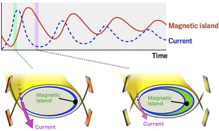
Predator and prey: The upper graph shows the predator-prey relationship between the magnetic island and the bootstrap current. The bottom diagrams show the situation in the Large Helical Device when the current is at its maximum (left) and minimum values. (Courtesy: National Institute for Fusion Science)
If fusion reactors are ever to provide energy commercially, they will have to operate non-stop for extended periods of time – something they are not currently able to do. Researchers in Japan report having taken a step in that direction after discovering a new kind of oscillation triggered by magnetic fields used to confine a reactor’s ultra-hot plasma. By comparing that oscillation with the fluctuating populations of predators and prey sometimes seen in nature, they reckon it should be possible to remove the variation and stably isolate the plasma.
Many physicists engaged in fusion research work on reactors known as tokamaks. These rely on a set of circular magnetic coils positioned at regular intervals around a doughnut-shaped hollow vessel to confine a plasma of light nuclei within the vessel so that the nuclei can fuse and release copious amounts of energy. The most ambitious such device is the ITER reactor currently being built in the south of France, which is designed to generate ten times the energy it consumes.
However, tokamaks have an Achilles heel – their inability to operate in a steady state. The circular coils are more closely spaced on the inside of the doughnut than they are on the outside, which causes the plasma to drift outwards. Overcoming this involves setting up a second field by using a transformer to induce a current in the plasma, but this is an inherently pulsed operation that yields energy in bursts.
Diverting heat
Reactors known as stellarators are designed to overcome this problem by using more complex-shaped magnetic coils to in effect move some of the reactor’s inner magnetic field to the outside. As such, they avoid the need for an induced current. Yet these devices – like tokamaks – still have to remove waste heat from the fusion reactions. They do this by magnetically directing plasma particles to “divertors”, which transfer the heat to cooling water. But although made from thermally-resistant materials, these components can only withstand a certain heat flux for so long before they start to degrade.
In the latest work, Tatsuya Kobayashi and colleagues at the National Institute for Fusion Science in Toki sought to limit this flux through the process of detachment. This involves neutralizing the plasma before it touches the surface of a divertor, by lowering the plasma’s local temperature enough that its constituent ions and electrons recombine. The temperature can be lowered in several ways, but in this case the idea was to use additional coils to create what are known as magnetic islands in the field close to the divertors. These cool parts of the plasma by virtue of separating them from the rest of the hot plasma.
The researchers carried out the work using the institute’s Large Helical Device, a stellarator that confines plasma along a helix. Having added ten pairs of coils to the top and bottom of the reactor, they heated and compressed deuterium gas in the reactor chamber. A little over 3.5 s into the experiment the plasma became detached from the divertors’ graphite surfaces, at which point oscillations were observed. This was a fluctuation in the size of the magnetic islands and other plasma parameters at a rate of about 40 Hz, which caused the plasma to detach and reattach at that frequency.
Predator and prey
To better understand what caused the oscillation, the researchers turned to a so-called predator-prey model. This was originally designed to explain very distinctive fluctuations in the numbers of certain pairs of animal types in the wild – in which one animal preys on the other. These fluctuations involve a repeating cycle of fairly steep rises and falls in the numbers of both prey and predator – the latter lagging slightly behind the former. The idea is that the predator depends for its survival on that one source of food, and in consuming it becomes more numerous – until the food source starts to run out. Its numbers then dwindle, allowing the prey to repopulate – and the cycle starts again.
Such models have previously been applied to other areas of science, including astronomy and indeed plasma turbulence. But Kobayashi and team found that it can also be used to explain their observed divertor oscillations. The predator in this case is the magnetic island while its prey is a “bootstrap current” set up in the plasma by the bouncing motion of confined electrons. As the current ramps up it expands the island, which increases resistivity at the edge of the island. This in turn lowers the current and causes the island to shrink. At that point the current starts to rise again, and another cycle begins.
Running the model, the researchers found what they describe as “good qualitative agreement” with the experimental results. They admit that the model may be a bit simplistic – pointing out, for example, that it yielded an oscillation frequency of only around 20 Hz. But they reckon it should be possible to bring the numbers in line by making changes such as using non-linear functions to account for the very sudden switching between detached and attached states. Once they have done that, they might then be able to use it to work out how to tune the relevant parameters so that the plasma remains detached from the divertors for extended periods without its temperature and stored energy declining.
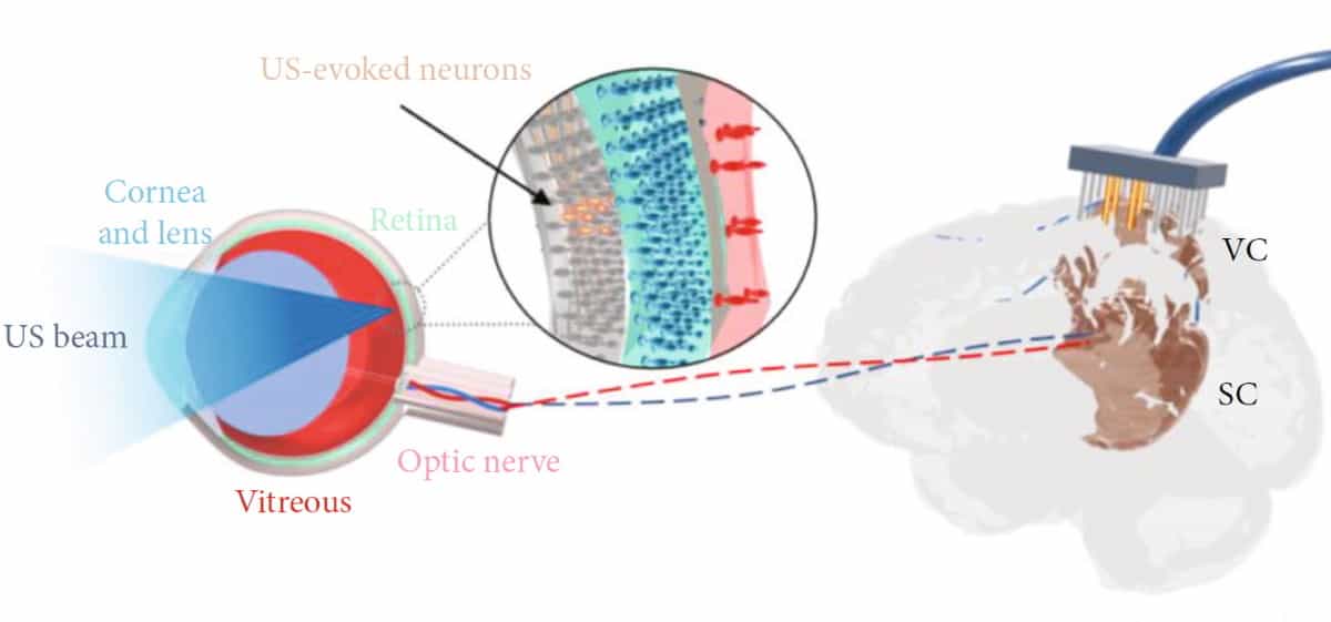
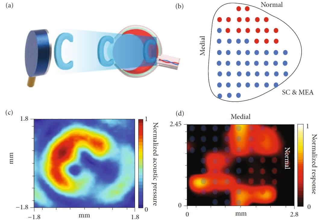
The ultrasound stimulation system: Acoustic waves targeted at the retina excite the retinal neurons, which generate neural signals that are transmitted through the optic nerve to the brain. Brain activity is recorded using a multielectrode array. (Courtesy: CC BY 4.0/BME Frontiers 10.34133/2022/9829316)
Retinal degeneration, in which the light-sensitive photoreceptors in the eye deteriorate and lose function, is a major cause of blindness worldwide. But even though the retinal cells have lost sensitivity to light, the neural circuits connected to the brain are often well preserved. This provides an opportunity to restore vision by bypassing the damaged photoreceptors and directly stimulating the retinal neurons.
Visual prostheses that restore sight via electrical stimulation of retinal neurons have already been developed and used successfully in patients. But such devices are invasive and require complex implantation surgeries. Instead, researchers at the University of Southern California propose the use of non-invasive ultrasound to activate these neurons. Reporting their findings in BME Frontiers, they demonstrate that stimulating a blind rat’s eyes with ultrasound activated small groups of neurons in the animal’s eye.
“This is a step towards a non-invasive retinal prosthesis that works without invasive eye surgeries,” says corresponding author Qifa Zhou in a press statement. “Special glasses with a camera and an ultrasound transducer are intended to give blind and partially sighted people a new view of the world.”
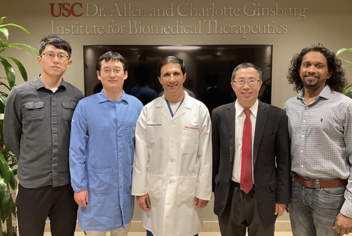
The USC researchers (from left to right): Gengxi Lu, Xuejun Qian, Mark Humayun, Qifa Zhou and Biju Thomas. (Courtesy: Department of Ophthalmology, USC)
Proof-of-concept
The rationale for using ultrasound is that the sound waves exert mechanical pressure on neurons in the eye, activating them to send signals to the brain. Zhou and colleagues first examined the impact of ultrasound stimulation in normal-sighted rats, using a 3.1 MHz ultrasound transducer with a focal depth of 10 mm. They recorded neuron activity using a 32-channel electrode array (150 μm spacing) inserted into the animal’s visual cortex or superior colliculus (SC), the brain area connected directly to the optic nerve.
The researchers measured the rats’ responses to light and ultrasound stimulation, observing comparable neuron activities from both stimuli. These findings in normal-sighted rats indicated that ultrasound can provide an alternative way to stimulate the retina.
Next, the team investigated a rat model of retinal degenerative blindness. Neural signals recorded in 16 blind rats showed that ultrasound could stimulate the retinal neurons. As expected, no light-evoked visual activity was achieved. The induced neuron activities in blind rats were generally weaker in amplitude and duration than those observed in normal-sighted rats.
In contrast to light-evoked neuron activity, the response to ultrasound stimulation can be modified by the beam parameters. The researchers assessed the effects of varying the ultrasound intensity (acoustic pressures from 1.29 to 3.37 MPa) and duration (from 1 to 200 ms). They observed that both the amplitude and duration of neuron activity increased with increasing ultrasound intensity. The amplitude did not vary for ultrasound durations of 10 ms or longer, but the response duration did increase with increased ultrasound duration.
Towards clinical transfer
One important requirement for a visual prosthesis is that its user can see sharp images. To quantify the spatial resolution of ultrasound-evoked neuron activity, the researchers reconstructed the response across the SC surface while moving the transducer. Activated SC regions had spatial resolutions ranging from 161 to 299 μm, with this variation likely due to the curvature of the retina. The average spatial resolution was 250 μm – comparable to that of the first FDA-approved retinal prosthesis, the Argus II.
The temporal resolution (frame rate) of ultrasound stimulation is another important factor, as it determines whether a prosthesis can provide smooth vision of moving objects. Consecutive 20 s stimulations delivered at various frame rates revealed that frame rates of up to 5 Hz generated stable neuron activity, while a higher frame rate (10 Hz) could potentially suppress the firing neurons. They note that this suppression was caused by neuron saturation rather than neuron damage.
Pattern generation: A C-shaped beam is projected onto the retina (a) and a 56-channel MEA placed over the entire SC surface (b). The hydrophone-measured acoustic field (c) and the MEA-recorded neuron activities (d) show the C-shaped pattern. (Courtesy: CC BY 4.0/BME Frontiers 10.34133/2022/9829316)
Finally, to validate the technique’s ability to generate visual patterns, the researchers designed a 4.4 MHz helical transducer that projects ultrasound onto the retina in the shape of the letter “C”. Using a 56-channel electrode array placed over the entire SC region, they observed the same C-shaped pattern of neuron activities in the SC.
“These results represent a step towards non-invasive retinal prosthesis development using ultrasound,” conclude co-first authors Xuejun Qian and Gengxi Lu. “The in vivo demonstration of visual restoration in blind rats suggested that ultrasound opens a new avenue for the development of a novel non-invasive retinal prosthesis.”
“For the next step, we are working on several deeper investigations,” Lu tells Physics World. These include ultrasound stimulation with a higher centre frequency to provide better spatial resolution and lower stimulation threshold; use of a 2D ultrasound array to dynamically generate different stimulation patterns; and behaviour tests to show how an awake animal responds to the ultrasound stimulation, in addition to the neuron recording.
Texas-based Nanoscope Technologies plans to license this patent-pending ultrasound stimulation technique and provide support for future experiments. If these studies are successful, the team predicts that the technology could be translated into human clinical trials within the next three to five years.
The source of fast radio bursts (FRBs) first detected in 2020 is likely to be located within a dense cluster of ancient stars, according to astronomers led by Franz Kirsten at Chalmers University of Technology in Sweden. This comes as a surprise because current theories suggest that FRBs are emitted by neutron stars called magnetars, which are not expected to be present in clusters of ancient stars.
First observed in 2007, FRBs are short, intense pulses of radio waves of unknown origin. Because FRBs are unpredictable, they have proven notoriously difficult to study. However, since 2007 hundreds of FRBs have been observed, allowing astrophysicists to develop models that predict that FRBs come from magnetars – which are neutron stars that have extremely strong magnetic fields.
To gain further insights into the origins of FRBs, the team focused on a repeating FRB source called FRB 20200120E, which was first observed in 2020 in the constellation of Ursa Major. They used the European Very Long Baseline Interferometry Network, which is a system of multiple radio telescopes located primarily in Europe and Asia. They also used data from the Karl G Jansky Very Large Array in New Mexico.
Big surprise
By using large arrays of telescopes, the team was able to pinpoint the source of the FRB to the M81 galaxy, which is about 12 million light-years away. The big surprise is that the bursts appear come from a dense globular cluster populated by older stars, which is a region where magnetars are not expected to be. This is because magnetars are thought to be created by the violent demise of younger, massive stars that can no longer support themselves under their own gravity.
The researchers believe that their observations suggest that magnetars could also be formed by older stars. This could involve an ancient white dwarf in a binary system collapsing into a neutron star after it has accreted material from a companion star. Alternatively, two ancient compact stars could merge and then explode in a “kilonova” that could leave behind a magnetar.
Crab pulsar
In a separate study led by team member Kenzie Nimmo at the Netherlands Institute for Radio Astronomy, researchers found that some of the FRB 20200120E bursts flicker on very short timescales of a few tens of nanoseconds and are very powerful. This is reminiscent, they say, of signals that come from the Crab pulsar in the Milky Way. This is a rapidly rotating neutron star that emits pulses of radiation including radio waves.
As a result, the team says that FRB 20200120E bridges the gap between the signals seen from young pulsars and magnetars in the Milky Way and the more powerful bursts emitted from distant extragalactic FRBs. This suggests that these phenomena share a common magnetically-powered emission mechanism that can operate over a wide range of timescales and luminosities.
Kirsten and colleagues report their observations in Nature, while Nimmo’s team report in Nature Astronomy.
Rapid advances in imaging techniques based on computed tomography (CT) have underpinned a revolution in treating cancer patients using radiotherapy. An X-ray scan lasting just a few seconds yields an accurate 3D visualization of the patient’s internal anatomy, and delivers the critical information needed by medical physicists to calculate the optimal dose distribution for treating the tumour. Ongoing advances in CT technologies have enabled clinical teams to access higher quality images that have enabled more precise targeting of the tumour while minimizing the damage to healthy organs and tissues.
However, conventional CT images can sometimes lack the contrast needed to clearly distinguish between different types of soft tissue. That makes it difficult for radiation oncologists to precisely define the size and shape of the tumour, and to contour the nearby organs, tissues and blood vessels that need to be protected from ionizing radiation. Another notable limitation of the standard CT scans conventionally used in radiotherapy departments is that they only provide anatomical information, and so are unable to reveal functional processes that might provide some additional insight for treatment planning.
For cases that require enhanced soft-tissue contrast or functional information, CT is often combined with other imaging modalities such as magnetic resonance imaging (MRI) or positron emission tomography (PET). While Siemens Healthineers offers MRI and PET solutions that have been optimized for radiotherapy and for imaging patients in the intended treatment position, in some situations such complementary imaging methods may not be available. For making clinical decisions, particularly when critical organs are at risk – such as in the head-and-neck region, chest or the abdomen – improved soft-tissue contrast is crucial.
Now, however, a novel method that can improve the quality of CT images is starting to make a difference in radiotherapy clinics. Called dual-energy CT, or DECT, the technique acquires CT images from two different X-ray spectra rather than one. Conventional CT captures images using a single X-ray beam, which has a spectrum of photon energies with an average of around 70 keV and a typical peak energy of 120 keV. The image contrast of each material depends on how well it attenuates X-rays, which in turn depends on energy.
“At standard CT energies, most of the soft tissue we’re trying to image has very similar attenuation coefficients,” explains Jainil Shah, a research professional and R&D collaboration scientist at Siemens Healthineers. “That means that most of the organs look very similar when they are visualized on a CT image.”
Routes to dual-energy CT in radiotherapy
DECT mitigates this problem by generating images from X-ray scans taken over two different energy ranges. Since the first experiments that showed the potential of dual-energy CT in the 1970s, several different approaches have emerged for acquiring DECT images – and each one has its advantages and disadvantages. The simplest way is to scan the patient twice at two different energies, a technique known as “Dual Spiral” or “Twin Spiral”. Such consecutive scanning can offer excellent image contrast, since it allows for a wide separation between the two spectra. Because any movements of the patient between the two scans can introduce errors, a non-rigid image registration between the two images is performed automatically during post-processing to account for and compensate for any changes in position. “That makes Dual Spiral DECT most suitable for non-moving regions such as the brain and the head and-neck,” comments Shah.
Other techniques capture the two spectra simultaneously, recording all the information in a single scan and limiting the patient’s exposure to X-ray radiation. One option offered by Siemens Healthineers is to split the X-ray beam using a filter in the scan direction, creating two separate beams with different average energies. Such TwinBeam technology offers a wide field-of-view, but the use of a filter limits the spectral separation and therefore the image contrast that can be achieved.
The third option available from Siemens Healthineers is a CT scanner that exploits two X-ray sources operating at different energies, each one coupled to its own detector. This Dual Source approach offers better spectral separation than TwinBeam technology, and therefore sharper images for treatment planning, as well as more X-ray power in each of the separate beams. The field-of-view is slightly smaller because the equipment needs to accommodate two separate X-ray tubes, which needs to be considered when imaging larger regions of the body.
These DECT systems are already routinely used in radiology clinics for diagnostic imaging, while ongoing improvements to the scanners and the software have made it much easier for radiotherapy centres to integrate the technique into their clinical workflow. “The scan can be acquired by any technician in the clinic, and all the information needed by the radiation oncologist is generated automatically,” explains Shah. “Clinical workflows can be set up in the software to automatically perform additional post-processing and image reconstruction from a single scan.”
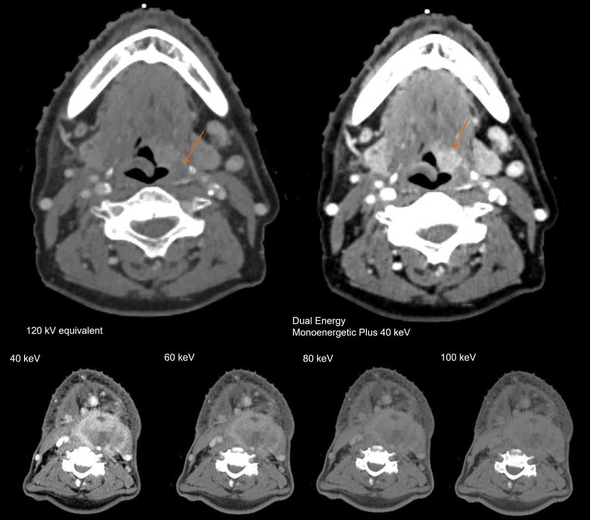
Capturing X-ray spectra with two different energy distributions makes it possible to reconstruct an image at any single energy. This yields a series of virtual monoenergetic images (VMIs), also called Monoenergetic Plus – at energies ranging from around 40 keV up to 190 keV – that can be used to optimize the soft-tissue contrast. “The energy can be easily changed with a slider bar in the software,” explains Shah. “The radiation oncologist can decide which energy provides the best contrast for contouring organs.”
Compare and contrast: A VMI of the head-and-neck region at 40 keV (top right) reveals detail that cannot be seen on a normal CT image (top left). VMIs at energies ranging from 40 to 100 keV (bottom, left to right) show that the 40 keV reconstruction offers the best image contrast. (Courtesy: Hospital del Mar, Barcelona, Spain)
Shah says that dual-energy CT is also able to provide some functional information about dynamic processes inside the body, such as perfusion within the lungs or the uptake of iodine in different organs and blood vessels. As an example, capturing X-ray spectra over two different energy ranges makes it possible to determine the material composition, since the attenuation of X-rays in each material is dependent on energy.
“That means you can do things like remove bone from the image, or distinguish between fat and liver tissue,” says Shah. “From the material composition you can predict the electron density of the material (Rho image), which is the key information that is used for dose calculations in radiation therapy.” For proton therapy, meanwhile, Siemens Healthineers offers a specific reconstruction called “DirectSPR” that calculates the stopping-power ratio from dual-energy CT.
Into the clinic
Ongoing advances in technology and software are now enabling medical teams to integrate dual-energy CT into their clinical practice.
Beth Bradshaw Ghavidel, Emory University, USA – DECT for head-and-neck patients
Beth Bradshaw Ghavidel, one of the lead medical physicists at Emory University, says that TwinBeam DECT is primarily used for head-and-neck patients, in which VMIs at higher energies can help to remove the artefacts that arise from metal objects inside the body, such as dental fillings (DECT is compatible with iterative metal artefact reconstruction, iMAR). “It is easy to set up the desired dual-energy CT workflows on the scanner and allow automatic post processing,” says Bradshaw Ghavidel. “Depending on what CT scans are needed, the dosimetrist can select specific studies for import. At this time, we have not needed to alter our clinical workflow for additional imaging studies.”
Lili Chen, Fox Chase Cancer Center, USA – DECT for intracranial target delineation and rectal tumours
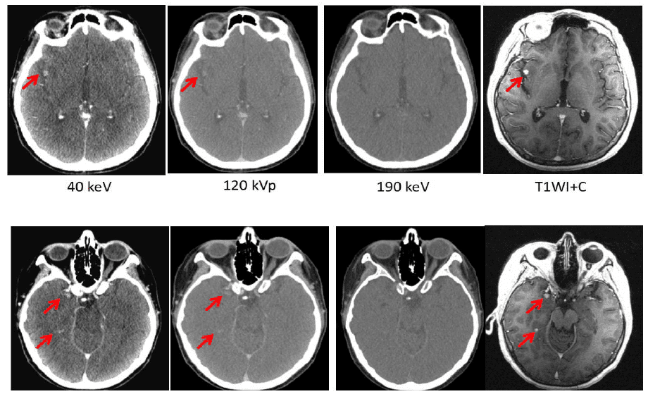
Lili Chen at Fox Chase Cancer Center has also been exploring the potential of dual-energy CT to boost the image quality for different disease sites. When imaging the head and neck, she found – like Bradshaw Ghavidel – that a VMI at 190 keV offers an effective way to reduce the artefacts caused by dental fillings and iodine uptake in the soft palate. Chen has also compared DECT images of brain tumours in 34 different patients with those taken with MRI. When an iodine contrast agent was used, she found that a VMI at 40 keV can reveal metastases in the brain that are not detected by conventional CT, or by VMIs at higher energies. What’s more, in this research study the brain tumour volume derived from the 40 keV DECT image was comparable to that obtained from MRI.
Low-energy benefit: Lili Chen at Fox Chase Cancer Center compared brain metastases when imaged by (from left to right) a dual-energy VMI at 40 keV, a conventional CT image, a VMI at 190 keV, an MR image with T1 weighting. The 40 keV image with a contrast showed similar brain metastasis volume compared to the T1 weighted MR image with contrast. (Courtesy: Lili Chen, Fox Chase Cancer Center)
“Meaningful clinical differences were found on CT images with contrast, with the 40 keV image delineating the tumour much more clearly than images taken at other energies,” Chen comments. “Our results suggest that dual-energy CT with contrast may be used for intracranial target delineation in radiotherapy treatment planning.” Analysis of a rectal tumour also revealed obvious differences between images taken at different energies, with the one taken at 40 keV clearly showing the necrotic area of the tumour and indicating that cancerous cells had spread to the adjacent seminal vessels.
Xiaofeng Yang, Emory University Hospital – DECT image contrast for contouring head-and-neck cases
Also at Emory University, Xiaofeng Yang has been working with Bradshaw Ghavidel and other colleagues to investigate whether dual-energy CT can improve the precision of a deep-learning model they have developed for automatic organ segmentation. In a recent research study, they designed a neural network that exploits DECT data to automatically segment 19 organs-at-risk in the head and neck, and trained the model using manual contours produced for 66 patients with carcinomas in different disease sites. Automatic contours generated with the DECT-based model were then compared to manual contours produced by a physician, as well as to those obtained using the same model developed at Emory University Hospital with conventional CT data. “DECT-based segmentation of organs-at-risk has the potential to facilitate the current head-and-neck cancer radiation therapy workflow in treatment planning,” concludes Yang.
George Noid, Medical College Wisconsin (MCW), USA – DECT in clinical routine and for tracking the effect of treatment
George Noid, a medical physicist at MCW, says that dual-energy CT is now in routine clinical use for almost all cancer patients, particularly those needing treatment in the abdomen or chest. “We use the VMI reconstructions to enhance the image contrast,” he says. “As well as abdominal and thoracic patients, we’ve found it really useful for pre-operative breast cancer and other rare disease sites in the abdomen, such as the adrenal glands.”
One particularly gnarly problem that Noid hopes to address in the future is to improve the images used to plan the treatment of pancreatic cancer. “We want to deliver as much radiation as possible to the pancreatic head, but the major limiting factor is the amount of radiation that we can deliver to the adjacent duodenum,” he explains. “That makes it clinically important to accurately define the edge between the pancreatic head and the duodenum.” In a recent research study, Noid and colleagues compared conventional CT data with dual-energy CT scans of 10 patients being treated for pancreatic cancer, and in each case the image contrast was enhanced by injecting iodine-based contrast media into the patient before the scan. They found that the image contrast was boosted by a factor of 2.8 for the VMI at the lowest possible energy of 40 keV, while another important indicator of image quality, the contrast-to-noise ratio, was also maximized at this energy. Images from other treatment sites, including the liver, breast and thymus, also showed that the tumours were more clearly visible at 40 keV than at higher energies.
Noid is also investigating whether quantitative data extracted from dual-energy CT images could be used as an indicator of how well a patient is responding to treatment. “It has been shown that the aggressiveness of pancreatic cancer is correlated to the extracellular volume (ECV) fraction, which can be calculated from a DECT scan,” he explains. In a recent research study, which won the Best in Physics award at AAPM 2021, Noid and colleagues used DECT images acquired at weekly treatment sessions to calculate the ECV fraction. For 12 pancreatic cancer patients, the study revealed a correlation between the ECV fraction and the concentration of a cancer antigen found in the blood, suggesting that regular DECT scanning could be used to track the effect of treatment. “That offers the potential of stratifying your patient’s risks based on calculations of the ECV fraction,” Noid explains. “That information could help to drive your clinical decisions, such as giving a higher dose to a patient who has a more aggressive disease. We are not yet using that in our clinical workflow, but that’s the idea.”
Noid is confident that DECT has the potential to deliver more quantitative data that in the future could be used to analyse the properties of the tumour. “Unlocking that information would be clinically very useful,” he says. “We’re starting to see that DECT has the power to access that functional information, and I think there’s much more we can do.”
More information about dual-energy CT is available in the Dual Energy CT Cookbook from Siemens Healthineers
Note that the results by Siemens Healthineers’ customers described herein are based on results that were achieved in the customers’ unique setting. Since there is no “typical” hospital and many variables exist (eg. hospital size, case mix, level of IT adoption) there can be no guarantee that other customers will achieve the same results.
In this webinar, we will be covering the new features released in the latest version of RadCalc (7.2.2). These include new workflow features for intelligent automation with Eclipse Scripting and RadCalcAIR that delivers your report in Aria with a single click of the script within the Eclipse TPS. We will also be introducing LAP Academy.
Learn how you can evaluate the treatment performance of your patient’s full course of treatment automatically with the implementation of our Treatment Performance Profiles. This TPP provides a single click method for computing your EPID dosimetry plan dose. We will also give a sneak preview on what we are working on for the next release.
Also, we would like to address a valuable topic: the importance of reviewing change logs and upgrading as new versions are released to ensure the highest safety and quality in the intended patient plans. We will discuss a recent example and our ability to quickly fix our unique plan comparison tool.
Carlos Bohorquez, MS, DABR, is the product manager for RadCalc at LifeLine Software, Inc, part of the LAP Group. An experienced board-certified clinical physicist with a proven history of working in the clinic and medical device industry, Carlos’ passion for clinical quality assurance is demonstrated in the research and development of RadCalc into the future.