From geckos’ feet to Formula 1: how surface science underpins our world

I have to come clean about something. I thought I understood the science of surfaces. As a physicist working on nanomaterials for light-emitting diodes (LEDs), solar panels and sensors, I literally spend my days playing with polymers. The devices I build are multi-layered structures, where you have to really know what’s going to happen when you deposit one layer on top of another. For example, if the light-emitting layer of an LED doesn’t stick well to the electrodes below, then the pixels on our mobile phone and TV screens won’t light up. So, this being the field I work in, I thought I had a pretty good grasp of surface science. But within the first few pages of Sticky: the Secret Science of Surfaces by physicist and author Laurie Winkless, my confidence in my knowledge of surface science started to come unstuck.

It has been four years since Winkless’ first book, Science and the City, introduced the public to the science of the metropolis. While her enthusiasm and charm are apparent throughout both books, Sticky feels like the more grown-up sibling. Rather than providing a superficial overview of all the relevant scientific topics, Winkless takes the time to unpack those she is most interested in. The writing, including the technical descriptions, feels effortless, benefiting from her years of experience in science communication.

Unlike many popular-science books, Sticky is not just a disparate list of facts about forces. Every chapter, from “A gecko’s grip” to “Break the ice” is a story, masterfully assembled into an accessible, clear and highly engaging manuscript. Winkless’ writing is informed by discussions with a diverse group of scientists and engineers, and the unparalleled excitement of discovery is evident in every interaction. Alongside these interviews, the author provides an enticing (but not overwhelming) level of detail, introducing readers to the scientific method, peer review and even the politics of scientific theories. The science of the sport of curling, for example, is a hotly debated topic that has divided physicists for the past 30 years and fuelled “Broomgate” – the 2015 controversy over whether a new hi-tech broom was changing the fundamentals of the sport too much.

Winkless doesn’t shy away from complicated topics, such as fluid dynamics, organic chemistry and geophysics, but instead makes them relevant to her audience. The topic itself is ubiquitous by nature, so there’s something in the book to pique the interest of everyone from academic researchers to fans of Formula 1. For scientists, the properties of surfaces are an essential consideration. Double-sided Sellotape (of the appropriate stickiness) is required for nanoscale imaging using atomic force microscopes; carbon tape is crucial for mounting samples inside vacuum chambers; and Kapton tape is necessary for soldering. The “Scotch tape method”, by which tape is stuck to graphite and peeled off again, taking tiny fragments with it, paved the way for the graphene revolution. I’d even go as far as saying I’ve never met an experimental physicist who doesn’t rely on Blu Tack, whether that is to hold together pieces of equipment, secure a substrate to a stage or safely package samples to send to collaborators.

But adhesion principles also show up in everyday life and culture far beyond the lab, from the frescoes of Italian Renaissance art to non-stick frying pans and swimwear that helps you glide through the water more easily. So, while a truly brilliant read for scientists, I think Sticky appeals to a very broad audience. Irrespective of your level of expertise, Winkless’ language is not remotely patronising. The analogies are well thought through and the examples are appropriate. In fact, she somehow manages to make the mundane beautiful: “curling is a curious ballet of stones, brooms and ice”. For the super-keen, Winkless provides a comprehensive reading list (apparently just a handful of the 900+ references she read when researching the book) for further study.

My favourite chapter explores the gecko’s impressive ability to cling to even very smooth surfaces, which Winkless describes as the “smartest on-off adhesive in the world”. Little did I know that the attempt to unravel the mechanisms behind this creature’s grip has spanned two centuries, provided fuel for entire research careers and inspired a lot of questionable hypotheses.

Working out the hierarchical adhesive systems of geckos has presented a truly interdisciplinary research effort that is inspiring the design of futuristic robots and reusable adhesive tapes

The first theory – that geckos cling to surfaces through suction from the lamellae in their feet – was debunked a century after it was proposed, when it was shown that geckos maintain their grip even at vanishingly low air pressures. Next came the “climber’s boot” hypothesis: that densely packed hairs called setae on the gecko’s toes act as tiny hooks that grip onto bumps and grooves. This proposal was dismissed when experiments revealed that geckos can firmly attach to ultra-smooth surfaces with very low surface roughness. More theories followed, involving electrostatics (think sticking a balloon on a wall after rubbing it on your hair) and friction. It turns out that geckos stick to surfaces through a complicated interplay of many physical phenomena including Van der Waals forces and nanoscale networks of setae known as spatulae. Incredibly, they can detach their feet six times faster than you can blink. Working out the hierarchical adhesive systems of geckos has presented a truly interdisciplinary research effort that is inspiring the design of everything from futuristic robots to reusable adhesive tapes.

It will no doubt surprise fans of friction that there are still so many unknowns – such as how exactly curling works, or how we can quantify the slipperiness of swimmers – that continue to perplex scientists. So although this research field has existed for hundreds of years, it remains a lively and cutting-edge one. When I picked up Sticky, I wasn’t sure what I was in for, but I remained glued to every page.

  • 2021 Bloomsbury £16.99hb 336pp

Wireless implant uses optogenetics to control spinal cord activity in mice

Optogenetics – a technique that uses light to control the activity of neurons or other types of cell – has revolutionized our ability to manipulate and discern the mechanisms underlying brain function. Spinal cord activity underpins control of movement and several other basic physiological functions. But compared with the brain, optogenetics in the spinal cord presents a series of challenges that require the development of new light-delivery technologies.

For instance, implanting optical fibres is not a straightforward task, as the spine undergoes continuous displacement during natural movement. Therefore, one design challenge is the positioning of light-delivery sources over the very surface of the dura mater, the outer membrane that surrounds and protects the soft and dynamic spinal cord. Targeting deep intraspinal neurons presents additional obstacles. Light rapidly scatters when penetrating biological tissues. High irradiance, meanwhile, may result in local tissue heating due to light absorption, which can affect neuronal responses.

To overcome these challenges, and find a way to efficiently deliver light to the spinal depth of interest, researchers at the Swiss Federal Institute of Technology (EPFL) have developed a compliant, wireless-controlled optoelectronic implant customized for optogenetic studies of the spinal cord. They describe this novel implantable system in Nature Biotechnology.

NeuroRestore group

Technology features

The new implant technology encapsulates miniaturized LEDs that can be switched on and off on the surface of a mouse’s spinal cord. A miniature head-mounted, wireless platform powers these micro-LEDs and performs customized on-chip processing to control light stimulation trains. This enables modulation of light pulses in real time, for instance, in response to the animal’s muscular activity or routine physiological signals. The wireless electronic circuit can control the duration and intensity of the emitted light with high precision.

In contrast to wire-based systems usually used for this type of research, wireless optoelectronic systems allow for unrestricted photostimulation of target neurons in the spinal cord of mice under untethered and ecological conditions.

In one key aspect of this work, the researchers, led by Grégoire Courtine and Stéphanie Lacour, used the micro-LED arrays to take advantage of the evolving library of experimental opsins (light-sensitive proteins activated by specific wavelengths) to target a broad range of cellular mechanisms.

They designed the micro-LED array to shift the emitted blue light toward a desired wavelength, enabling photostimulation using a broad spectrum of light, including red-shifted wavelengths that penetrate further into the spinal cord.

To optimize LED positioning and avoid tissue heating, the team ran simulations and performed in vivo recordings to quantify heating within the spinal cord for different levels of irradiance and cycles of photostimulation.

The researchers designed the new soft optoelectronic implant to be resilient and adaptable for long-term implantation and to meet the demanding mechanical dynamics of the spinal cord in freely behaving adult mice. Using a hybrid approach combining stiff LEDs and elastic interconnects, they created miniaturized implants that wrap around the surface and move along with the spinal cord.

The team’s research has potential to pave the way for the development of therapeutic optogenetic applications. The ability to control the activity of spinal cord neurons with light could allow doctors to reduce pain and improve autonomic function.

Even though it may still be some time until these implants can be used clinically, Courtine finds optogenetics “revolutionary” and is eagerly anticipating further developments in biointegrated optoelectronic implants.

Photonic band structure created in electrically driven OLED microcavities

A photonic crystal emitter has been fabricated by stacking multiple organic light emitting diode (OLED) microcavities into a single structure. It was created by a team led by Matthew White at the University of Vermont in the US, that was able to demonstrate that the degree of interaction between the cavities depends strongly on the different layers employed, meaning that both the colour of light emission and the band structure of the devices are highly tunable. This paves the way towards electrically driven photonic crystals that allow further control over the emission of OLEDs. The full study is described in Nature Communications.

OLEDs have received a large amount of attention in recent years due to their numerous applications in display screen technologies and efficient low-cost lighting. They are also easy to integrate into other technologies and structures.

White and colleagues have placed semi-transparent mirrors on either side of an OLED to form a microcavity – that is, a cavity with a length at the order of a micron. By varying the distance between the two mirrors, the length of the cavity and therefore the emission wavelength (colour) of the OLED can be finely tuned, even without changing the organic material.

Communicating OLEDs

The team then created a stack of N microcavities separated by semi-transparent mirrors to make a device with a photonic band structure analogous to that in a photonic crystal. If the mirrors are much thicker than the penetration depth of light, no communication between the cavities is possible and they act as N separate cavities. Conversely, if the mirrors are infinitely thin, the N cavities will act as one cavity with a total length Nd, where d is the length of each individual cavity.

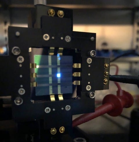
Blue emission from an OLED microcavity

If the mirror thickness lies between these two extremes, the system acts similarly to that of the extended cavity, but with small perturbations in the electric field caused by the internal mirrors. This results in a hybridization of the energy states, forming a photonic band structure that is similar to a photonic crystal or hybridized orbitals in molecules. This hybridization alters the energy of the states and therefore affects the OLED emission.

The researchers have shown that the band structure is heavily dependent on the properties of the stack. For example, the internal mirrors used in the stack alternate between silver and aluminium. This symmetry doubles the unit cell size so that it consists of two cavities with a semi-transparent internal mirror and total length equal to 2d.

Tuning the band

As aluminium has a shorter penetration depth and higher losses than silver, the aluminium mirrors primarily control the length of the unit cell and therefore the energy separation of the hybridized states. The researchers have shown, however, that the presence of the silver mirror perturbs the states, decreasing the energy gap between the two sub-bands. Therefore, by altering the thicknesses of the different mirrors, it is possible to control the size of the band gap, the separation of the energy states and the total bandwidth of the OLED stack.

“The effect is the result of structural colours so we can use green organic emitters, which are known to be more stable, to produce any desired colour combination”, says White. In this way, it will be possible to “tune the emission from OLEDs to include broadband white light, narrow bandwidth single peaks, or multiple peaks forming a low-fidelity frequency comb”.

Help me if you can: founding a business doesn’t have to be lonely

In 1978 the Silicon Valley entrepreneur Nolan Bushnell was fizzing with ideas for new businesses. He had just left Atari, where he had masterminded the creation of Pong – one of the earliest and most successful video games – and many other pioneering tech products. But one of the biggest problems with start-up companies, Bushnell realized, was the sheer amount of “bullshit housekeeping stuff” involved.

His solution was to create Catalyst – believed to be the world’s first hi-tech business incubator. “My idea was that I would fund [the businesses] with a key,” Bushnell told Fast Company in 2017. “And the key would fit a lock in a building. In the building would be a desk and chair. They would sign their name 35 times and the company would be incorporated. So in 15 minutes, they would be in business working on the project.”

The idea was not entirely new. The first formal business incubation service that I can find details of dates back to 1959 in the US, but Catalyst was geared specifically to the demands of hi-tech companies. So as well as providing fledgling firms with office space, IT systems and legal and accounting services, Catalyst offered laboratory space and most importantly money and support – in fact, Bushnell was often chief investor and chairperson of each firm’s board.

Catalyst was designed to help those companies get through the initial challenges of starting up. Within a year, it had funded 10 businesses, many of which were very much ahead of their time. They included Etak, which released an in-car computer navigation system in 1985 – fully four years before the GPS network was available for civilian use. Parts of the press ridiculed the product (it didn’t help that the symbol of the car on the driver’s screen looked uncannily like the ship from Atari’s Asteroids game) but sat-navs are now a feature of almost all modern cars.

Catalyst folded but the blueprint for business incubation had been set

James McKenzie

Catalyst created around 20 companies, but it unfortunately over-invested in some of them, which therefore took a lot longer than expected to generate a return. Eventually, in 1986, Catalyst folded but the blueprint had been set and by 2006 there were more than 1400 incubators of all kinds in North America, up from only a dozen in 1980. A report that year from the US National Business Incubation Association indicated that US incubation programmes had helped more than 27,000 companies, together employing over 100,000 people and generating annual revenues of $17bn.

Global growth

The idea quickly spread across the world as more and more start-ups saw the benefits of lab space, help, advice and access to capital. In 1997 there were around 25 incubation environments in the UK, but by 2017 that number had risen to almost 400, according to a report from the Department for Business, Energy and Industrial Strategy.

With hi-tech incubators and accelerators usually needing specialist facilities, many are located at (or linked to) universities and other national labs in areas where there are lots of similar companies with a local talent pool. The Institute of Physics also has a business accelerator at its new headquarters, giving small firms access to affordable office space in central London and letting them hook into the IOP business and member networks.

As well as many business incubators, the UK now in addition has the Catapult Network of technology innovation centres. These have been set up over the last decade in the wake of a 2010 government report into technical innovation. Written by Hermann Hauser – the physicist-turned-entrepreneur and honorary fellow of the IOP – he reckoned that the UK has a science capability second only to that of the US, but was falling short on translating scientific ideas into leading positions in new industries. “Technology and Innovation Centres can help solve this shortcoming and give Britain the lead in some of the most promising technologies leading to new industries with transformational economic impact,” he wrote.

The Catapult Network, which is part of Innovate UK, now has nine technology and innovation centres spanning more than 40 locations across the country. These centres cover everything from compound semiconductors, renewable energy and high-value manufacturing to satellites, energy systems and digital technology. They provide high-quality facilities for hi-tech businesses of all sizes and have a support programme for start-ups and small-to-medium-sized enterprises (SMEs). Since 2013, more than 8000 start-ups and SMEs have been supported. Innovate UK also offers grants to support its industrial strategy, with various competitions running throughout the year.

So whether you are an early-stage firm, a pre-seed innovator, or a more established company, there are many networks and resources to support your innovation journey. A good place to start is the Knowledge Transfer Network, which has experts in many areas on tap. There is also Innovate UK Edge – part of UK Research and Innovation – which has business advisers who can provide advice and introductions. In addition, it can carry out a grant-funded audit of your firm’s intellectual property. It even has an “investment readiness” programme as well as a showcase called Pitchfest to help you improve your pitch to investors.

There is now more help than ever for new hi-tech start-ups

James McKenzie

I know from personal experience that starting a new hi-tech firm is not easy and many physicists have great ideas but are unwilling to take the plunge. But there is more help on offer than ever before: through business incubation, catapult networks, grants and business advice. So why not have a go? Whether it’s energy, medicine, quantum tech or new materials, there are plenty of opportunities.

From whole-organ to cellular resolution: synchrotron X-ray images reveal COVID-19 lung damage

The ability to image human tissue at scales from an entire intact organ down to individual cells is key to advancing our understanding of health and disease. To meet this challenge, a European research collaboration has developed a new imaging technology known as hierarchical phase-contrast tomography (HiP-CT), using X-rays from the Extremely Brilliant Source (EBS) at the European Synchrotron Radiation Facility (ESRF).

The EBS is the first high-energy (6 GeV), fourth-generation synchrotron source. It provides the brightest source of X-rays in the world, with the spatial coherence required to resolve faint density contrasts at high resolution. Using the EBS, HiP-CT can perform non-destructive 3D scanning of an entire human organ and then zoom down to the cellular level.

“The ability to see organs across scales like this will really be revolutionary for medical imaging,” says first author Claire Walsh from University College London. “As we start to link our HiP-CT images to clinical images through AI techniques, we will – for the first time – be able to highly accurately validate ambiguous findings in clinical images. For understanding human anatomy, this is also a very exciting technique, being able to see tiny organ structures in 3D in their correct spatial context is key to understanding how our bodies are structured and therefore how they function.”

Walsh and colleagues have used the new synchrotron X-ray tomography technique to scan donated human organs, including lungs from a COVID-19 donor, reporting their findings in Nature Methods.

High-resolution scanning

Using the EBS test beamline, BM05, the researchers developed the specialized sample-preparation, scanning and reconstruction approaches required for HiP-CT. They designed a scanning geometry that reduces sample dose (to avoid tissue damage), optimizes the detector’s dynamic range, reduces artefacts and suppresses beam hardening.

For imaging, organs are fixed, partially dehydrated and stabilized in agar–ethanol in a jar. The team recorded reference scans of a jar of agar–ethanol mounted on top of the sample to provide a background that can be removed during image reconstruction. This process eliminates low-frequency background variations and enables extreme off-axis local reconstructions.

Synchrotron X-ray imaging of human organs

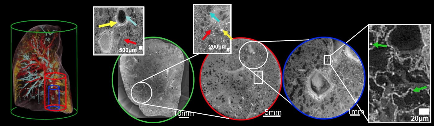
The HiP-CT scans are performed hierarchically, typically starting at 25 μm/voxel over the whole organ, then followed by magnified imaging of selected volumes-of-interest (VOIs) at 6.5 and 1.3–2.5 μm/voxel. They team assessed the performance of the HiP-CT technique by scanning an intact human lung. The estimated image resolutions were 72±3.4, 18.3±0.6 and 10.4±0.17 μm, for images recorded at 25, 6.5 and 2.5 μm/voxel, respectively.

To assess the consistency of scans at various depths and distances from the organ centre, the researchers analysed images of two high-resolution VOIs. They found minimal differences in mean intensity or image quality between the two, suggesting that HiP-CT can achieve high-resolution scanning in any region of the lung with consistent quality.

Next, they imaged five intact donated human organs – brain, lung, heart, kidney and spleen – performing HiP-CT at 25 μm/voxel to provide a structural overview of each organ, followed by multiple higher-resolution scans of selected VOIs. The 25 μm/voxel scans clearly identified macroscopic features such as individual lobules in the lung, for example, and the four chambers of the heart and associated coronary arteries.

The higher-resolution scans successfully visualized functional units in the organs, as well as imaging certain specialized cells. In the brain, for example, HiP-CT revealed layers of the cerebellum and individual Purkinje cells. Lung images showed the intralobular septa and septal veins, as well as terminal bronchi and bright cell-sized objects identified as pneumocytes and/or alveolar macrophages. Images of the heart showed bundles of cardiac muscle fibres comprising individual cardiomyocytes, while epithelial tubules were evident in the kidney, and red and white pulp was seen in the spleen.

COVID-19-related lung damage

The team also used HiP-CT to investigate structural changes in the lung tissue of a patient who died from COVID-19-related acute respiratory distress syndrome. Lung slices imaged at 25 μm/voxel contained high-intensity regions in the lung periphery, consistent with clinical radiology findings.

COVID-19-related lung damage

Scanning VOIs at 6 μm/voxel revealed heterogeneous parenchymal damage, with some secondary pulmonary lobules displaying greater deterioration than others. The team performed higher magnification images of a more affected lobule at 2 μm/per voxel. The scans revealed how severe COVID-19 infection remodels the finest blood vessels in the lungs, causing blood to shunt between the capillaries that supply oxygen to the entire body and those that feed the lung tissue itself. This cross-linking prevents the patient’s blood from being properly oxygenated – a process that was previously hypothesized but not proven.

“By combining our molecular methods with the HiP-CT multiscale imaging in lungs affected by COVID-19 pneumonia, we gained a new understanding of how shunting between blood vessels in a lung’s two vascular systems occurs in COVID-19 injured lungs, and the impact it has on oxygen levels in the circulatory system,” explains Danny Jonigk from Hannover Medical School.

The researchers emphasize that HiP-CT will continue to evolve alongside advances in synchrotron technology. Completion of a new ESRF beamline next year is anticipated to provide several key advances.

“The beam [will be] much larger and higher energy, hopefully enabling scanning of a whole torso at 20 micron voxels, while still being able to zoom locally to one micron. The increased beam size enables increased speed for the same sample size,” project lead Peter Lee tells Physics World. “We hope to form a hub to help us populate the Human Organ Atlas with a statistically significant number of organs in health and disease.”

  • All image data from this study are publicly available in an online Human Organ Atlas.

RadCalc adds in vivo dosimetric verification, intelligent automation for independent QA

Customer-driven innovation and continuous improvement are once again front-and-centre in the latest release of RadCalc QA secondary check software, a suite of widely deployed quality-assurance (QA) tools that provides medical physicists and dosimetrists with fully automated and independent dosimetric verification of their radiotherapy treatment planning systems (TPS). Top billing in RadCalc v7.2 is the commercial introduction of 3D EPID-based functionality to underpin measurement-based patient QA and in vivo dosimetric verification. Meanwhile, speed and workflow efficiency remain ongoing priorities for the RadCalc development team following the integration of a range of customization features for intelligent automation into RadCalc AIR, the software’s control centre for automated data import and reporting.

With the release of v7.2, RadCalc’s portfolio of calculations now encompasses secondary dose checks, with the use of point-dose analysis, 3D Monte Carlo or 3D Collapsed-Cone Convolution Superposition algorithms to identify clinically relevant deviations within the entire patient volume; EPID for pre-​treatment dosimetry, in which the software reconstructs the dose from the delivered pre-treatment plan on the patient’s original planning CT (giving a direct comparison with both the intended dose from the TPS and RadCalc’s 3D dose second check); and EPID for in vivo dosimetry to reconstruct the dose delivered during treatment, yielding a direct comparison on the actual delivered dose reconstructed on the original planning CT to evaluate intrafraction changes in the patient.

“We are not aware of any other product on the market able to match RadCalc’s in vivo 3D capability using EPID-based dosimetry,” claims Craig Laughton, CTO and co-founder of the RadCalc software portfolio, part of LAP’s growing QA product line in radiotherapy. “In the clinic,” he adds, “RadCalc 7.2 enables the medical physics team to compare the whole dose volume versus the original treatment plan, measuring what is actually being delivered in vivo to the patient during radiation therapy.”

Dynamic systems

Such insights are especially powerful given that patients are dynamic systems, always in flux rather than steady-state. Between treatment sessions, for example, patients can gain and lose weight; their stomach, bladder and bowel contents change; their organs may shift, rotate or deform; and their tumours may shrink, move or rotate.

Those changes can be problematic for traditional radiotherapy regimes that rely on a single CT snapshot of the patient at the start of treatment, with most clinics currently limited in their ability to track geometric deformations in patient anatomy over time. Put another way: a treatment plan attuned to the initial simulation can quickly become suboptimal as radiotherapy progresses over the course of many fractions typically spread across a month or more.

Craig Laughton

“Our EPID-based module is going to pick up any changes in dose delivery over a course of multiple fractions, triggering a conversation between the clinical physicist and radiation oncologist to understand what’s happening inside the patient,” explains Laughton. “That dialogue could ultimately mean reimaging and replanning of the patient – another important step towards a more personalized approach to radiation therapy.”

What’s more, RadCalc’s 3D EPID module also has an enabling role to play in supporting the latest hypofractionated and ultrahypofractionated radiotherapy schemes, in which an increased dose per fraction results in significantly fewer overall treatments over a compressed timeframe. The goal, as always, is enhanced targeting accuracy and enhanced dose distribution accuracy – minimizing collateral damage to adjacent organs at risk and critical structures – over a course of treatment running to just one or a few high-dose fractions.

“With a hypofractionated treatment scheme,” notes Laughton, “you need to know if something’s not right straight away – for example, in the case of a machine error or incorrect patient set-up. You can’t afford to wait a week, because in a week the treatment’s done and it’s too late.”

Automate to accumulate

More broadly, intelligent automation remains a long-term fixture on the RadCalc development roadmap, giving medical physicists the power to optimize processes versus their own clinical requirements – customizing which DICOM tags to trigger actions, for example, and new layouts for cleaner workflows. “While we see user-defined automation as a key differentiator,” notes Carlos Bohorquez, RadCalc product manager, “automation is ultimately all about patient safety and minimizing human errors and the burden on clinical resources associated with manual QA tasks.”

Carlos Bohorquez

According to Bohorquez, the v7.2 release incorporates significant customizations to the RadCalc AIR import and reporting tool to streamline workflows in the clinical environment (e.g. the import/export of clinical protocols; also specific tolerance criteria for each treatment plan based on any DICOM tag the user’s TPS is able to export). “It’s an ever-evolving process to provide clinicians with true and faithful automation for existing and emerging radiotherapy technologies,” he explains.

That’s certainly true for RadCalc’s new 3D EPID module, which imports the necessary EPID data/image files for processing before sending to the Collapsed-Cone dose engine to calculate the dose. “The EPID-based solution needs further automation so that the physics team can use it on a routine off-line adaptive basis, eliminating manual interventions within the RadCalc UI,” explains Bohorquez. “That work will be the focus of our next release in a few months’ time, increasing the clinical and operational impact of this functionality.”

Another priority for the RadCalc development team is to tap into the scripting features of leading TPS offerings. “In effect,” Bohorquez concludes, “these are changes that we will implement outside of RadCalc to make the software’s automation tools even more powerful to the end-user in the clinic.”

Laser-free trapping of heavy molecules opens an alternative route to new physics

The quest for physics outside the Standard Model often takes place at major accelerator facilities like CERN’s Large Hadron Collider or huge underground detectors for neutrinos, dark matter and other exotic particles. Researchers in the Netherlands have now opened an alternative front in this quest by developing a new laboratory-scale technique for trapping heavy neutral molecules. Such molecules are considered ideal candidates for detecting beyond-the-Standard-Model asymmetries in the electron’s electric dipole moment (eEDM), but previous methods were not capable of confining them. The technique therefore gives physicists a fresh set of tools for finding new physics.

Standard methods used in eEDM searches involve performing high-precision spectroscopy on atoms or molecules that are first slowed and then trapped with lasers or electric fields for the duration of the measurement. The problem is that finding new physics may require trapping molecules that are too heavy to be confined with lasers. Electric fields, for their part, can only trap heavy ions, rather than neutral atoms or molecules.

It’s a trap!

A new method can now be added to this list thanks to researchers at the University of Groningen, who developed it in collaboration with colleagues at Vrije Universiteit Amsterdam and Nikhef, the Dutch particle-physics institute. The researchers begin by creating molecules of strontium fluoride (SrF) via a chemical reaction that takes place inside a cryogenic gas at a temperature of around 20 K. These molecules have initial velocities of 190 m/s, compared to around 500 m/s at room temperature.

The molecules then enter a 4.5-metre-long device called a Stark decelerator in which alternating electric fields act to slow and then stop them. The SrF molecules remain trapped for 50 ms, after which the researchers analyse them using a separate laser-induced fluorescence detection system. Such measurements reveal fundamental properties of the electron, including the eEDM, that can then be checked for any asymmetry.

The heavier the better

These SrF molecules are around three times heavier than other molecules previously trapped using similar techniques, notes Steven Hoekstra, a physicist at Groningen and lead investigator on the research. “Our next step is to trap even heavier molecules, such as barium fluoride (BaF), which is one-and-a-half times heavier than SrF,” he says. “This type of molecule is even better for measurements on the electron dipole. Basically, the heavier [the molecule], the better these measurements will become.”

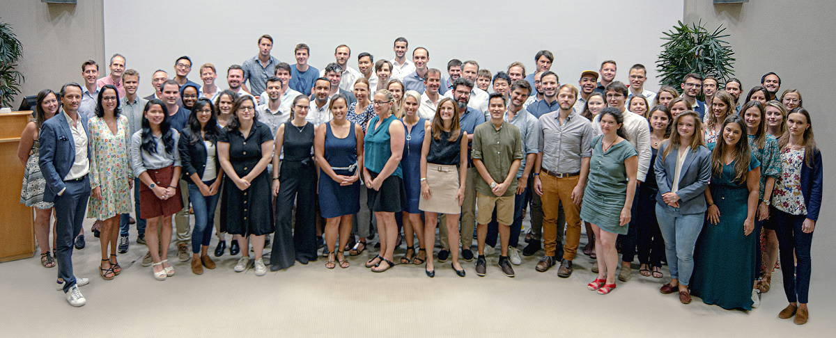
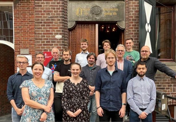
Photo of members of the NL-eEDM team standing on the stairs outside their laboratory

Trapping heavy molecules has other applications beyond eEDM measurements. One example might be to study collisions between molecules at low energies, under conditions similar to those found in space. Measurements on slowly moving molecules could also yield deeper insights into the quantum nature of their interactions. At high enough densities, the molecules’ so-called dipole–dipole interaction, which depends on their orientation relative to each other, makes a big difference in how they interact. These types of measurements offer opportunities that are not possible with stationary atoms, which do not interact in this way.

Complex and chiral

As a next step, Hoekstra says that he and his colleagues, who report their work in Physical Review Letters, plan to increase the sensitivity of their measurement setup by upping the intensity of their molecular beam. “We are also thinking of trapping more complex molecules, such as BaOH or BaOCH3,” he tells Physics World. “Additionally, we could use our technique to study asymmetries within chiral molecules: those that have a left and right-handed version.”

Ben Sauer, a physicist at Imperial College in the UK who was not involved in the current study, describes the result as the culmination of about 20 years of research on molecule deceleration. He predicts that it will have a big impact on precision measurements of the eEDM, where the resolution of the measurement is directly proportional to the time available to interrogate the molecules. As for wider applications, Sauer says: “I can see it being applied to some special cases. I think the limit is that there is a lot more interest in light molecules than heavy ones, since most of chemistry takes place at the top of the periodic table. But it is really good for physics investigations.”

Solar-powered harvesters could produce clean water for one billion people

One billion people could access safe drinking water using devices that use solar energy to condense water from the air. That is the conclusion of a team of researchers in the US led by Jackson Lord at X, The Moonshot Factory, who have developed a new tool for assessing the global potential for water harvesting. Their tool could soon help researchers to design completely off-grid water sources, suitable for use in local communities in many parts of the Global South.

The lack of access to safely managed drinking water now affects some 2.2 billion people worldwide. Addressing this serious problem using existing technologies is a key part of the United Nation’s sustainable development goals – with the organization declaring that everyone should have access to five litres of safe drinking water every day.

This could be achieved in some regions using atmospheric water harvesters (AWHs), which draw clean liquid water out of humid air. There are several different types of AWH, and Lord and colleagues focused on the solar-driven, continuous-mode AWH (SC-AWH). In such a device, heat from sunlight drives warm, humid air through a heat exchanger where it cools and releases water via condensation. Because a SC-AWH operates during the day when relative humidity tends to be low, it has a low efficiency and it had not been clear which locations worldwide are suited for its use.

Geospatial tool

Now, Lord’s team has created a geospatial tool called “AWH-Geo” for assessing the global potential for water harvesting. Based on a Google Earth Engine, the tool uses data from ERA5: a database containing a vast number of historical climate observations going back to 1979. To assess the varying outputs of atmospheric water harvesting in different regions, AWH-Geo considered a location’s sunlight irradiance, its relative humidity and its average air temperature. In addition, the tool accounted for annual variations in these parameters.

The team also looked at the global distribution of people without access to safe drinking water, using data from the World Health Organization and UNICEF. Combining this with AWH-Geo output, the researchers showed that atmospheric water harvesting could realistically provide five litres of  safe drinking water for some one billion people worldwide.

This is based on the use of a hypothetical SC-AWH with a harvesting area of 1 m2. The team calculates that such a device could yield 0.2–2.5 litres of water per kilowatt hour of primary solar energy when operating at a relative humidity range of 30% to 90% and average air temperature of 20 °C.

The researchers are now developing such a device, and with technological improvements they believe it could provide cost-effective, completely off-grid access to high-quality drinking water for many communities in the Global South. The researchers will now continue to use AWH-Geo to guide the development of new types of water-harvesting devices – with the ultimate aim to bring the UN’s goal for clean water a step closer.

The research is described in Nature.

Theory of teapot dribbling is complete at last, solar panels host astonishing microorganisms

Pouring from a teapot

Cast your mind back to 2009 and you might remember how physicists in France devised a way to end the trauma of tea dribbling down the underside of the spout of a teapot when you are pouring a brew. They found that the surface of the spout affects the flow of the liquid and that the best option to create dribble-free pouring is to give the spout a thin layer of a “super-hydrophobic” coating – a material that strongly repels water.

Now researchers in Austria and the UK say they have formulated a “complete theory” of the dribbling teapot effect – one that considers the inertia, viscous and capillary forces at play when a drop forms at the edge of the teapot spout that then wets the underside of it. By carrying out experiments – presumably involving lots of tea drinking – they confirmed their theoretical analysis, finding that the liquid trickles down the underside of the teapot spout when poured slowly but not when poured at a faster rate. The end of tea-stained tablecloths may be in sight. If you’re fast enough, that is.

In some locations, the build up of dust and biological materials can seriously degrade the efficiency of solar panels. Now, researchers at the São Paulo Research Foundation in Brazil have discovered that some microorganisms are well adapted to thrive in the harsh conditions found on the surface of solar panels. What is more, they say that these tough life forms could be used in a wide range of applications including sunscreen and pigments for textiles. Indeed, they believe that the resilient organisms could even be co-opted to help keep solar panels clean.

Astonishingly, the team found very similar microbiota on panels in locations as disparate as Brazil, California, Spain, the Arctic and Antarctic. About 90% of the organisms belong to two genera of bacteria. More recently they have identified a yeast that is present on panels in cooler climes. This organism has tensoactive properties – which means that it reduces the surface tension of water. This, say the researchers, could make the yeast useful for creating new detergents and antimicrobial agents.

Record-high thermal conductivity anisotropy comes with a twist

A simple twist has achieved a record-high anisotropy in thermal conductivity, reports an international group of researchers at the University of Chicago, University of Illinois at Urbana-Champaign, Cornell University and the Chalmers University of Technology. Their versatile approach could be used to cool nanoelectronic devices internally.

As described by Moore’s law, the number of transistors in integrated circuits is doubling every two years. But as computers become faster, the heat they produce also increases. To tackle this problem, one needs materials with high thermal conductivity, and even better materials with thermal properties that are anisotropic. Anisotropic materials have high thermal conductivity in some directions and low thermal conductivity in other directions. This means that they can simultaneously dissipate heat from a hotspot (local overheating) in directions with high thermal conductivity, while providing thermal insulation in other directions.

Layered materials

In this latest research, van der Waals crystals were used as starting materials. These are materials such as graphite and molybdenum disulphide, which comprise atomically thin layers that are stacked together. They combine strong in-plane bonding with the weak cross-plane bonding, which allows for the easy separation of the layers.

The team, lead by Jiwoong Park, David G Cahill and Paul Erhart, showed that by utilizing the anisotropy of van der Waals crystals and interlayer rotation, the thermal conductivity in the stacking direction can be suppressed to that of twice the thermal conductivity of air.

“Our nanomaterial film behaves as if it were 2D,” says Shi En Kim, who is a graduate student in molecular engineering at the University of Chicago. “Heat flows through the sheets as sluggishly as if through the air. That’s quite surprising, considering what we have here is a fully dense solid.”

High and ultralow thermal conductivity

Van der Waals materials combine strong in-plane bonding with weak bonding between planes. However, it is hard to change the conductivity of only one axis. It is known that twisting the two layers of graphene by a certain angle produces novel electronic properties. In the same line, researchers have incorporated twists in the single-layered molybdenum disulfide nanosheets by stacking monolayers of molybdenum disulfide randomly, leading to symmetry breaking in the cross-plane direction but preserving the perfect crystalline order in the in-plane direction.

They were thus able to preserve the high in-plane conductivity of a pristine bulk crystal, but the crystalline mismatch of the layers leads to increased phonon scattering, and ultra-low thermal conductivity, in the cross-plane direction. Currently, the ratio between the in-plane and cross-plane thermal conductivity of the material is a record high, reaching 900. This beats the previous record of 340 set by pyrolytic graphene.

The material was tested in a real-life application by being used as a heat-spreader for gold electrodes, which are a proxy for the nanowires that run through modern-day portable electronics. One gold electrode was covered with the molybdenum disulfide layers, while the other was not. When an electrical current was passed through the electrodes they warmed up due to Joule heating. The team found that the pristine electrode deteriorated quickly from overheating. The electrodes covered with molybdenum disulfide, however, were still functioning due to the cooling effect coming from the effective heat dissipation in the in-plane direction

The authors insist that this approach is not limited to molybdenum disulfide but could use many different 2D materials. Due to the infinite variability of possible combinations of 2D materials, this breakthrough opens a huge area of future research where the thermal conductivity of materials can be finely tuned.

The research is described in Nature.

Copyright © 2026 by IOP Publishing Ltd and individual contributors