

High-spec open-source microscopy for all

Six years ago, on a Friday afternoon, I (Richard Bowman) made a microscope focusing stage with the £400 3D printer in our lab. What started as a moment of idle curiosity quickly snowballed and pushed aside my “day job” research, to grow into so much more. Today, the OpenFlexure Project – developing a 3D-printed laboratory-grade motorized microscope to analyse samples and detect diseases – has a global community of users and developers that spans hobbyists, research scientists, entrepreneurs and clinical researchers.

All of them are supported by a core team spread around the globe – at the universities of Bath and Cambridge in the UK, along with the Tanzanian engineering company BTech (Bongo Tech & Research Labs, formerly STICLab) and the Ifakara Health Institute. We are currently working towards medical certification in Tanzania, and exploring commercialization options in various parts of the world. This growth has largely come about because of our commitment to openness and reproducibility – and the OpenFlexure project is just the beginning of what open-source hardware enables.

Complementary to open-source software, the open-source hardware movement aims to allow people all over the world to make, modify and share hardware for scientific use. Such technology could be particularly appealing for education and training purposes, where expensive professional equipment is particularly prohibitive. More essentially though, for researchers and medical professionals in the Global South, open-source hardware built and specified by themselves could be a game-changer.

Optical microscopes are an essential tool, both to detect disease in clinics, and for scientific research in general. But commercial high-performance microscopes are expensive – usually selling for tens of thousands of pounds – and are hard to maintain. Our most basic 3D-printed microscope, in contrast, can be built for as little as £15 (the cost of the printed plastic, a camera and some fastening hardware). Our top-end version – including a microscope objective and an embedded Raspberry Pi computer – would cost a couple of hundred pounds. If you visit openflexure.org, you can download, print and assemble the latest version of the OpenFlexure microscope. The fully automated microscope is highly customizable, with a number of options readily available for optics, camera and control, including motorized sample positioning and focus control.

Setting the stage

A key feature for any microscope is the ability to precisely position samples and probes – so it must have a precise and stable translation stage to focus the microscope, and move to the right area of the sample. This is a ubiquitous challenge when designing apparatus, and is one reason why research microscopes cost so much. Indeed, machining a fine translation stage that doesn’t wobble or stick requires smooth surfaces, hard materials and precise dimensions.

We found that simply 3D printing the stage designs used in most microscopes results in poor performance. That’s because printed plastic is soft, the surface finish is usually rough, and the printed object often doesn’t match its nominal dimensions exactly. Instead, our design exploits the flexibility of the plastic by using a deformable mechanism to move the sample. The “flexure hinges” that give the project its name are used in many precision instruments, but are usually made of metal and have a limited range of motion. The greater deformability of plastic, and the ability of 3D printers to create intricate shapes, mean our flexure stage can move a relatively long way: 12 × 12 × 4 mm.

The initial publication of the microscope in 2016 focused on its mechanics (Review of Scientific Instruments 87 025104). At that time, there were a number of low-cost and/or open-source microscope projects that provided a good optical solution, but there were far fewer easily manufactured solutions for the mechanics of such a microscope. We characterized our microscope’s drift over time; how repeatable the stage was when moved by stepper motors; and how linear the motion was. By all those metrics, our microscope compared well to others costing orders of magnitude more.
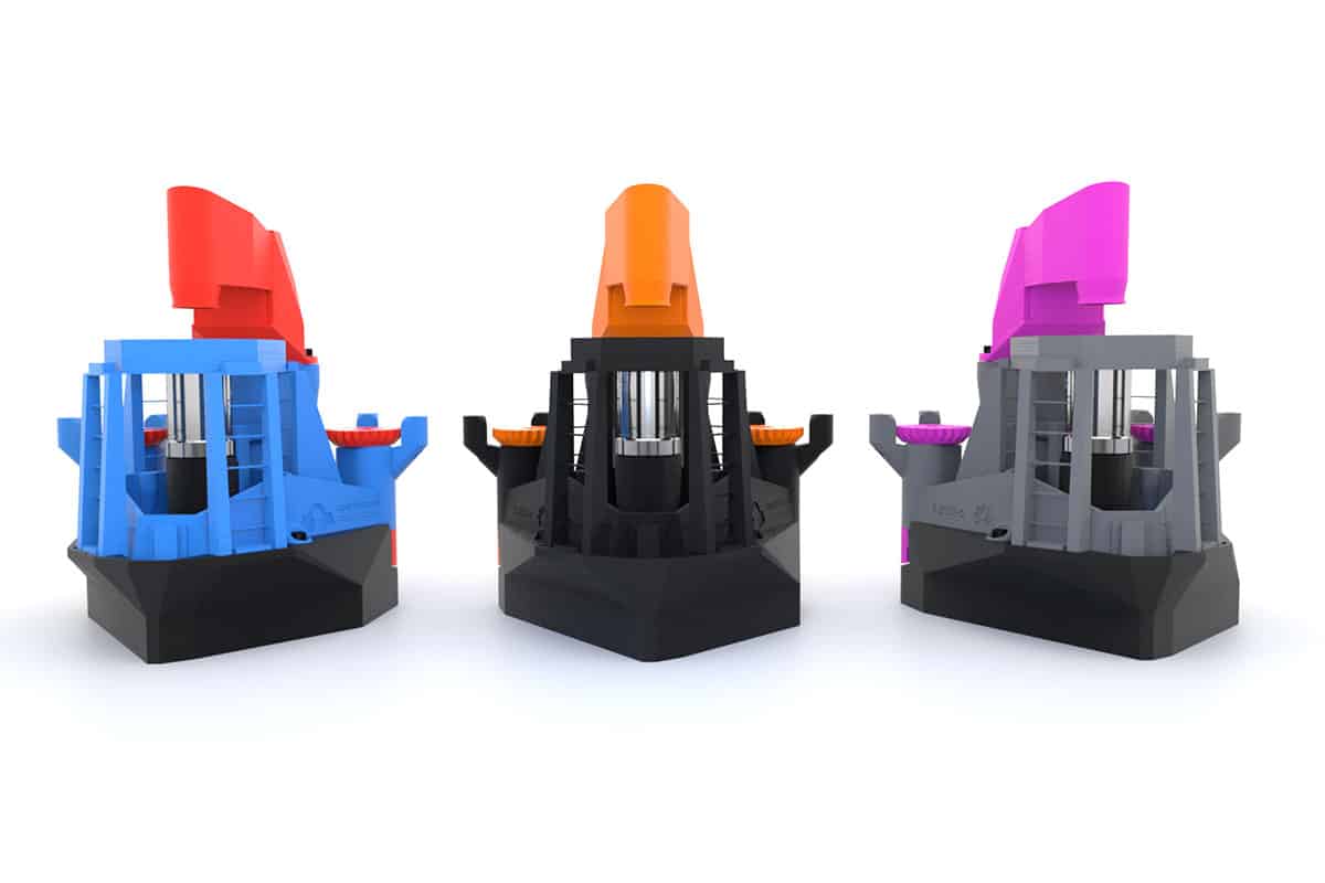
D rendering of the OpenFlexure microscope

By the time we’d completed this work, the OpenFlexure microscope was already starting to be noticed by others. A team in Cambridge picked up the OpenFlexure design and turned it into OpenScope – a programmable open-source microscope. Also, the ability to do low-cost timelapse imaging meant the microscope was the perfect base for the first generation of water-testing technology used by WaterScope – a company Richard co-founded in 2015 to provide low-cost methods for testing water quality in the Global South.

Now that we had active users, they started requesting features and improvements. In particular, we improved our optics options, which used conventional microscope objectives in addition to the inverted webcam lens that enabled most of the early work. The OpenFlexure microscope was then a capable lab prototype, able to take good quality images and even move the stage automatically. However, it was very much a lab prototype. The interface involved obscure keyboard commands with little documentation; there were loose wires and circuit boards; and the whole thing was usually packed inside a shoebox padded out with blue kitchen roll.

Building equitable partnerships

In 2018 we were funded by the UK’s Engineering and Physical Sciences Research Council and the National Institute for Health Research to develop and evaluate the microscope for malaria diagnostics – a project run in collaboration with Ifakara Health Institute and BTech in Tanzania. A central theme of this project is local manufacturing, rather than shipping microscopes from the UK to Tanzania, so that the microscopes used at the Institute can be built a couple of hours’ drive away, at BTech.

We believe this local manufacturing is crucial, as the World Health Organization estimates that nearly 70% of donated medical equipment in sub-Saharan Africa is out of service or not in use usually because authorized service engineers, proprietary consumables and spare parts are not available locally (Med. Biol. Eng. Comput. 49 719). Our approach means BTech can provide this missing infrastructure, thereby making a sustainable impact as well as creating skilled jobs. Ultimately, releasing the hardware under an open-source licence is a way to share ownership and control of the project, which we feel is key to building equitable partnerships between groups with very different economic contexts.

Working in collaboration, we improved the design to make it more acceptable in a hospital lab – for example, adding a better interface and enclosing the electronics. Together with BTech, we made it easier to manufacture too, adding 3D-printed tools for tricky steps, simplifying the bill of materials, and taking more care over the specification of the components. In particular, our initial prototype was very hard to reproduce as it required specific components that are expensive to order or re-order and are typically only found in well-funded labs. The current version of our microscope (Biomedical Optics Express 11 2447) is a capable lab microscope with options for high-resolution bright field, fluorescence and reflection imaging.
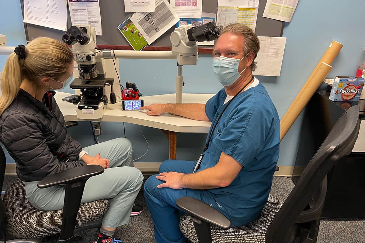
Pathologist Daniel Rosen using the OpenFlexure microscope

Over the three and a half years that our malaria project has run so far, we have collected terabytes of images of blood smears, to evaluate the microscope and train machine-learning algorithms to understand the images. We have also identified and fixed many more issues that only came to light when we had multiple microscopes running long-term in realistic scenarios, in the field. When we scan a blood smear by taking a grid of images spread over the sample, for example, those images can now be evaluated on the fly, so that we can repeat any measurements that are out of focus. This approach of making the instrument smarter, so it can self-correct and become more reliable as well as more capable, is a key theme in the OpenFlexure project.

Open-science test drive

In tandem with our funded research focused on malaria, we have worked to build up a community around the microscope. Most project communication was initially done on the software-development platform GitLab. While GitLab is still where all our designs are managed, last year we set up an online forum that opened up the project to many more people. The forum is a much easier place for users and contributors to engage with the project without having to navigate the complicated, software-focused interface of GitLab.

Our community now includes scientists and engineers from all over the globe – from both physical and life science backgrounds – as well as hobbyists, teachers, members of community groups, and even companies using parts of the hardware or software in their own products. Our best estimate is that there are hundreds of microscopes that have been manufactured “in the wild” without our direct involvement. That means the OpenFlexure project is a useful testbed for many aspects of how to create and share a piece of open scientific hardware.
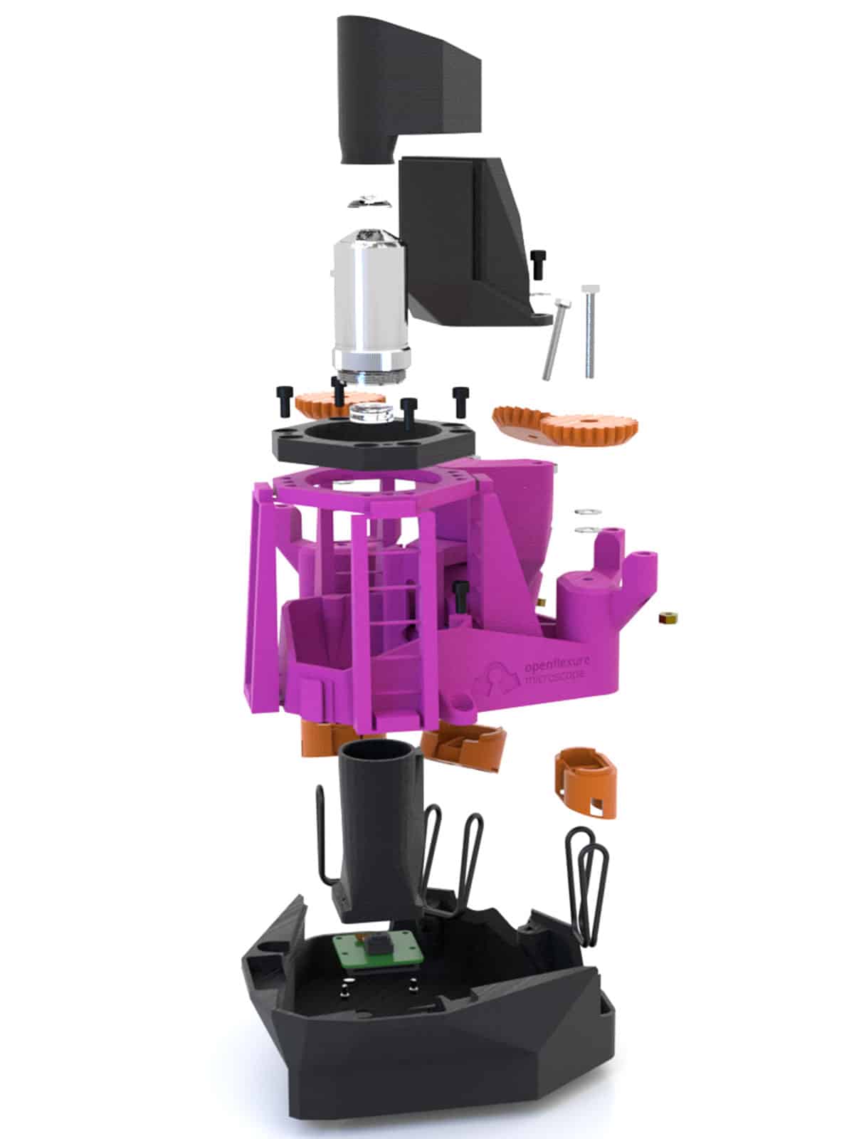
An exploded render of the OpenFlexure microscope

The single most important thing we have learned about how to allow a piece of lab apparatus to be replicated exactly is the importance of good documentation. Our build instructions have gone through numerous complete rewrites and lots of minor updates – and there is still a long way to go. It’s not uncommon for replications of scientific experiments, even ones that intend to be fully open, to rely on direct communication between research teams. While this is less work for both teams the first time a project is replicated, it doesn’t scale well. Relying on word of mouth also means that crucial know-how never gets written down, which means the scientific record isn’t enough to reproduce an experiment once the researchers involved have moved on.

The growth of the OpenFlexure community has forced us to work in ways that scale better. Well-honed instructions are a good starting point, but moving all the questions and answers about the project from spoken or e-mailed conversations into an open, searchable forum means that not only the core instructions, but also a great deal of associated know-how is now available for others to learn from. It’s great to see more and more researchers adopting a similar approach, and we try very hard to make sure that what we’ve learned from this project is made available so that others can learn from our experience.

On the open record

One aspect of sharing a project openly that’s not often talked about is the emotional journey of opening up. The decision to release a project openly might seem simple, but there are many worries that often stop academics from fully sharing their work. Many scientists, for example, feel unable to share details before their work is published, in case they are “scooped”. We too did not initially share the first version of the OpenFlexure microscope online – only doing so when the preprint of our first paper was uploaded to arXiv – but we have since moved to sharing projects much earlier. Doing so eliminates worries about who we share ideas with, and more fully realizes the benefits of open working (The Design Journal 10.1080/14606925.2020.1859168). Sharing designs as they evolve actually makes us less concerned about others “stealing” our work, as there is a verifiable, public record of who did what in our repositories. This makes it much harder for someone to claim the work as their own, than if we kept all of our work a secret until we’re ready to publish.

One aspect of sharing a project openly that’s not often talked about is the emotional journey of opening up

The OpenFlexure microscope adapted for the WaterScope project

The deeper worry, however, is usually that releasing the project openly will diminish the creators’ ownership and control of it; meaning they don’t receive full credit for the work – be it financial, or through authorship and citation of published articles. This is a reasonable concern, but our experience is that the benefits far outweigh the downsides. By and large, people who make use of open designs are happy to give credit where it’s due. After all, citing the design properly is a small effort to repay something useful, and the scientific community is increasingly paying attention to good practice around open science.

While there may be people and projects who have not credited their use of our work properly, this is more than made up for by the large network of collaborators who do recognize its value. That network is far bigger than it would be if we had taken a less open approach, and indeed it is much larger than we could support if we had to deal with requests individually rather than through open, reusable platforms like the forum.

Experimental ecosystem

As scientific instruments go, the OpenFlexure microscope is more of a workhorse tool than a state-of-the-art microscope existing in only a few labs. However, it has been extended to do super-resolution and phase imaging, and can be connected to another open-source toolbox – the UC2 modular optics system – for even more customizable use.
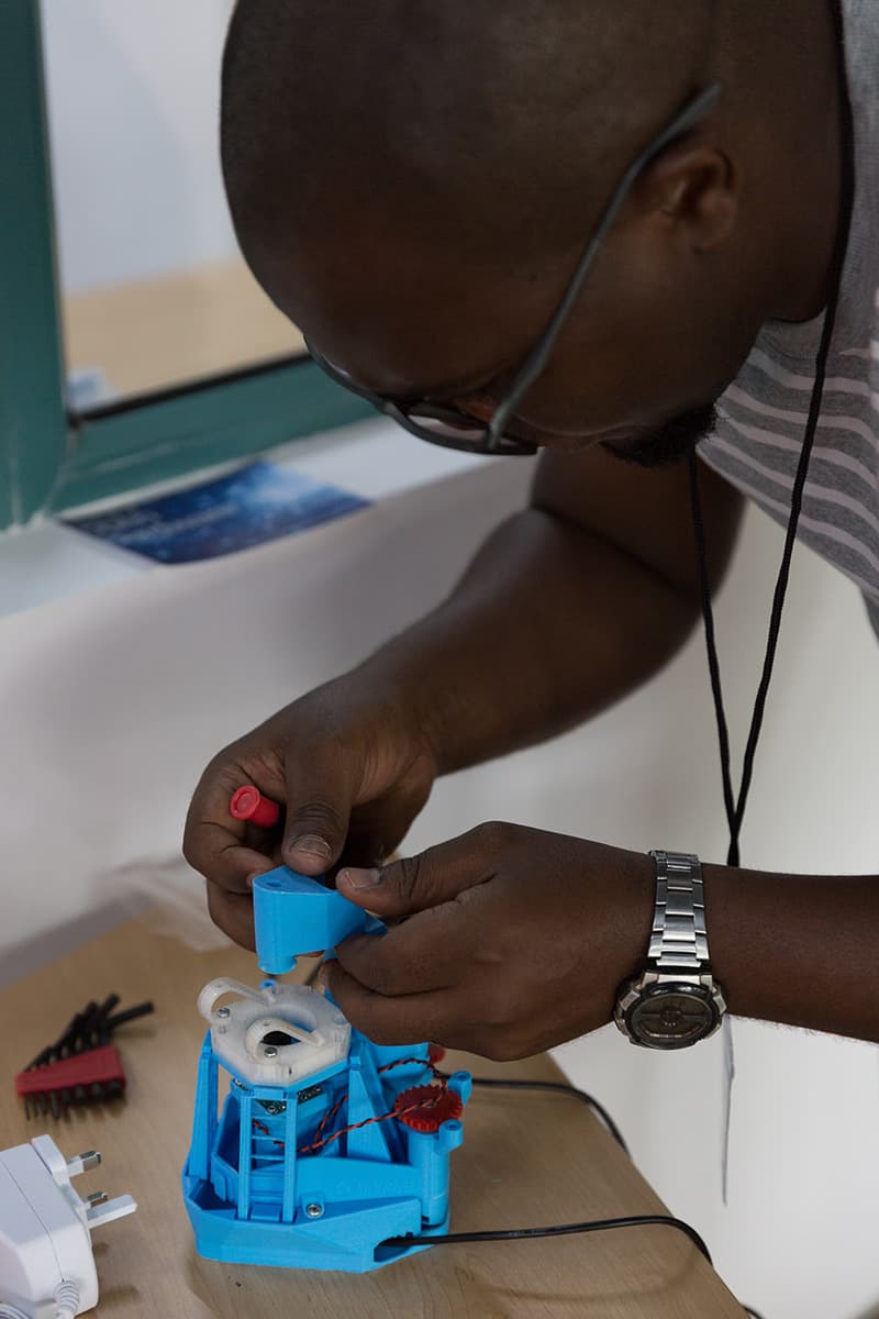
Valerian Sanga

One of the reasons it has been extended so frequently is its ease of replication: it is simple and inexpensive enough for other labs to replicate on a whim, without feeling the need to justify their efforts with a new publication or apply for a research grant to pay for it. A big part of our vision for the project is that it can support an ecosystem of experimental techniques that can be fully replicated at moderate cost. Our hope is that this will lead to experimental science becoming more transparent, being repeated more often, and ultimately improving the quality and trustworthiness of our experiments.

The tools and working practices we develop to support the sharing of instrument designs need active, frequently replicated projects like ours for their development, but are intended to scale well to more expensive and specialist projects. This is reflected in CERN’s White Rabbit Project – a multi-laboratory, multi-company collaboration to develop new technology for control and data-acquisition systems that covers hardware, gateware and software; or the mesoSPIM initiative, which focuses on open-source light-sheet microscopes (Nature Methods 16 1105).

Support and funding in the long run

Looking to the future, one of the biggest obstacles to achieving a long-term, sustainable impact with a project such as ours is supporting the team over a number of years. We are fortunate to have been funded for five years through the Global Challenges Research Fund (GCRF) – part of the UK’s aid budget that funds partnerships between UK scientists and their counterparts in low and middle-income countries. Building the trust that is needed for a productive, equitable partnership takes a long time, as does learning to work around the many differences in culture and working practices between different countries and institutions.

The project so far has been a great success, but recent cuts to the UK aid budget mean that future funding for our work, as well as hundreds of other projects, is now scarce and the phase 2 funding for our pan-African network was cancelled earlier this year. The GCRF represents a significant investment from the UK, and if we are serious about achieving a meaningful impact with this kind of work we must make sure that we have a plan in place to support its translation out of academia. In the UK, this support often comes from companies that will commercialize the work, supported by government grants.

If we are serious about achieving a meaningful impact with this kind of work we must make sure that we have a plan in place to support its translation out of academia

In the spirit of “sustainable development”, we want to avoid leaving our African partners dependent on a UK company, as this would undermine many of the key benefits of an open, distributed project. However, start-up capital and support for small businesses is much harder to come by in countries with fewer economic resources. Transferring work from academia into charitable organizations is also harder, and much less well supported, than simply patenting and licensing technologies in a way that entrenches the inequalities already present in the world’s economic system.

On an individual level, this project could not have succeeded without postdoctoral researchers on short-term contracts who have devoted several years to building relationships, sharing knowledge and documenting projects to get ready for real-world use – often at the expense of chasing prestigious publications that would further their career. The recent aid cuts, and the consequent slashing of the GCRF programme, means that these incredibly valuable and committed people will now have to find jobs elsewhere. This comes at a huge personal cost to the people involved, but it also means that their skills, knowledge and partnerships are lost.

There’s a great deal of anger from the GCRF research community that has flourished in recent years, and problems will continue even if the UK does reinstate aid funding to meet its legal aid budget commitment of 0.7% of gross national income. We are pursuing other ways that our project and others like it can continue to make progress, for example by creating a foundation that can help open projects move towards medical certification, and are keen to collaborate with others to bring this about. We believe that open-source hardware truly has the potential to revolutionize the way we do science – from building instruments and developing educational tools, to launching clinical applications and growing global partnerships.

Ultrafast photon detectors enable reconstruction-free medical imaging

Many medical imaging techniques rely on a mathematical process called tomography, which reconstructs data recorded in one or two dimensions into three-dimensional image volumes. Positron emission tomography (PET) – which images the body using an injected radiotracer that emits positrons as it decays – offers the unique possibility of reconstruction-free 3D imaging. This is because PET can also localize the signal source by exploiting the time difference between detection of the two photons created when the positrons annihilate with electrons in the body.

Today’s state-of-the-art PET systems have a timing resolution of around 210 ps that, based on the speed of light, translates to a spatial localization of 3.15 cm. As such, image reconstruction is still required to create accurate images. But if this timing resolution could be improved, the tomography step could be eliminated completely.

A collaborative team from the University of California, Davis and Hamamatsu Photonics has now developed radiation detectors with an average coincidence timing resolution of 32 ps. The researchers – also from University of Fukui and Kitasato University – have used these detectors to create the first experimental cross-sectional medical image that doesn’t require tomography, a technique they call direct positron emission imaging (dPEI). They report their findings in Nature Photonics.

Ultrafast detection

First author Sun Il Kwon and colleagues achieved dPEI by combining three technology innovations. Firstly, they exploited the Cherenkov luminescence emitted when annihilation photons interact in the detector. If the detector material has a high refractive index and high atomic number, this interaction creates electrons with sufficient energy to produce Cherenkov photons.

To convert this prompt optical signal into an electronic signal, the researchers developed a photosensor based on microchannel plate photomultiplier tubes (MCP-PMTs), which have extremely high single-photon time resolution (SPTR), approaching 20 ps. They integrated the Cherenkov radiator (lead glass) with the photocathode in the MCP-PMT to eliminate any optical boundaries and increase the chances of detecting the small number of Cherenkov photons. Finally, they employed convolutional neural networks (CNNs) to predict the timing information for detected events from the detector waveforms.

To assess the performance of the new detector, the team measured the signal from a 22Na point source placed between two Cherenkov-radiator-integrated MCP-PMTs. The set-up detected Cherenkov photons with an average timing resolution of 32 ps, allowing annihilation events to be localized with a spatial precision of 4.8 mm.

Experimental set-up

The researchers then tested the system using three test objects filled with the PET radiotracer 18F-FDG: an image quality phantom; a spatial resolution phantom; and a 2D Hoffman brain phantom representing 18F-FDG distribution in a slice of human brain. To capture the signals, the detector pair was translated linearly over the width of each object. The system then created images directly from the measured data, without using any tomographic reconstruction algorithm.

The image quality phantom has a uniform background activity with two 8-mm diameter voids filled with air and with water, all of which could be clearly visualized. The dPEI image of the resolution phantom resolved 3 mm rods, demonstrating a spatial resolution of 4–5 mm. The dPEI image of the larger 18.4-cm diameter brain phantom faithfully captured its detailed structure, with a spatial resolution of around 4.8 mm, suggesting that the method could be scaled for human imaging.

The two-detector set-up, with its average timing resolution of 32 ps, produced images with a similar spatial resolution to that achieved by diagnostic PET scanners. The team note that these initial experiments used long acquisition times and high levels of radioactivity. The signal collection efficiency could be increased, however, by replacing the lead glass radiator in the MCP-PMTs with a higher atomic-number radiator, increasing the radiator thickness, and tiling multichannel detectors to increase geometric coverage.

Such an upgraded system would enable shorter acquisition times and/or lower radiation doses. For example, these three changes could reduce the data acquisition time for the brain phantom image from 24 h to roughly 1 min. In addition, multidetector configurations that cover the entire imaging volume-of-interest would remove the need for detector translation and allow dynamic radiotracer imaging.

The team is already working to implement some of those advances. “We are testing new detectors that have the scintillator bismuth germanate (BGO) integrated in the MCP-PMT, rather than lead glass,” senior author Simon Cherry tells Physics World. “This leads to a significant efficiency improvement because BGO has a higher atomic number; it also produces more Cherenkov light because of its higher refractive index.”

Huge haul of fast radio bursts hints at astrophysical origins

The greatest number of fast radio bursts (FRBs) ever to be observed from a single source has been reported by an international team of astronomers. Led by Di Li and Pei Wang at the Chinese Academy of Sciences, the group used the Five-hundred metre Aperture Spherical radio Telescope (FAST) in China to detect more than 1600 radio bursts from an object called FRB 121102 within a span of just two months. The observations allowed them team to carry out the first detailed statistical studies of the FRB phenomena – shedding new light on their astrophysical origins.

In 2007, two astronomers at West Virginia University detected an extremely rapid burst of radio waves, just under 5 ms in duration. Since this discovery, hundreds of similar FRBs have been detected in separate studies. Most FRBs are detected as single events, but in a small fraction of cases, FRBs have been shown to repeat. This has allowed astronomers to roughly identify the galaxies where these events originated.

Compelling mystery

Although they are relatively weak once they reach Earth, astronomers know that the bursts involve the release of large amounts of energy – a year’s worth of the Sun’s total energy output over the span of just a few milliseconds, for example. A variety of theories have been put forward to explain FRBs, including merging black holes, magnetized neutron stars, and exotic defects in the early structure of the universe. So far, however, no one theory has emerged as being more plausible than any other, and today, the astrophysical sources of FRBs remain a compelling mystery.

Over 47 days between August and October 2019, Li and Wang’s team detected a record-breaking 1652 FRBs originating from FRB 121102, which appears to be located in a dwarf galaxy about three billion light-years from Earth. This is the first known repeating FRB and it was discovered in 2021.

High activity

The FAST tally of 1652 events is greater than all previous observations of FRB events combined. At one point of particularly high activity, the astronomers recorded as many as 122 bursts in a single hour.

Such an extensive sample size allowed the team to do a detailed mathematical analysis of the properties of the bursts. This revealed key differences between lower- and higher-energy FRBs. Contrary to some theories, the analysis of FASTS’s data revealed little repetition in the bursts over timespans ranging from just 1 ms to 1000 s. This lack of periodicity challenges the idea that FRBs may originate from single rotating objects, such as neutron stars.

In addition, the fast rate of bursts within the event implies that FRBs can be generated extremely efficiently – discrediting the idea that their triggering mechanisms requiring large amounts of energy. Altogether, the team’s discoveries place tighter bounds on existing FRB theories and could bring astronomers a step closer to uncovering their enigmatic origins.

The research is described in Nature.

Scatter imaging during lung stereotactic body radiation therapy

Want to learn more on this subject?

Due to breathing motion, lung tumours move during stereotactic body radiation therapy (SBRT). Scatter imaging, which collects photons scattered out of the radiation therapy beam, is a potential technique for real-time tracking of lung tumours.
In this webinar, presented by Kevin Jones, the scatter imaging method is characterized through simulation, phantom experiments and analysis of clinical patient scatter images.

Kevin Jones, has published a paper on this topic in Physics in Medicine & Biology.

Want to learn more on this subject?

Kevin Jones is a medical physicist in the department of radiation oncology at Rush University Medical Center in Chicago, Illinois, USA. He is board certified by the American Board of Radiology. His research focuses on developing novel imaging techniques to guide radiation therapy.


MR-guided focused ultrasound delivers antibody therapy directly into the brain

For the first time ever, researchers have delivered an antibody therapy drug directly into the brain to target breast cancer metastases. A team at Sunnybrook Health Sciences Centre in Toronto used MR-guided focused ultrasound (MRgFUS) to non-invasively and temporarily open the blood–brain barrier (BBB), enabling the monoclonal antibody trastuzumab to reach specific areas of the brain. Writing in Science Translational Medicine, the researchers describe the first visual confirmation that focused ultrasound can improve delivery of targeted antibody therapy across the BBB.

The BBB consists of a thin layer of cells that protect the brain from toxins, viruses and bacteria, but which also blocks the delivery of therapeutic drugs. Trastuzumab, an antibody therapy that helps the immune system fight cancer cells, is used in conjunction with chemotherapy and radiation therapy to treat breast cancer metastases in the brain. However, it is 100 times larger than typical compounds able to enter the brain across the BBB.

This first-in-human trial suggests that MRgFUS provides a safe and effective method to deliver drugs across the BBB. It sets the stage for the possibility of delivering both established and novel therapies for numerous brain conditions that otherwise cannot gain access to the brain.

Early findings

Principal investigator Nir Lipsman, director of Sunnybrook Research Institute’s Harquail Centre for Neuromodulation, and colleagues report the results from the first four patients participating in the ongoing Phase I clinical trial. All patients had Her2-positive breast cancer with brain metastases, and had previously received whole-brain radiation and/or stereotactic radiosurgery, plus chemotherapy, to treat progressive intracranial and stable systemic disease.

To perform BBB opening, the team used Insightec’s ExAblate, a helmet-like focused ultrasound device containing over 1000 transducers that converge ultrasound waves onto discrete points in the brain. The pulsed FUS was delivered in conjunction with a continuous intravenous infusion of microbubble ultrasound contrast agent and trastuzumab-based therapy.

MR/SPECT images

The trastuzumab used in the study, developed by Raymond Reilly from the University of Toronto and colleagues, was radiolabelled with 111In. Use of this radiotracer-infused trastuzumab allows direct visualization of trastuzumab distribution in single photon emission computed tomography (SPECT) images.

The team delivered a total of 20 outpatient treatments combining transcranial MRgFUS with standard of-care trastuzumab injection, with an average treatment time of 138 min. Patients had a SPECT scan prior to treatment, 4 hr after treatment and 48 hr later. The researchers performed additional SPECT scans 30 and 90 days following the final treatment and continue to monitor the patients.

The SPECT images showed trastuzumab precisely targeting the tumours. After BBB opening, the researchers could directly visualize increased SPECT signal within the sonication volume for all lesions treated. MR images revealed that the patient’s brain tumours decreased in size by between 7% and 31%, at up to four months after treatment. The procedure caused no serious adverse effects. Lipsman reports that three of the patients remain stable, and that one patient died from progressive, non-intracranial systemic disease.

The researchers note that one potential advantage of MRgFUS over other physical and biological approaches to brain drug delivery is the high degree of spatial and temporal control that it provides. MRgFUS enables selective targeting of single or multiple brain lesions, which can be located at the extremes of the brain or in deep eloquent central regions of the cortex that control bodily function.

“This study adds the brainstem, cranial nerve nuclei and cerebellum to the list of regions that can be safely and precisely targeted with MRgFUS, all areas in which radiation and surgery may be limited,” the authors write. Also, because MRgFUS can be combined with diverse therapeutic agents, therapies with less side effects can be used, improving treatment tolerability and patient safety.

Future outlook

This study is continuing to enrol patients, with the goal of performing a 10-patient trial. “Completing our current trial is an important first step,” Lipsman tells Physics World. “It will help inform what the design of a larger trial would look like, what kinds of outcomes we should be measuring, and how to optimize a study for safety and feasibility. The goal is to determine what role focused ultrasound will play in the management of brain cancer patients. For that, creating partnership and collaborations with other institutions will be key.”

Sunnybrook is a Focused Ultrasound Center of Excellence, the only Canadian site recognized as such by the Focused Ultrasound Foundation, which is helping to fund this study. Lipsman says the team is investigating several aspects of using focused ultrasound in brain cancer.

“We want to determine whether peripheral biomarkers, or so-called liquid biopsy, can be enhanced following BBB opening, to help with less invasive approaches to diagnosis and treatment monitoring,” he explains. “We also will conduct more comprehensive tests looking at the safety of BBB opening, including what impact, if any, we are having on important inflammatory markers and other features of the BBB.”

“Importantly, we are interested also in streamlining the procedures, making them faster, more efficient and more comfortable for patients,” he adds. “Enhancements in imaging technology will aid in targeting, and improvements in hardware and software will give us even better control of microbubbles and ultrasound transducers, to control just how much of the BBB is open and hence how much of a therapeutic can be delivered. All of these advancements are underway and will be rolled out in future trials that are now under active development.”

Superconductivity appears in novel hydride phases

Researchers in China and Russia have discovered superconductivity in two new phases of a hydride material at pressures much lower than those needed to stabilize other recently-discovered hydride superconductors. The work will aid the search for lower-pressure and potentially room-temperature superconductors.

Superconductors are materials that conduct electricity without any resistance. Many materials display superconductivity when they are cooled to low temperatures, and the phenomenon was first observed in 1911 in solid mercury, which has a superconducting transition temperature Tof 4.2 K. The search for superconductors that operate at warmer temperatures – perhaps even room temperature – has been on ever since. A material that remains superconducting at ambient temperatures would hugely increase the efficiency of electrical generators and transmission lines, as well as simplifying existing applications such as superconducting magnets in particle accelerators and MRI scanners.

Step closer to holy grail

Physicists came a step closer to this holy grail in the 1980s and 1990s with the discovery of high-temperature superconducting copper oxides, which have Tcs between 30–133 K. It wasn’t until 2015, however, that the maximum critical temperature took a major leap forward with the discovery that hydrogen sulphide has a Tc of 203 K when compressed to pressures of 150 GPa.

While materials that only superconduct at such extreme pressures have few (if any) practical applications, studying them may offer a path to discovering new compounds that superconduct at milder temperatures and pressures. For this reason, the hydrogen sulphide result sparked a flurry of interest in solid materials containing hydrogen atoms bonded to other elements. Since then, several other hydrogen-rich superconductors have been made in the laboratory. In 2019, researchers reported breaking hydrogen sulphide’s record with lanthanum decahydride (LaH10), which they found to have a Tc of 250–260 K. And in 2020, another group reported observing a Tc of 288 K in the C-S-H system at around 275 GPa.

Superconductivity in cerium hydrides

The team, led by Tian Cui, Xiaoli Huang and Wuhao Chen from Jilin University in China together with Artem Oganov and Dmitrii Semenok of Russia’s Skolkovo Institute of Science and Technology (Skoltech), took a slightly different approach. In 2019, members of the team synthesized a new cerium hydride with the formula P63/mmc-CeH9. Each cerium atom in this material is enclosed in a H29 cage in the atomic hydrogen sublattice, and it had previously only been studied theoretically. This same team has now found superconductivity in novel phases of both CeH9 and another newly-synthesized material, CeH10.

The researchers note that cerium hydrides are remarkable materials, as they are stable and display high-temperature superconductivity at lower pressures (about 0.8 million atmospheres) than other so-called “superhydrides”. As such, they say that these compounds “serve as an ideal starting point to further study the mechanism of superconductivity in these fascinating compounds, and design other superconductors, stable at even lower pressures”.

In their previous work, the Skoltech-Jilin researchers found a close relationship between the location of elements within the periodic table and the superconductivity of hydrides made from these compounds. They now believe that this relationship may also apply to materials other than hydrides. “Take La and Ce, for example – they are neighbours in the periodic table and indeed both form high-temperature superconductors,” Oganov explains. “However, there are differences: LaH10 superconducts at higher temperatures, while CeH10 is stable at lower pressures.”

Now that the binary hydrides have been well-studied, the Skoltech-Jilin team says that it will focus on achieving higher-temperature superconductivity at lower pressures in ternary hydrides. “We know which elements lead to higher-temperature superconductivity and have begun to learn which ones lead to stability at lower pressures,” Semenok says. “These are the main notes, but it takes imagination to combine them into a melody.”

The present work is detailed in Physical Review Letters.

How to prepare for the post-pandemic jobs market

As offices, labs and workplaces begin to reopen, it is clear that the COVID-19 pandemic will have a lasting effect on ways of working across industry and academia. It might even alter the careers landscape, in terms of the numbers and types of opportunities available. But the key factors dictating whether physics students will get the jobs they want after university remain broadly the same: access to work experience and professional-skills development, engagement with careers support and informed career-decision processes.

These points are central to the debate about the role of higher education and the growing need for universities to demonstrate that their graduates are getting good, well-paid jobs. An important way of doing so is through career-oriented activities that help them develop “work-ready” skills and learn how they can “fit” their degree to the workplace. Such activities should be designed with the current and future jobs market in mind.

Labour market data suggest that before COVID-19, graduate vacancies that were “hard to fill” included programming, software development and engineering-related roles. The good news for physicists is that these are all career areas that they typically go into. But the reason why these roles are hard to fill, according to a report by the graduate careers organization Prospects, is that applicants often lack relevant technical and practical skills, including advanced problem-solving related to a specific situation, complex numerical or statistical understanding, and role-specific specialist skills. So it’s important that physics students make the most of the practice that their degree offers them to improve in these areas.

While these job opportunities will remain, the growing trend towards online and virtual working might prompt employers to change their priorities when assessing candidates. According to Prospects, businesses want to adapt by placing greater importance on digital skills like effective online communication and the ability to work autonomously using various online platforms. Employers are also increasingly valuing creativity, critical thinking, interpersonal communication and leadership abilities. So if you’re a physics student, developing these competencies, as well as more technical skills, could help you stand out in future applications.

Employer input

Even companies that haven’t been affected much by the pandemic so far are reconsidering their criteria in hiring decisions. This was a point that came up at a one-day physics careers education webinar held last year by the White Rose Industrial Physics Academy and the South East Physics Network – two organizations that support physics students in their transition to graduate-level work. During the webinar, a panel of representatives from defence firms AWE, BAE Systems and Ultra Energy, along with deep-tech company MeVitae said that, although COVID-19 has had a minimal impact on their businesses, it will affect their future needs. That’s largely because it has accelerated the trend towards flexible working. Recruiters will therefore place even more emphasis on qualities such as adaptability, resilience and high-level communication skills.

To ensure that graduates become competent in these areas, it is essential that employers tell universities what sort of people they are looking for, and that institutions adapt their degree programmes accordingly in response. For example, Keele University’s chemistry department worked with industrial partners to redesign its degree course after receiving feedback that its graduates had limited reporting skills.

To teach scientific report-writing to first-year students, the department introduced a new assessment process featuring iterative assessment-feedback cycles. In the first term students analyse an example report, deconstructing it into the constituent parts from the introduction through the experimental phase to the conclusion. They also compare this with “real” published scientific papers.

In the second term, students draft a full lab report and take part in a peer-review workshop and group discussion. This approach enables students to improve their teamworking skills and self-awareness, as well as their scientific reporting. We think such collaborations are vital to help universities understand the changing needs of industry and shape their programmes so that students have the abilities to thrive in future job markets.

What employers can do

One factor that can hinder graduates’ confidence when searching for jobs is a lack of certainty around what employers expect from them. Recruiters can address this by doing the following:

  • Ensure job adverts are clear and specific, as physics students can be put off applying for jobs where information is unclear, for example being asked to apply “as soon as possible”. A higher proportion of physics students have certain disabilities, such as dyslexia, autism/social communication disorder, and struggle with ambiguity
  • Be aware that female students are less inclined to apply for jobs if they don’t feel they have all the skills/experience outlined in the job description
  • Use inclusive language in job adverts. Textio is one handy example of a tool to check adverts meet acceptable standards for inclusivity and clarity

The graduate perspective

But it is not only employers whose decision-making will be altered by the lasting impact of COVID-19; graduates themselves might start to make different careers decisions too.

We know that for many graduates, finding a job in a specific location is more important than finding a particular type of job. After analysing 1.87 million records of students who graduated from UK universities between 2011 and 2017, Alastair Buckley of the University of Sheffield found that physics graduates behave in the same way as other graduates in terms of their work mobility. Approximately 85% of all students choose to study and later find jobs within 100 km of their domiciled address (the address they state as their permanent address, which, in most cases, is their parents’ home). Furthermore, around 65% of all UK graduates, including physicists, end up working within 20 km of their domiciled address.

There is a particularly noticeable divide between the north and south of England, with very few students who are domiciled and study in the south going on to work in the north or vice versa. One exception is London, which sucks graduate talent from the rest of the UK.

This idea that many graduates prioritize location in their career decisions is sometimes called “emotional geography”, and relates to the level of connection they feel towards their home, as well as the influence of family and social networks, and future life plans (see “There’s no place like home” October 2019). One study carried out by researchers at the University of Leeds investigated this through interviews with eight undergraduate physics students completing a physics degree between the years 2016 and 2018. The students were interviewed three times throughout their final year of university and once again six months after graduating.

These interviews showed that, when making career decisions, students think about their individual situation, accounting for both personal and professional elements, and that these don’t necessarily lead them on a linear path from university to a graduate job. This supports the idea that “emotional geography” plays a significant part in many physics graduates’ decision-making, and therefore needs to be taken into account when offering them career support. One way that physics departments could do this is by looking at the skills that local employers need and designing their curricula to help students develop those skills.

However, it could be that businesses reduce office space post-COVID, and that a higher proportion of employees remain working from home. In this case, London might become less of a draw as there will be more opportunities to work remotely. This could enable job-seekers to place more emphasis on the specific role they are looking for, and lead to a more geographically distributed graduate workforce. Nevertheless, the extent to which businesses permanently increase their home-working practices remains to be seen.

What universities can do

Students face many challenges in transitioning from university to employment, but there are lots of ways in which physics departments can help them to prepare for the world of work. Here are some practical examples:

  • Engage students with employability activities through timetabled modules – preferably accredited
  • Ensure students build a portfolio of experiences from first to final year to provide them with evidence of personal development and a tangible track record of performance
  • Maintain alumni contact through student mentoring
  • Understand and minimize the additional invisible barriers to employment facing disabled, BAME and female students
  • Challenge academics on their perception of professional skills and their value
  • Build students’ confidence and help them understand what transferable skills they have and how they can be applied in different job markets
  • Make students aware that employers don’t all want extroverts. They value diversity and students with different abilities
  • Make students realize that employers don’t expect them to hit the ground running, but rather encourage them to learn from failures
  • Help students understand that having technical knowledge is not enough. They also need to demonstrate in an application and interview that they have the ability to apply their technical knowledge, often a key quality to set them apart from others

The “science ego”

Another interesting finding from the physics students who were interviewed in the Leeds study is that they all were very confident that their degree would enable them to easily find a job after graduating. Like all physicists, the Leeds students strongly identified with being “a physicist” and were often told that many career opportunities would be waiting for them when they had finished their studies. In other words, physics students possessed what the authors of the study called a strong “science ego”.

But this strong ego can sometimes work against physics graduates, because not all possess the additional skills and attributes needed to make a successful transition from academia to the world of work. In fact, having a science ego can reduce the perceived need to engage with career-development and job-seeking activities. To put it bluntly, physics students are told they can do anything but often don’t know where to start looking. Students who then struggle to find work can be left confused when turned down for roles and this can lead to them experiencing a lack of confidence and a feeling of “imposter syndrome”.

Evidence also suggests that a “science ego”, along with a lack of confidence or awareness of their skills, can cause many physics students to avoid work-based learning opportunities, such as summer internships and years in industry, which are an important way of developing work-ready skills.

Fortunately, we feel there is a lot that physics departments can do to address this problem and all the other challenges discussed above, from incorporating timetabled employability activities within degree courses to setting up alumni mentoring programmes. Employers can also help students transition to the world of work, not only by communicating their needs to universities, but also by understanding how to write job adverts in such a way that all qualified graduates feel confident to apply. This emphasizes the importance of continuous dialogue between industry and universities to ensure that people leaving higher education are prepared to enter the world of work, whatever that might look like.

3D printing makes a smaller, lighter cold atom trap

A team led by physicists at the University of Nottingham, UK has created a 3D-printed magneto-optical trap (MOT) capable of holding more than 2 × 108 rubidium atoms at temperatures a fraction of a degree above absolute zero. The demonstration shows that 3D printing, which is more formally referred to as additive manufacturing (AM), can meet the demands of highly precise cold-atom experiments, potentially paving the way for portable quantum devices based on this technology.

The starting point for cold-atom experiments is to to trap and cool as many atoms as possible in the smallest possible space. Once this is accomplished, it then becomes feasible to study the atoms’ quantum behaviour, which is only observable at very low temperatures. This behaviour can then be controlled to create some of the most accurate measurement devices in the world, including atomic clocks. A MOT is a key component in this process, bringing together the laser light and magnetic fields that slow the atoms and pin them in place.

Additive manufacturing for optical systems

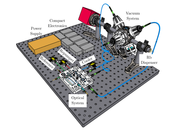
To construct their 3D-printed MOT, which they describe in PRX Quantum, the Nottingham researchers worked with colleagues at a specialist AM consultancy, Added Scientific Ltd. The process starts with a computer design such as the one shown below. The design is then transformed into a set of instructions that a printer uses to construct a physical object layer-by-layer. The printed components can thus benefit from a mathematically optimized design, making them smaller and lighter than conventionally manufactured components.

The new design incorporates several AM elements, including the mounts that precisely direct laser light into the vacuum chamber containing the atoms (see image). The vacuum chamber itself was built using AM techniques, which further assists in trapping the atoms as it allows careful control of the direction of the magnetic fields that hold the atoms in place. Vacuum chambers built using this technique also have a mass of just 245 g, compared to commercial chambers of up to a kilogram, making them desirable for portable applications beyond the laboratory. They are quick to manufacture, too, meaning that long delivery times for highly specialized designs could become a thing of the past.

As well as making components smaller and lighter, the AM process also introduces new capabilities. The positioning of the laser light, for example, is controlled and regulated via a unit known as the spectroscopy and power distribution apparatus. The adjustment-free AM mounts in this unit enable researchers to insert optical equipment into pre-aligned slots instead of having to fine-tune the setup manually – an advantage that anyone who has spent time in a lab painstakingly aligning optical components will welcome.

One big challenge when constructing optical systems is ensuring that the system is stable and the laser light remains aligned. The new AM design addresses this by reducing the number of times the light gets reflected before it enters the vacuum chamber. With fewer reflections, the beam’s position has less chance of deviating and not reaching the vacuum chamber. Another benefit of the AM approach is that the design contains only enough material to hold the components in place. This reduces the system’s overall weight whilst keeping it cool enough to perform the experiment.

The inside of the chamber also incorporates advances. Whereas traditional MOT designs generate magnetic fields by running current through coils of wire, using energy continuously, the Nottingham team showed that it is possible to trap atoms with an array of permanent magnets instead. Borrowing a technique widely used in the field of medical physics for MRI machines, they generated the required magnetic field by optimizing the configuration of the magnets.

System performance and outlook

These AM components combine to produce an MOT that can capture 2.5 × 10⁸ rubidium atoms, demonstrating that 3D-printed parts can be successfully integrated into cold atom experiments. “Our approach enables rapid fabrication of complex optical systems while also improving long-term stability,” says team member and Nottingham PhD student Somaya Madkhaly. She adds that AM technology offers even greater scope for miniaturization and improved system performance in the future, with possible strategies that include further optimizing the magnetic field generation to increase trapping capabilities and the automation of the electronic systems.

“3D-printing has become increasingly popular in recent years as it allows direct implementation of intelligent designs and algorithm-based improvements,” Madkhaly says. “In the future, quantum optical installations or vacuum chambers can be used to create new standards using our AM technology.”

Using RadCalc to verify Gamma Knife treatment plans

Want to learn more on this subject?

Gamma Knife is widely used for a variety of intracranial pathologies from benign and malignant tumours to vascular malformations. With any high-dose radiotherapy, it is necessary to utilize an accurate second check of the treatment plan.

The RadCalc Gamma Knife module is an accurate, efficient second check of the Leksell Gamma Planning system.

RadCalc performs point dose verification calculations for various Gamma Knife versions and the Leksell GammaPlan (LGP) planning system. RadCalc computes the dose and percent difference for each target utilizing proprietary TMR data, OAR data, and source position information. It utilizes a virtual machine to perform the dose computation – no physics setup is necessary. RadCalc’s virtual machine automatically selects the data based upon the type of plan.

This webinar will review the workflow and benefits of using the RadCalc Gamma Knife module to verify treatment plans.

Want to learn more on this subject?

Kathie Carrington is director applications and training at LifeLine Software, Inc. Company of LAP Group. She has more than 25 years radiation oncology experience, having positions from radiation therapist, dosimetrist and department director. She joined Lifeline Software in 2012 and has been connecting radiation therapy departments with software that increases productivity and safety ever since.

How fast does sound travel through 2D materials? It depends on how their layers stack

A new ultrasound technique that measures the strength of atomic bonds within two-dimensional (2D) materials, as well as the weaker forces between layers, has shown that the velocity of sound within these materials depends on the layers’ stacking arrangement. The technique, which was developed by researchers in the UK, could be used to craft “designer” 2D compounds by stacking layers in different ways while monitoring their bonding forces and studying how these relate to the materials’ physical and chemical properties.

2D materials such as graphene and metal chalcogenides are made up of stacked layers, or sheets, just one atom thick. While the sheets are bonded to each other only weakly, via van der Waals (vdW) forces, the atoms within each sheet form extremely strong covalent/ionic bonds. These dramatically different bonding strengths make it possible to exfoliate, or peel off, perfect single layers of these materials from bulk samples. Indeed, this was how graphene was first isolated from bulk graphite in 2004.

Techniques that can measure the strength of these atomic bonds and vdW forces in a non-destructive way are, however, lacking. This restricts scientists’ ability to explore various unusual phenomena (such as capillary condensation, the quantum anomalous Hall effect and even room-temperature superconductivity in monolayer sheets) that make 2D materials so promising for next-generation electronics. What is more, previous measurement efforts have produced conflicting results.

Technique similar to medical ultrasound

Researchers at the University of Nottingham and Loughborough University have now developed a technique that uses fast laser pulses to generate and detect tiny, transient strains in the crystalline lattice of indium selenide (In2Se3). This unique approach, backed by theoretical analyses by Alexander Balanov and Mark Greenaway at Loughborough, makes it possible to measure both the strong covalent bonds and weak vdW forces in the different phases of In2Sewithout damaging the material, says team member Wenjing Yan, a physicist at Nottingham.

Yan goes on to explain that the technique works in a similar way to medical ultrasound, but at a much higher, sub-terahertz, frequency. It involves sending a “pump” laser pulse just 120 femtoseconds long into flakes of In2Se3 to generate coherent phonons (quantized sound waves) that then travel through the material, interacting with its atomic bonds. The properties of these phonons reveal information about the strength of the atomic bonds, and they are measured by a second “probe” laser pulse with picosecond time resolution.

The technique is non-invasive because the laser pulses merely deform the crystal slightly, rather than destroying it. Indeed, the system may be considered as a sequence of springs: by knowing the speed of sound from measurements and how these springs respond to the deformation, the researchers can extract the relative strength of the covalent forces between the atoms and the vdW forces between the layers. “If we apply so-called density function theory with the help of high-performance computers, we can numerically estimate these forces for different stacking configurations and suggest how to tune the elastic, electric and even chemical properties of different polymorphs of vdW materials,” Greenaway explains.

Different phases, different properties

The researchers chose to study In2Se3 because it has a range of technological applications, including solar cells, photodiodes, ferroelectric field-effect transistors, and non-volatile memory elements. It also exists in several phases, denoted α, β, β′, γ and δ, each with a different crystalline structure.

The α- and β-In2Se3 phases are particularly interesting for materials designers. The α-phase is ferroelectric, with different ferroelectric properties for its hexagonal (2H) and rhombohedral (3R) arrangements in which the individual (quintuple) layers are stacked in an AB and ABC arrangement, respectively. In contrast, β-In2Se3(3R), which forms when α-In2Se3 is annealed, is not ferroelectric, and behaves as a superconductor at high pressures.

The researchers, who report their work in Advanced Functional Materials, found that sound waves travel at very different speeds in these different phases. Yan compares the team’s findings to the differences between pancakes and Yorkshire pudding. “Both foods are made from the same mixture: egg, flour and milk, but their different cooking processes give them different structures and properties,” she explains. “Although this is obvious in the macroscopic world, finding such differences in nanostructured materials due to subtle differences in vdW forces is surprising and exciting.”

Copyright © 2026 by IOP Publishing Ltd and individual contributors