Taking the helm Dava Newman, who invented the BioSuit for lunar and Martian exploration, will take over from Joi Ito as head of the Massachusetts Institute of Technology’s Media Lab. (Courtesy: NASA/Bill Ingalls)
An expert in the human exploration of space has become director of the Massachusetts Institute of Technology (MIT) Media Lab. Dava Newman, is professor of astronautics at MIT, took over the post on 1 July, becoming the first woman to do so. She replaces Joi Ito who resigned in the wake of last year’s scandal over MIT’s acceptance of grants from financier and convicted child-sex trafficker Jeffrey Epstein.
Newman received her Bachelor’s degree in aerospace engineering from the University of Notre Dame in 1986 that was followed by a PhD in aerospace biomedical engineering from MIT in 1992. After a year at the University of Houston, she joined MIT in 1993, and has been a member of the faculty ever since.
Newman is best known for her invention of the BioSuit, which she designed for future exploration of the Moon and Mars. She has served as the principal investigator on four space missions as well as being NASA’s first female deputy administrator from 2015 to 2017.
“Leading the Media Lab is a dream for me, and I can’t wait to help write the next chapter of this uniquely creative, impactful, compassionate community,” Newman said in a statement. “I plan to start by doing a lot of listening and learning to tap into everyone’s creativity.” In a statement, Hashim Sarkis, dean of the school of architecture and planning, noted that Newman “stood out for her pioneering research, wide range of multidisciplinary engagements, and exemplary leadership”.
The Media Lab has some 400 faculty, researchers, students and staff. Since its founding in 1985, the Media Lab has promoted an interdisciplinary culture and has 22 research groups working in robotics, smart prostheses, cognitive enhancement, innovative learning and music. MIT says it identified 60 candidates for the director role, of which 13 were interviewed.
Newman will replace Ito, who resigned in September 2019 following revelations that the lab and he had secretly accepted financial support from Epstein, who had been convicted of child sex-trafficking in 2008. Epstein was convicted of other sex-related crimes more recently and died in his jail cell in August 2019.
Carlo Rovelli’s new book Helgoland is, ostensibly, a journey into quantum physics. The story is told linearly, beginning with a small group of men whose work cemented its most foundational ideas, and ending with a meditation on how quantum mechanics provides insight into human neuroscience. It is a slim volume, coming in at under 170 pages. For this reason, one might imagine that it doesn’t contribute much to the already extensive literature for general audiences on quantum mechanics, which includes the recent book Einstein’s Unfinished Revolution: the Search for What Lies Beyond the Quantum, by Rovelli’s good friend (and my PhD co-adviser) Lee Smolin.
But Rovelli isn’t just giving a history lesson or background. He arrives with an agenda, and one that is tied to the reasons why quantum theory continues to capture the public’s attention. Ideas like wave–particle duality intrigue the general public, because they are so wildly unintuitive, while the best physicists in the world are still unsure of how to interpret their meaning.
Here, Rovelli’s Helgoland takes a specific position, introducing the reader to the “relational interpretation”. In short, the relational interpretation insists that the quantum state of a system depends on the observer, and it is a concept that Rovelli has helped to formalize and convert into an area of active research. It is only natural, therefore, that he will want to advertise it. Certainly, the reader gains insight into the ideas that influence Rovelli’s thinking. There is a longer-than-expected discussion of the ideas of Vladimir Lenin as well as Aleksandr Bogdanov. Given that both were thinkers who heavily influenced the Communist Revolution in Russia, it was surprising to see them brought into a text on quantum mechanics.
Elsewhere, he discusses the philosophies of second-century Mahāyāna Buddhist scholar Nāgārjuna, suggesting that ideas from outside of Europe have something to offer as we try to understand this fantastical theory of the subatomic world. Probably more than most readers, I am sympathetic to the entangling of physics and philosophy, and I was intrigued by this. Even so, in the end it felt more like an aside than a real attempt to decentre European analytic frames.
In fact, Rovelli’s book ultimately operates as yet another book about Great (White) Men. This is not to say that it is poorly written. Importantly, one of the blurbs on Helgoland says, “Physics has found its poet.” Indeed, Rovelli can write a sentence. This might seem like a pedantic comment, but the sentence is the most important part of prose, and good prose is composed of good sentences. Though I read the book in translation, the successful handiwork of Erica Segre and Simon Carnell, Rovelli’s voice – as I recall it from hearing him talk at conferences I attended during graduate school – comes through. It is passionate, excited and charismatic – much as I remember him.
Even so, the absence of women as real, complete human beings in the story is highly noticeable. This is not because I expected Rovelli to go out of his way to include the research of women who have made contributions to quantum physics. Intuitively, I knew not to expect this. What I did not see coming was that, in the act of setting the scene, Rovelli would do things like casually mention that Schrödinger “made no secret of his fascination with pre-adolescent girls” (p21). This particular passage goes on to detail the impact that Schrödinger’s sex life had on his academic positions.
While I believe in the value of sharing the whole picture of who someone was, rather than valourizing dead scientists as perfect heroes, nothing in Rovelli’s text problematizes Schrödinger’s behaviour. Instead, it is mentioned almost as a curious aside. Schrödinger still gets to be great in the end, with an equation named after him that every university physics student in the world learns.
Elsewhere, Rovelli describes how a poem cannot help him survive, though “a young woman might fall in love with my romantic soul” (p146). Over and over again, rather than being ignored, women are invoked in the text as objects and/or attendants to the journey of a Very Important Man. There’s a kind of old-school masculinity embedded in the narrative as well, for example through the glorification of Bogdanov’s death due to a blood exchange experiment. “Right up until the end, he had the courage to experiment, the courage to exchange and share, and the dream – and practice – of brotherhood” (p115). It feels like a political statement in 2021 to write a book that is about a theory of the whole universe and then gender it so intensely.
For those who like to understand how Rovelli thinks about the physical theories that he has devoted his life to developing, Helgoland will be an interesting addition. There’s a way in which it can feel like it meanders and/or is throwing lots of things at the wall to see what sticks. But that’s alright – that, for many of us, is the practice of theoretical physics.
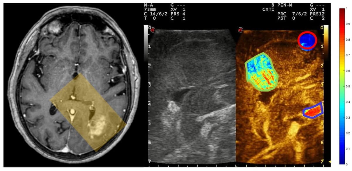
Left: MR image showing a glioblastoma lesion; the yellow rectangle corresponds to the ultrasound images on the right. Centre: standard ultrasound image. Right: contrast-enhanced ultrasound image showing the microbubble density in three regions-of-interest: artery (blue outline); tumour (green outline); and white matter (red outline). (Courtesy: CC BY 4.0/Sci. Rep. 10.1038/s41598-021-91252-w)
Contrast-enhanced ultrasound delivered using microbubbles can readily differentiate between different brain regions and structures, as well as various types of tumours, researchers from Italy and the US have reported. The findings of the study – which the team says is the first large-scale quantitative analysis of microbubble-enhanced cerebral ultrasound images – could open up new avenues for ultrasound-based imaging and therapies.
Formed by encapsulating gas within a flexible lipid, polymer or protein shell, microbubbles, as their name suggests, have diameters in the range of one-to-four microns. When injected into the body intravenously and then subjected to a sonic field, these tiny bubbles vibrate and collapse, producing distinctive harmonic signals that can be used to distinguish them from surrounding tissues, allowing their use as an ultrasound contrast agent.
In addition, when combined with focused ultrasound, microbubbles can be used to produce various biological effects, including reversibly opening the blood–brain barrier and easing drug delivery. But they also come with the risk of cavitation-induced tissue damage. Understanding how microbubbles end up distributed within the body is therefore important for both accurate imaging and safe therapeutic applications. For the brain, however, such knowledge has been somewhat lacking.
To address this, neurosurgeon Francesco Prada of the Fondazione IRCCS, Istituto Neurologico Carlo Besta in Milan and his colleagues performed contrast-enhanced ultrasound imaging of the brains of 19 patients while they were undergoing surgery to remove tumours. Following each procedure, they quantified and compared the changing distribution of microbubbles across the target area for key structures – specifically, arteries, tumours and white matter, as mapped by a trained neurosurgeon.
The researchers found that there were clear differences in microbubble distribution across the brain, with arteries showing the earliest and largest enhancement of the structures studied, followed by tumours and then regions of white matter. Specifically, the team’s analysis revealed that artery, tumour and white matter structures had peak microbubble intensities of 0.584, 0.436 and 0.175 units, respectively.
“Understanding cerebral microbubble distribution holds tremendous potential to improve both image guidance during surgical procedures and planning ultrasound-mediated therapies,” Prada tells Physics World. “Performing quantitative analysis of such phenomena will provide a sharper understanding and also will allow data sharing and ensure replicability of the results,” he explains, noting that such work will help pave the way towards safer procedures for patients.
“Performing brain surgery allows one to literally ‘open’ an acoustic window to the brain,” Prada adds. “This is an opportunity that should be exploited to its best, not only to improve a single procedure, but to gather pivotal data that will change our understanding of how the brain and its diseases function.”
“This work nicely demonstrates how ultrasound imaging in conjunction with microbubbles injection provides quantitative and highly informative parameters on different brain tissue,” comments Mickael Tanter, a medical physicist from the French National Institute of Health and Medical Research who was not involved in the present study. “The ability of contrast-enhanced ultrasound imaging to discriminate white matter, parenchymal tissue and intra-tumoural tissue with portable imaging scanners will surely make ultrasound the method of choice for per-operative imaging during brain surgery in the coming years.”
Jens Eyding – a neurologist from the Gemeinschaftskrankenhaus Herdecke in Germany, who was also not involved in the present study – notes that contrast-enhanced ultrasound imaging of the brain has long been regarded as having potential for use during tumour resection, especially given that shifts in brain mass following a craniotomy can render inaccurate surgical guidance derived from a pre-operative MRI scan.
“I absolutely agree on the need for more research on perfusion patterns of different brain tissue,” he says. “A prospective and systematic study to characterize specific parametric perfusion patterns of different tissue types may help to improve the significance of surgical guidance by means of ultrasound.”
With their initial study complete, the researchers are now looking to explore the potential of contrast-enhanced ultrasound-guided and microbubble-mediated therapies as a cheaper alternative to MR-guided focused ultrasound. Prada has also patented a cranial implant that can allow transcranial direct ultrasound imaging and therapy.
Researchers at the Beijing National Laboratory for Condensed Matter Physics and Institute of Physics, Chinese Academy of Sciences, have found evidence for an unusual superconducting state in CsV3Sb5, a so-called Kagome metal that exhibits exotic electronic properties. The finding could shed new light on how superconductivity emerges in materials where phenomena such as frustrated magnetism and intertwined orders play a major role.
Kagome metals are named after a traditional Japanese basket-weaving technique that produces a lattice of interlaced symmetrical triangles. Physicists are interested in this configuration (known as a Kagome pattern) because when the atoms of a metal or other conductor are arranged in this fashion, their electrons behave in unusual ways.
An example is frustrated magnetism, which occurs when electrons are “not happy to live together”, observes Ludovic Jaubert, a condensed-matter physicist at the University of Bordeaux in France who was not involved in the present work. In frustrated materials, not all interactions between electron spins can be satisfied at the same time, which prevents the spins from ordering themselves on long length scales. This failure has significant consequences for the material’s properties: if water behaved like this, for example, it would never freeze.
First Kagome metal and a new family
In 2018, researchers at the Massachusetts Institute of Technology (MIT), Harvard University and the Lawrence Berkeley National Laboratory created the first Kagome metal in the laboratory. The material in that work was an electrically conductive crystal consisting of layers of iron and tin atoms arranged in a Kagome lattice pattern, but the discovery of a whole family of Kagome materials with the chemical formula 𝐴V3 Sb5 (where 𝐴 = K, Rb, Cs) came hot on its heels.
These newer Kagome materials behave like conventional superconductors at temperatures below 2.5 K. Under these conditions, their electrons form correlated electron (or Cooper) pairs that carry current without any resistance. However, researchers suspected that the electrons in these compounds might also pair in unconventional ways.
Two-stage-like transition
To investigate further, groups led by Xiaoli Dong, Jinguang Cheng, Jiangping Hu, Hong-Jun Gao and Zhongxian Zhao made a series of magnetic and electrical measurements on single crystals of CsV3Sb5. The researchers began by recording the material’s X-ray diffraction pattern to confirm that it was indeed arranged in a Kagome lattice pattern. They then determined that it became superconducting at around 3 K by measuring its magnetism and electrical resistance as it cooled. This superconducting transition temperature is slightly higher than that observed in previous studies, and Dong and colleagues also noted that the transition to the superconducting phase was not as sharp as expected. Instead, they found that the tell-tale (diamagnetic) signal for superconductivity onset set in gradually below around 3.5 K before dropping abruptly below around 2.8 K.
The team then measured the sample’s angular-dependent magnetoresistance within the plane of the Kagome lattice and identified a twofold rotational symmetry in the mixed state (that is, a state that contains both superconducting and non-superconducting phases). Below 2.8 K, they found that the orientation of this twofold symmetry displays a peculiar twist by an angle of 60o, which is characteristic of the Kagome geometry.
Finally, the researchers measured the sample’s magnetoresistance in two directions – perpendicular to the plane and across it – as they applied magnetic fields of varying strengths (up to 8 Tesla) to it. They did this to obtain the material’s upper critical field, which is defined as the magnetic flux density that would completely suppress superconductivity at 0 K.
They found that this field is higher across the plane than perpendicular to it, such that the ratio of the two at 0 K is very large. This result, say the researchers, is best explained if the material is a quasi-two-dimensional multiband superconductor – a type of superconductor that may display one or several phase transitions with increasing temperature from or to chiral superconducting states. These transitions can occur when two or more electronic bands are involved in the superconducting transition.
“Our result, together with other observations, suggests that the superconducting state in CsV3Sb5 comes about partially due to electron-electron correlations,” Dong tells Physics World. “Other effects such as spin-orbit coupling and chiral flux phases may also be present in this multiband Kagome system.”
The researchers, who report their work in Chinese Physics Letters, are now planning to investigate the exotic normal and superconducting states in this multiband system using microscopic spectroscopy techniques.
Cardiac positron emission tomography (PET) with the radiotracer 18F-FDG is used to measure the health of the heart muscle, and can detect tissue damage and scarring following a heart attack. The quality of the PET images, however, can be impaired by respiratory and cardiac motion, reducing their diagnostic accuracy. A study headed up at King’s College London has shown that simultaneous PET–MR scanning combined with a single image reconstruction framework enables the comprehensive assessment of cardiovascular disease with improved PET image quality.
The benefit is twofold: MR-derived respiratory information enables motion correction, while MR-based anatomically guided image reconstruction suppresses noise and increases contrast in the images. This latter technique, which had previously only been demonstrated in brain PET (which doesn’t suffer from physiological motion), significantly improved PET image quality compared with alternative reconstruction methods, as the research team reports in the Journal of Nuclear Medicine.
Cardiac PET with 18F-FDG is usually performed to monitor the heart in patients diagnosed with coronary artery disease, to determine the extent of infarcted areas and evaluate myocardial damage produced by previous coronary blockages. The exam also helps to determine whether coronary stenting or bypass surgery is more appropriate for a patient.
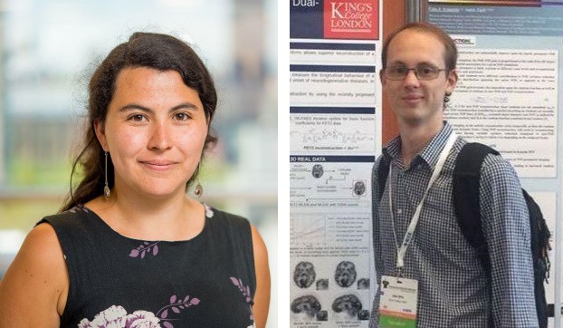
Co-principal investigators Camila Munoz (left) and Sam Ellis.
Co-principal investigators Camila Munoz and Sam Ellis and colleagues developed a single cardiac PET–MR image reconstruction framework, which uses MR-derived information to allow both motion compensation and anatomical guidance within the reconstruction of cardiac PET images. The framework resolves previous image degradation issues, such as artefacts, blurring and noise, by utilizing MR-based attenuation map alignment, respiratory motion correction, cardiac gating and MR-guidance image reconstruction.
Images are acquired using a cardiac imaging protocol designed for simultaneous diagnostic PET and coronary MR angiography (CMRA). The framework improves myocardial PET image reconstruction by aligning attenuation maps to the end-expiration respiratory position, using the CMRA images as reference to reduce attenuation-induced artefacts. It also incorporates MR-derived motion information into a motion-corrected PET image reconstruction, and uses the high-contrast, motion-corrected 3D CMRA images for anatomically guided PET image reconstruction, suppressing noise while preserving quantification performance.
Clinical assessment
To evaluate their framework, the researchers imaged five cancer patients without known or suspected cardiovascular disease. This enabled them to quantify the effect of each of the aforementioned improvements upon the final image quality. They then used the same protocol to image 10 patients with symptomatic coronary artery disease, chronic total occlusion of a relevant coronary artery and evidence of wall motion abnormalities.
Munoz, Ellis and colleagues applied a variety of image reconstruction techniques to the PET datasets and assessed the resulting image contrast and noise. They measured contrast as the contrast recovery coefficient between the left-ventricular myocardium and blood pool, and calculated noise as the standard deviation of voxels within the myocardium.
After comparing various established PET image reconstruction approaches for the two patient groups, the researchers report that each of the approaches incrementally increased contrast in the images, although this was usually at the cost of increased noise. However, their proposed framework incorporating MR-guidance further improved contrast while reducing noise. Notably, the myocardium-to-blood pool contrast increased by 143% on average compared with conventional uncorrected, non-guided PET images. Anatomical guidance reduced image noise by 16.1% compared with non-guided reconstruction.
While these findings are promising, further improvements might be needed to reduce noise to a clinically acceptable level. The researchers suggest that this can be achieved with better MR-based guidance information or by modelling the uncertainties of guidance information.
“Our results suggest that using MR information to correct for patients’ physiological motion and perform patient-specific denoising may allow for a more accurate reconstruction of the underlying distribution of PET radiotracer, in our case, 18F-FDG. This could give clinicians confidence that the cardiac PET images they are reading are a faithful representation of patients’ physiology, without artefacts from motion, image noise or other reconstruction artefacts,” explain Munoz and Ellis.
“In particular, in our results, we observed an improved depiction of myocardial viability defects, represented as an increased contrast-to-noise ratio between defect and surrounding healthy tissue,” they add. “We think that this could result in an increased diagnostic sensitivity to myocardial defects in clinical practice, by enabling the detection of smaller defects and improving delineation of the extent of the diseased tissue. However, further research is required to study the impact of these improvements in clinical decision making.”
Munoz and Ellis also point out that novel radiotracers besides 18F-FDG have recently been introduced for myocardial PET–MR imaging. “Some of these, such as 18F-NaF, generally produce very poor-quality PET images due to both physiological motion and the low radiotracer uptake from the biological processes in question,” they explain. “We believe that our motion-corrected, MR-guided PET reconstruction could prove especially useful in these situations, aiding in bringing these state-of-the-art radiotracers into clinical practice.”
During my childhood I received abuse on a daily basis, whether it was about my disability, my non-binary dress sense or the colour of my skin. Such discrimination and prejudice prompted me to continue into higher education to study science rather than to go straight into employment. Academia, it seemed, offered shelter from the frightening outside world and looked to be so promisingly full of allies and forward-thinking people.
But I was wrong. The solidarity of some “allies” turned out to be showmanship. While at first they seem to be engaged in helping marginalized people, the main beneficiaries of their actions are all too often themselves. I dub these people “alliars” – those who support well-meaning attempts for equality only to use it to enhance their own reputation and careers. For all the rainbow flags they display for everybody to see on their Twitter profiles or office doors, they show little concern whether, for example, their trans and genderqueer co-workers have access to unisex toilets in the workplace. Or they write articles about the legacy of slavery in academia and the need for decolonization but use this so-called “championing of diversity” not to empower their Black and Asian colleagues but to push ahead of them in the promotional queue instead.
Harmful action
Such alliars should not be confused with those “performative” allies on social media who are all talk and no action. At worst performative allies are reward-seeking and delusional, sharing what “must” be done by others without ever considering what they should change in their own world. Alliars, on the other hand, offer duplicitous talk and harmful action – they know what needs to change but any progress must be on their own terms.
Indeed, alliars may encourage those of us who belong to marginalized groups to “open up” and describe the discriminatory experiences and incidents of prejudice that have shaped our lives, so that they can “share our pain” and “understand how we feel”. Yet they cannot begin to comprehend our lived experiences and nor do they want to. They promise to listen and work together, but there is no togetherness.
Alliars commodify our anguish and take full credit for all the diversity initiatives to carefully craft a favourable public image of themselves. This cultivated image is not simply designed to boost their own careers but also to shield them from scrutiny. How could they possibly be linked to racism, ableism or sexism when they are so selflessly committed to the diversity cause?
Alliars are also quick to forget that improving diversity starts at home. What use is superficially lobbying for change at international level when they cannot even extend small acts of goodwill and compassion to ethnic minority, LGBTQ+ and disabled colleagues in their own faculties or departments? In this way, alliars are no better than the openly prejudiced folks outside academia – unkind and intolerant.
Alliars may once have been well meaning but after years of working in academia, they could gradually have become immune to institutional racism, sexism and ableism. Or perhaps they genuinely believe that they are superior and have more insight into matters of social injustice than its victims. It is possible that they condone and are complicit in discriminatory policies and practices because they fear that an influx of diverse researchers into academia will jeopardize their own careers and positions of power.
By comparison, genuine allyship is like much of the academic research process itself – it is dynamic and involves constant change and adjustment rather than glamour, publicity and self-promotion. Real allies are those who realize that applying a rigid, universal approach to supporting marginalized people does more harm than good. To offer meaningful support and be a true ally requires forming relationships and building trust. It involves tailoring the support to suit the specific needs and struggles of marginalized individuals, rather than viewing them as members of a homogenous club that needs appeasing.
Worse still is repeating impressive rhetoric in public, while in private treating marginalized colleagues as inferior outsiders. Spending the weekend parroting adages on social media like “the system is flawed for minority people” does little to improve their prospects or situation. What does help, however, is using a working hour to write a reference to help a marginalized colleague make a strong job application.
Difficult task
Just like starting to write a new research paper, learning a coding language or figuring out a new piece of scientific equipment, genuine, compassionate allyship is sometimes messy, often difficult and always time-consuming. But an ally using their privilege to empower and change another’s life for the better is a reward that is worth the effort every time. Some unoppressed people, however, find it difficult to admit that they may be wrong and need to do things differently. That is especially the case when it comes to diversity work, where emotions run high, there are many grey areas and personal integrity is at stake. This is why real allies can be so difficult to find for marginalized academics.
It follows that trying to navigate the wonderland of academic science, with its miscellany of allies and alliars, can be disorientating, unnerving and isolating. Someone who identifies as under-represented, in a minority or different in any way, must be cautious. Try not to naïvely believe that alliars are the saviours that they claim to be. Look for people who want to listen to your worthwhile ideas and opinions. Look for people who want to work with you, not instead of you and certainly beware of those who look to work against you. Look for people who match the virtual grinning emoji with an actual, lasting smile. Because friendly public faces are no guarantee of “friends” in the academy.
The image for this article was updated on 9 July 2021 as the original image was inappropriate.
A new explanation for why thermal systems subjected to extreme heat fluxes display far lower temperature fluctuations than expected has been developed by researchers in France. By adding two additional components to a widely-used theorem for predicting fluctuations, Alex Fontana, and colleagues at ENS de Lyon have explained a key discrepancy between theory and experimental observations of silicon cantilevers exposed to extreme temperature gradients. Their discovery could improve high-precision experiments and the design of tiny machines.
As physical systems exchange heat with their surrounding environments, their temperatures will naturally fluctuate over time. These variations may be minuscule, but they can have a significant impact on the operation of microscale mechanical devices and the outcome of high-precision experiments. This has created a practical need to predict the extent of thermal fluctuations in different environments. To do this, physicists use a powerful tool called fluctuation-dissipation theorem (FDT).
Currently, it is often assumed that systems are in thermal equilibrium with their environments – absorbing exactly as much heat as they emit. Instead, out-of-equilibrium heat fluxes are common in many systems including living tissues and aging materials. According to the FDT, such systems should undergo higher temperature fluctuations than those in equilibrium. Yet in recent experiments, involving silicon cantilevers exposed to high temperature gradients, the mechanical vibrations induced by temperature fluctuations were far lower than the FDT predicted.
Extreme temperature gradient
In their study, Fontana’s team took the cantilever experiment to its extreme by cooling one end of a cantilever to cryogenic temperatures, while heating the other to just below its melting point. This induced a temperature gradient of 1700 K – the highest possible difference the system could sustain in a vacuum. The researchers discovered that while the amplitude of the vibrations did increase with the temperature gradient, they were far lower than the FDT predicted, given the average temperature of the system.
Fontana and colleagues successfully accounted for this discrepancy by introducing a simple extension to the FDT, containing two key aspects of mechanical dissipation. First, a “clamping loss” describes the mechanical energy lost at the cantilever’s base; and second, “distributed damping” accounted for the energy lost due to defects in the atomic structure of silicon along the length of the cantilever.
The team now hopes that by generalizing the FDT in this way for high heat fluxes, improvements could be made across a wide array of areas in research and engineering.
Better predictions could enable researchers to better describe the interaction between electromagnetic fields and living tissues; and improve the sensitivity of microelectromechanical devices. Elsewhere, they may even improve the resolution of the interferometers used in ground-based gravitational wave detectors – helping astronomers to better link atomic-scale detector movements to ripples in space–time.
Reducing treatment uncertainty Katia Parodi and her team are developing new techniques to improve the accuracy of particle therapy. (Courtesy: Christoph Olesinski, LMU Munich)
How would you describe Physics in Medicine & Biology?
PMB is one of the most established journals in the field of medical physics and biomedical engineering. Since its foundation in 1956, the journal’s focus has always been on the development and application of theoretical, computational and experimental physics to medicine, physiology and biology, with a major emphasis on biomedical imaging and therapeutic interventions.
What does the journal offer the medical physics community?
It provides an excellent forum to communicate cutting-edge research in the field. In the area of radiation therapy, for example, several of the most influential papers on the development of intensity-modulated radiotherapy were published in PMB. Recently, the journal awarded its citations prize, the Rotblat medal, to a study describing the first patient treatment using a high-field 1.5 T MR-Linac. In this case, PMB published a lot of the initial fundamental and experimental work, right up to this first application in a human. This is a good example of the broad scope of the journal.
What are your plans for the journal?
I am honoured and thrilled to have this opportunity to intensify my commitment to the journal, having served on its editorial board since 2013. The task will be to constantly identify and evaluate new trends in the field, to keep the journal scope up to date, and to offer the best services to our authors and readers. For example, we recently introduced a new format, called roadmap papers, which are invited visionary contributions showing where relevant fields are going. One of the first of these roadmap papers was dedicated to the 10 ps time-of-flight PET challenge – the quest to develop ultrafast time-of-flight PET.
What are the other hot topics emerging in medical physics?
A lot of new trends exploit physical phenomena for biomedical applications, including developments in acoustics, optics and Cherenkov imaging. We are seeing exciting work in nanotechnologies, used for contrast agents or as radiation sensitizers, as well many activities in detector development, such as improvements in technology for ultrafast PET and photon-counting detectors for X-ray imaging. There are also efforts to combine different imaging modalities using hybrid detector technologies, as well as integration of imaging with therapy. In radiotherapy, we see new frontiers under exploration such as FLASH irradiation and micro/minibeam technologies.
In all of these areas there is a new trend of applying artificial intelligence (AI). PMB will not aim to host the development of new AI algorithms, but it will be the journal’s goal to see how new applications, and optimization of existing AI algorithms, could impact what we are doing in the fields of biomedical imaging and therapeutic interventions.
Your own research includes range verification of particle therapy, what are you working on?
Particle therapy is still an emerging radiotherapy technique. Its main advantage is that you can better target energy deposition in the tumour, with less side-effects for healthy tissue and critical organs. But particle therapy also comes with disadvantages: it is highly sensitive to range uncertainties, or in other words, knowing exactly where the beam will stop in the patient. There are a lot of physics processes that one can try to exploit to visualize the stopping position of the beam inside the patient. These include nuclear reaction processes, which can be visualized with PET, prompt gamma imaging and thermoacoustic emissions.
These are all techniques that require a lot of research to make the instruments compatible with a clinical environment and sensitive enough to capture typically very low levels of signal. We are also developing techniques to model the signals and reconstruct images, and also to combine these advanced methods and integrate them into a possible future workflow.
Another interesting research area is to improve in-room imaging of the patient prior to treatment. One of the uncertainties in knowing where the beam stops is due to our limited knowledge of the properties of the tissue interacting with the beam. If you use standard X-ray CT imaging there are relatively large uncertainties. But if other techniques are employed, such as the emerging dual-energy or spectral CT, or even the ion beam itself, to create radiographic or tomographic (so called proton or ion CT) images, then you can reduce this uncertainty and make a far more accurate treatment plan.
Are these in clinical use yet?
PET for range monitoring has been explored clinically for many years by a few institutions, but without dedicated commercial devices, so mostly using research instrumentation. Prompt gamma imaging is now reaching the stage of clinical evaluation, but just using first prototypes, which may not yet fully exploit its potential. Methods such as thermoacoustic imaging are still at the development stage. As for proton imaging, a first prototype is close to reaching clinical application.
What else is your research group investigating?
We have a project funded by the European Research Council to develop a small-animal proton irradiation platform with integrated image guidance. I think this is really exciting because it has been shown that if you want to translate new therapeutic approaches to the clinic, it is good to first perform investigations on animals. However, it is even more challenging to precisely irradiate such small targets.
We are aiming to develop a portable platform that can be integrated in a particle therapy facility’s research room, using a dedicated beamline that will take the clinical beam and focus it to irradiate very small targets. The platform will also include proton imaging for use before treatment, and will integrate PET and thermoacoustic imaging to verify the proton beam range.
There is currently a lot of research into new effects of biology that could be exploited therapeutically. It would be great to have this type of small-animal irradiation platform to test new possible effects in vivo in small-animal tumour models. This could help us figure out better therapeutic approaches that could hopefully be translated to the clinic.
Cool heads and clear thinking are mandatory in the competitive world of front-line research as academic leaders strive for that winning – and sustainable – combination of visibility, recognition and impact that will set their physics programmes apart from the rest of the field. The calculus is simple enough: a virtuous circle in which targeted research funding attracts the brightest and best scientific talent, while the brightest and best, in their turn, go on to secure more (and bigger) research grants. Delivering that sort of win-win doesn’t come easy, though, and requires clarity of vision, clarity of planning and clarity of execution – and especially so for a fast-growing research programme like the City University of Hong Kong Department of Physics.
Although it only came into being in 2017, after the university chose to create distinct disciplinary specialisms from the former combined physics and materials science programme, the CityU Department of Physics demonstrates a unity of purpose and collective endeavour that suggest its ambitious goal – to create one of the leading centres of research excellence for physics in the Asia-Pacific region – is a realistic proposition over the medium term.
The department has already taken small, yet significant, steps towards that objective. In the latest Research Assessment Exercise, commissioned by the University Grants Council of Hong Kong, the CityU physics programme fared well, with an independent international panel rating 38% of its research output as four-star (i.e. “world-leading”). Even so, the growth trajectory is a steep one, with a target of around 30 physics faculty members on board by 2027 (versus a current staff cohort of 23). It’s also the intention that postgraduate numbers, currently at 79, will scale significantly over the same timeframe to around 150 PhD students (and the number of postdoctoral researchers and research assistants, currently 30, is forecast to triple by 2027).
While fast-track expansion brings its own unique hiring challenges – amplified by the ongoing uncertainties associated with Coronavirus pandemic – CityU’s Global Scholar Recruitment Campaign is geared to maximize the chances of success. Underpinning the CityU offer is generous funding – at national, university and department level – to enable incoming faculty to quickly establish new research groups, laboratories and regional collaborations. Within this wider institutional recruitment drive, CityU Department of Physics is currently seeking exceptional scholars at all levels – assistant professors, associate professors, professors and chair professors – and from all regions – whether that’s local institutions in Hong Kong, further afield in mainland China, or leading physics laboratories in Europe and the US.
International perspective
Among the recent arrivals at CityU is Danfeng “Denver” Li, who joined the faculty at the end of last year as an assistant professor of physics pursuing various research themes at the interface between condensed-matter physics and materials science. Specific areas of interest include atomic-scale fabrication of oxide heterostructures and nanomembranes, kinetic-based synthesis of unconventional quantum materials, low-dimensional superconductivity and oxide interfaces for emergent states.
Before taking up his CityU post, Denver Li spent much of the previous decade broadening his research experience in Europe and the US. In 2016, he completed his PhD at the University of Geneva, Switzerland, followed by almost four years as a postdoctoral researcher at Stanford University in California. It was at Stanford, in 2019, where Denver Li and his postdoctoral supervisor, Harold Hwang, led the experimental team that discovered the first nickelate superconductor – a breakthrough that initiated new lines of enquiry on a class of unconventional superconductors that had been a target of materials scientists for more than three decades.
Right now, as he establishes his own research team and laboratory at CityU, Denver Li reckons those formative years spent working across diverse scientific cultures – he’d previously completed his undergraduate training at Zhejiang University in mainland China and a Master’s degree at The Hong Kong Polytechnic University – have equipped him with a unique perspective that informs and shapes his approach to team leadership, student mentoring and research collaboration.
“What I noticed in Europe is that, for the most part, scientific research is still curiosity-driven,” he explains. “For young scientists, in particular, the emphasis is on thinking, discussing and solving interesting problems.” Yet Denver Li also thrived in the high-pressure US research environment, where he relished the competition to deliver new breakthroughs and to communicate and publish results at pace. “In modern science,” he adds, “I guess the optimum approach is to find a balance of these two models – delivering research impact while ensuring that scientists are still able to explore the questions that interest them. That’s certainly one of our big selling points here at CityU.”
At an individual level, adaptability is one of the key attributes that Denver Li is looking for in would-be recruits to his research team – and doubly so given the restrictions that all scientists are facing as a result of the pandemic. The shift from real-world conferences to virtual meetings is a case in point. “Building relationships in a Covid world is hard,” he explains, “so it’s more important than ever for young scientists to be proactive regarding the communication and promotion of their results.” These days, for example, Denver Li takes every opportunity that he can to speak at online seminars and workshops, allowing his peers and potential collaborators to “put a face” to the research he’s doing. “It’s not the same as meeting colleagues over dinner at a real conference,” he concludes, “but it’s the best we’ve got right now.”
The collective conversation
Adaptability is also a priority for Xiao Li, another early-career scientist with a decade of US research experience on his CV when he joined the CityU physics faculty as an assistant professor in January 2019. Xiao Li is mainly interested in novel states of matter that arise due to the interplay between topology, disorder and electron–electron interactions. His research activities span various aspects of non-equilibrium systems (such as many-body localization and quantum information scrambling) as well as the application of machine-learning techniques in condensed-matter physics.
Xiao Li: broadening his experience through cross-disciplinary conversations. (Courtesy: CityU Hong Kong)
Prior to CityU, Xiao Li’s career included a four-year contract as a postdoctoral research associate in the Condensed Matter Theory Center at University of Maryland (UMD), College Park, a posting that followed completion of his PhD at the University of Texas at Austin in 2014 (and prior to that an undergraduate degree at Peking University). During his time in the US, Xiao Li was struck by the opportunities available to young scientists to deepen their understanding of how science works – and, in his case, to learn from leading physicists working across a range of subdisciplines.
“They have big and diverse condensed matter groups in Austin and College Park,” he notes, “so there are always a lot of seminars and workshops in the schedule. Just listening to other scientists explain their research means you can gain insights about their methods or pick up ideas on how to present complex scientific ideas with clarity and flair.”
Another defining experience, says Xiao Li, is the collaborative and open working environment that he encountered as a UMD postdoc – a model that he and his faculty colleagues are now seeking to replicate at CityU. “One aspect I treasure from my time at College Park is that all of the postdocs in the Condensed Matter Theory Center make an effort to have lunch together at least once a week,” he explains. “It’s so valuable to talk informally with colleagues in this way – being able to discuss a research problem you’re stuck with, for example, and get fresh insights from graduate students working on often-unrelated research projects.”
For now, Xiao Li continues to build up his team and is particularly keen to engage prospective PhD students and postdocs who – like him – are eager to broaden their international experience and networks. “We are a growing physics department,” he adds, “and we have a well-articulated talent strategy that sees us bringing together a cohort of ambitious young scientists under the guidance of experienced, internationally established research leaders. It’s a synergy that works well.”
Junzhang Ma: bringing big-science insights from Europe to CityU. (Courtesy: CityU Hong Kong)
“Most of my scientific work in Switzerland was carried out at the SLS, a shared research facility with a very different working model to the typical university department,” Ma explains. During his time at SLS, Ma also learnt a lot about the enabling technologies and infrastructure of a big-science facility, supporting users from around the world with routine maintenance and problem-solving on the synchrotron beam lines and experimental stations. “I became a lot more independent as a researcher,” he adds. “Meanwhile, collaborating with an international cohort of scientists was great preparation for recruiting and leading my own research group here at CityU.”
Ma chose CityU as his next career step based on the offer of generous start-up funding, dedicated laboratory space and the freedom to set his own research agenda. That agenda spans experimental studies of the electronic structure of topological materials, superconductors, low-dimensional materials and correlated materials using synchrotron radiation (and a specific technique called angle-resolved photoemission spectroscopy). Still only in his early 30s, Ma has already contributed to several high-profile research breakthroughs, including the discoveries of the Weyl semimetal, three-component Fermion, hourglass Fermion and the fluctuated magnetic Weyl Fermion.
Another factor in Ma’s choice of CityU is the university’s close engagement with the Greater Bay Area initiative. This ambitious regional investment programme aims to transform the Pearl River Delta (encompassing Hong Kong, Macau, Shenzhen, Dongguan and nearby cities in Guangdong) into a science and technology powerhouse that, its backers hope, will ultimately rival Silicon Valley in California. Among a raft of big-science facilities under construction in the region, Ma is particularly excited about a next-generation synchrotron light source that’s scheduled to come online within the next decade.
“For now,” Ma concludes, “the priority is to build up my research group and cooperation here at CityU – likely recruiting two PhD students a year for the foreseeable. Longer term, I aim to establish collaborations with the advanced synchrotron source and other physics laboratories in the Greater Bay Area, as well as further afield in mainland China and around the world.”
CityU Global Scholar Recruitment Campaign
CityU Department of Physics is currently seeking applications from outstanding scholars for several open faculty positions. Core areas of interest include (but are not limited to): theoretical and computational physics; spectroscopy and imaging; low-dimensional systems and quantum materials; soft matter and biophysics; and atomic, molecular and optical physics.
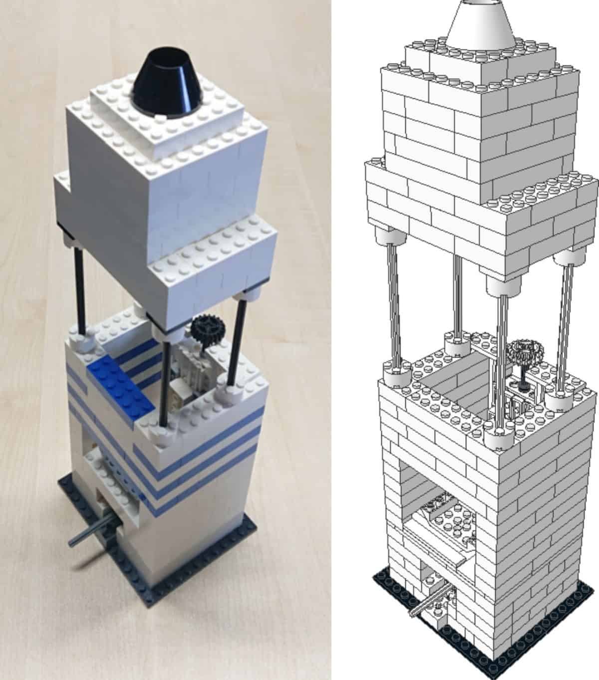
Simple design: the LEGO microscope (left) and a technical drawing of the instrument. The black eyepiece is at the top, and also visible is the black wheel that is used to adjust the position of the objective lens. (Courtesy: Timo Betz)
A fully functional modular microscope has been built using LEGO bricks and low-cost smartphone lenses. Designed by researchers, teachers and schoolchildren in Germany, the instrument is easy to build, yet it can resolve micrometre-sized objects such as individual living cells.
The idea for a LEGO microscope came to biophysicist Timo Betz (from the Universities of Göttingen and Münster) and his 10 year-old son one weekend while playing with LEGO – modular plastic building blocks that are beloved by children (and many adults) worldwide.
“My first reaction was that this is too hard, because of the precise movements and all the parts that are non-LEGO,” recalls Betz. “But my son came up with a series of great ideas on how to overcome the difficulties that I explained to him; he even figured out a way to use a LEGO light source to illuminate the samples.”
Junior author
By the end of the weekend, the father-son duo had built a prototype. However, even with assistance from Betz’s colleague Bart Vos, it took over a year to fine-tune the design, write construction plans and validate the instrument’s usefulness. Now, the trio has described the microscope in a paper published in The Biophysicist (with Betz’s son co-author, naturally).
The final design resembles a simple LEGO tower, but it hides some clever features (see figure). For example, to provide precise focus adjustment, the team had to “push the limits of the brick system” when designing the objective holder. This incorporates a gear rack with a gear worm screw that achieves approximately 3 mm of travel for every full rotation.
The only non-LEGO parts are the microscope’s lenses. For high magnification, these can be cannibalized from a low-cost replacement iPhone 5 camera module and then attached to a LEGO brick with transparent tape and a glass coverslip. Using these plastic iPhone lenses, the microscope can achieve 254× magnification.
Step-by-step building instructions
Most important to the creators, however, is that the microscope can be built and used in classrooms and homes around the world to learn about optics. To further this aim, they created build instructions and a step-by-step tutorial to guide people through construction while learning about the relevant optical characteristics of a microscope. Moreover, they enlisted eight 9–13-year-olds from a local Münster school who were tasked with building the microscope in an attempt to show how such a hands-on activity assists learning.
Given COVID-19 restrictions, the researchers could not see the children engage with the microscope activity in class. So instead, they created a kit that children could take home alongside a questionnaire to be filled out before and after playing with the kit. Answers revealed that the children’s knowledge of microscopy almost doubled after making the microscope.
Fun and informative activities
These children also conducted several experiments with their newly built microscope. These were suggested by the researchers as interesting, fun and informative activities. The experiments included watching crystallization in real-time as water evaporates from a thin film of salt solution; recording pigmentation changes to red onion epidermal cells exposed to an osmotic shock; and observing the movement of tiny swimming organism such as Artemia shrimp and water fleas.
This is not the first time a microscope has been made of LEGO. In 2013, a group of postgraduate students at the University of California, San Francisco designed a similar microscope called LegoScope. However, this instrument required a custom objective and 3D-printed parts.
Betz argues that the new LEGO microscope’s readily available and reusable parts “lower the barrier, especially for parents”, making it perfect for simply demonstrating the principles of microscopy.
“It was actually a lot of fun to develop this,” he says. “I just hope that children and their parents have a chance to realize here that even with simple tools, one can do amazing things.”
If you would like to build your own LEGO microscope, full instructions are available in English, German, Dutch and Spanish.