There is growing concern among some in the astronomy community that a mysterious flash of light that was seen coming from the direction of a distant galaxy may have simply been a glint from an artificial satellite orbiting Earth.
The target galaxy GN-z11 has a redshift of 11.09, which places it about 400 million years after the Big Bang. It is the earliest known galaxy and its place in cosmic history makes it an important object for astronomers to study because it can tell us about star-forming conditions in the early universe.
While observing the galaxy in December 2020 with the MOSFIRE spectrometer on the Keck I telescope in Hawaii, a team of astronomers led by Linhua Jiang of the Kavli Institute for Astronomy and Astrophysics at China’s Peking University got a surprise. MOSFIRE is a multi-slit infrared spectrometer and a flash of infrared light was seen in one of the slits targeting GN-z11. Describing the flash in Nature Astronomy, the astronomers conclude that it could have been redshifted light associated with a long-duration gamma-ray burst (GRB) from the ancient galaxy.
If their findings prove to be correct, it would be the first GRB to be seen in the early universe and could therefore harbour important information about the nature of early, massive stars and their environments.
Much closer to home
However, not everyone is convinced, with several researchers now claiming the flash was nothing more than light reflecting from a passing satellite. Although Jiang and team were careful to try and rule out satellites, some scientists believe they have underestimated the probability of detecting a satellite in the MOSFIRE observations.
“The satellite explanation is, at a minimum, thousands of times more likely than the GRB explanation,” says Charles Steinhardt of the Niels Bohr Institute at the University of Copenhagen.
Along with colleagues in Copenhagen and the University of Geneva, Steinhardt and colleagues set about looking to see whether previous MOSFIRE observations had also seen unusual flashes. From a search of 12,300 exposures, they found at least 27 other flashes, and attribute these to high-orbit satellites passing through the field of view.
“There are about 6000 known satellites and there are 41,253 degrees in the full sky,” says Steinhardt. He calculates the probability of a satellite passing over any given area of sky as being of the order of 10-3, or about one every seven square degrees. In comparison, he puts the probability of witnessing a GRB as between 10-8 and 10-10.
Around midnight
In response to these claims, Jiang’s team points out that low-Earth orbit (LEO) satellites are only visible around sunset and sunrise. The GN-z11 flash was seen around midnight, ruling out the satellites in LEO. A group led by Guy Nir, of the Weizmann Institute of Science in Israel, has suggested that the flash could have been a glint from a tumbling satellite in high orbit. Nir has himself seen these glints in his own observations, suggesting that they may have been confused in the past “with cosmic rays, fast-moving asteroids, or remain a mystery.”
Nir calculates that there could be 200 glints per day coinciding with the locations of galaxies on the sky. Jiang’s team counters this in two ways. One, that high-altitude or geosynchronous satellites would either produce an extended spectrum in the MOSFIRE observations as opposed to the compact spectrum of the GN-z11 flash, or not be visible in the direction of GZ-z11 from Mauna Kea. And two, it argues that Nir’s group has over-estimated the amount of sky taken up by galaxies, although Nir believes that even if his group has over-estimated, it is still not enough to make the GRB explanation more likely.
A potential culprit
However, not everything in Earth-orbit follows a predictable, deliberate orbit. A team led by Michał Michałowski at the Adam Mickiewicz University in Poland believes it has identified the object that could have caused the GN-z11 flash: an upper stage of an old Russian Proton rocket. Using Space-Track.org, the team showed that it passed as close as 18 arcseconds to GZ-z11 on the sky at the same time that MOSFIRE observed the flash.
Nir believes that Michałowski and colleagues have the right explanation, “There is certainly no need for a GRB, or even a glint off a rotating satellite, when there is a known piece of space junk right where the flash occurred.”
Crucially, points out Steinhardt, the GN-z11 flash displayed characteristics consistent with the solar spectrum, as though sunlight were being reflected. This is the same spectrum displayed by the 27 other MOSFIRE glint events that his team found in the archive data.
Laser beam could nudge space junk away
However, Jiang disagrees, telling Physics World that “we ruled out this possibility in our original analysis, and the line width [the length of the spectrum] is not consistent with what we observed.”
Counting the odds
Ultimately, says Steinhardt, it comes down to probability. If the probability of seeing a satellite in any given area of sky is on the order of 10-3, then for every thousand MOSFIRE observations, on average one satellite should be seen.
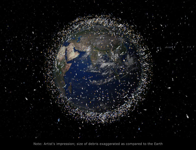
Satellites are being launched in greater numbers and keeping track of them all is becoming increasingly problematic. According to Nir, code can be written to remove known satellites from observations, and the scheduling of telescope time can be designed to avoid satellites, but incomplete tracking of satellites and space debris could lead to further contentious detections in the future.