The magneto-optical trap, or MOT, is the workhorse of cold-atom physics. Without this complex arrangement of laser beams and magnetic fields, the act of cooling atoms to just above absolute zero – and observing the quantum effects that emerge – would be nigh-on impossible. But before cold atoms can become part of a quantum sensor, quantum computer, or any other device that puts their quantum nature to practical use, this bulky old workhorse needs to become more like a pit pony: robust enough to do the job, yet much, much smaller.
Physicists at the US National Institute of Standards and Technology (NIST) have now taken an important step towards this goal. Led by William McGehee, the researchers used flat, lithographically-produced optics to create a MOT optical assembly just 15 cm long. While this is still too big for a practical cold-atom-based device, it is significantly smaller than the dinner-table-sized sprawl of ordinary MOTs, and a sign of how integrated photonics enables new designs. “Ultimately, what we’re trying to develop is something that is not just a small version of a laboratory experiment,” McGehee says. “You have to find different ways of doing the same things.”
Components working together
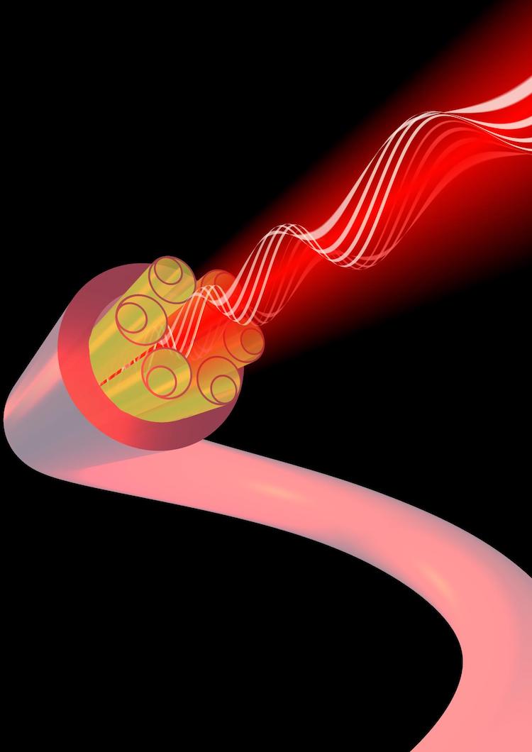
Like all MOTs, the NIST team’s device uses beams of precisely-tuned laser light to cool atoms within the region where the beams overlap. Unlike traditional MOTs, though, the beams in the new mini-MOT are generated, shaped and directed using planar optical elements. First, light from a laser is coupled into a nanophotonic waveguide on a photonic integrated circuit (PIC). Because the PIC’s output beam has a diameter of just 140 µm, and laser cooling is inefficient for beam sizes smaller than a few millimetres, the next task is to make the beam bigger. The NIST design does this with an optical metasurface that expands the beam and gives it a uniform intensity across its width after a distance of 15 cm. At this point, the beam strikes a diffraction grating and splits into the three pairs of equal-intensity but oppositely-directed beams required for laser cooling.
While none of these optical components is new, James McGilligan, a physicist at the University of Strathclyde in the UK who was not involved in the NIST study, is impressed by the way they work together. “The overlapping of these technologies is by no means straightforward,” he says. Although physicists have made progress on miniaturizing other elements of the MOT, McGilligan notes that the optical delivery system “remained a bulky and elusive component on the miniaturization checklist”. The NIST group’s planar-optics system is, he says, “a key step forward in cold-atom instrumentation”.
From mini to micro
To make a truly portable MOT, McGehee thinks the optical assembly will need to shrink still further, down to perhaps 1 cm. This would be tricky with the NIST team’s current setup, since it would mean that the laser beam’s intensity is no longer uniform in the region where it interacts with the atoms. McGehee notes that this would be “complicated” to fix. “With the appropriate simulation, you could figure out what you needed and build that, but it would take a couple of years,” he says.
Quantum sensor targets gravity and magnetism
Still, both he and McGilligan are confident that further shrinkage is on the cards. “It is highly likely that [this technology] will be adapted by research teams around the globe to aid in the miniaturization of their cold atom experiments,” McGilligan says. If combined with a compact vacuum system to isolate the atoms from their environment, he adds, the new optical assembly “has the potential to finally take cold-atom systems out of the lab environment and into chip-scale devices where their precision and accuracy can have the largest impact on our technological capabilities”.
The miniaturized optics are described in New Journal of Physics.
 The QUASAR™ GRID³ᴰ Image Distortion Analysis System is designed to evaluate MR and CT imaging data on Leksell Gamma Knife® platforms, including PERFEXION™ and ICON™.
 Joanne Tang is an application specialist at Modus QA. Having joined Modus QA after completing her BSc and MSc in medical biophysics at the University of Western Ontario, she is currently involved in customer application support of the QUASARTM GRID3D phantom.