With 80% of the world’s population at risk of water insecurity, there’s a clear need for a fast, simple and reliable way to test water for a wide range of contaminants. Julius Lucks and his team at Northwestern University believe they have the answer. Its name is ROSALIND – RNA output sensors activated by ligand induction.
In their recent study, published in Nature Biotechnology, Lucks and his team didn’t invent a sophisticated new technique. Instead, they took advantage of the elegant solutions that have evolved in nature to “taste” the water’s contents. This approach provides a straightforward positive or negative test for up to 17 different water contaminants, and it works in minutes.
Putting nature to a new use
The researchers’ approach is to take molecular machinery (including DNA, RNA and proteins) and reprogramme it for their own needs. In this case, the machinery comes from bacterial cells and uses the components that normally copy the DNA’s genetic code into a messenger form to be turned into the cell’s building blocks. Instead, the team re-engineered these molecular machines to work outside the cell for a new purpose.
Usually, DNA is copied when the code is needed by the cell. For this testing platform, that control is instead used to trigger DNA copying only when specific water contaminants are present. The water samples being tested are mixed in a tube pre-loaded with these molecular machines. When contaminants are present, the machines copy out a messenger form that lights up in the tube – giving off a signal that’s easily seen by the human eye. This allows ROSALIND to turn a contaminant we could never see into a light signal we can’t miss.
Importantly, the acronym ROSALIND also honours Rosalind Franklin, whose work was vital to the discovery of the DNA double-helix structure. According to Lucks, “her work essentially eventually enabled us to learn how to reprogram DNA to act in our technology”. Franklin’s 100th birthday would have been the same month that this research was published, he notes.
Taking technology where it is needed
Whilst current methods to test water require significant expertise and equipment – making them expensive – ROSALIND is different, according to Lucks. “We’re offering a technology that enables anyone to directly test their own water and know if they have contamination within minutes,” he explains. “It’s so simple to use that we can put it into the hands of the people who need it most.” The researchers have tested their method in the field, where it successfully matched the results of a gold standard laboratory test – flame atomic absorption spectroscopy – whilst being much quicker and more affordable.
Putting the technology into people’s hands doesn’t just mean making it cheaper, but also making it easier to use. Using the molecular machines outside of the cell allows them to be freeze dried, which keeps all of the components stable until they are needed. All the user needs to do is add a drop of water. Not only is this process simple, but the whole system is highly flexible. Currently, 17 different contaminants can be detected – including toxic metals, antibiotics and additives to personal care products – and ROSALIND could be easily updated to add more relevant contaminants in future.
Having previously tried to test water in his own home, Lucks was frustrated to find it a difficult or impossible task. “To ensure access to safe and clean drinking water, we need technologies that will allow easy monitoring of water quality,” he says. As the researchers move from creating their concept in the lab to developing this platform via a start-up company, Stemloop, Lucks is hoping that ROSALIND will soon bring water testing technology to the masses.
Graphene and other carbon materials are known to change their structure and even self-heal defects, but the processes involved in these atomic rearrangements often have high energy barriers and so shouldn’t occur under normal conditions. An international team of researchers in Korea, the UK, Japan, the US and France has now cleared up the mystery by showing that fast-moving carbon atoms catalyse many of the restructuring processes.
Graphene – a carbon sheet just one atomic layer thick – is an ideal system for studying defects because of its simple two-dimensional single-element structure. Until now, researchers typically explained the structural evolution of graphene defects via a mechanism known as a Stone-Thrower-Wales type bond rotation. This mechanism involves a change in the connectivity of atoms within the lattice, but it has a relatively large activation energy, making it “forbidden” without some form of assistance.
Using some of the best transmission electron microscopes available, researchers led by Alex Robertson of Oxford University and Kazu Suenaga of AIST Tsukuba found that so-called “mediator atoms” – carbon atoms that do not fit properly into the graphene lattice – act as catalysts to help bonds break and form. “The importance of these rapid, unseen ‘helpers’ has been previously underestimated because they move so fast and have been next-to-impossible to observe,” says co-team leader Christopher Ewels, a nanoscientist at the University of Nantes.
The breakthrough, Ewels says, came when they realized that these usually fast-moving atoms slow down when bound to existing defects in the lattice. “Our technique can be likened to those nature programmes on television in which cameramen often have a hard time filming some of the animals, which can be shy,” he explains. “They therefore sometimes set themselves up in hides next to a watering hole where they know the animals are sure to go, and that’s how they get their film footage.
“In our case, the mediator atoms shoot around too fast for our cameras. By instead imaging and watching pre-existing defects (the watering holes) that occasionally trap mediator atoms (which can linger near the defects for seconds, minutes or even hours), we are able to image them and observe how they influence defect restructuring.”
Since the images were blurred because of the speed of the processes involved, detailed theoretical modelling calculations were required to interpret them. These calculations were done at Seoul National University by Gun-Do Lee and his team and Ewels and his colleagues at the CNRS in Nantes.
The mediator atoms act as catalysts thanks to their reactive dangling bonds, Ewels explains. By bonding to defective sites and lowering the activation energy of reactions, they trigger a variety of bond-breaking and bond-forming processes. In some cases, the mediators become incorporated into the lattice, kicking out atoms that were already there and causing the ejected atoms to mediate further rearrangements (see image above). “These processes may occur in many other environments, from interstellar carbon chemistry to graphite moderators in nuclear reactors,” Ewels tells Physics World. The new work thus provides an important fundamental understanding of how graphene and related 2D materials structurally rearrange and repair themselves, he adds.
The researchers, who report their results in Science Advances, speculate that analogous mediator atom species may be present in other bulk materials. They now plan to explore this idea further. “We suspect similar process may underlie mechanical deformation processes of many 2D and 3D materials,” Ewels says.
The fundamental building blocks of human tooth enamel contain characteristic impurities that could contribute to their toughness, but also make teeth more vulnerable to decay. US researchers led by Derk Joester at Northwestern University made the discovery using two atomic-scale imaging techniques, which revealed distinctive distributions of impurities within the crystal structure of enamel. Their findings could lead to new ways to improve the health of our enamel, and to repair the damage created by tooth decay.
Coating the entire crown of the human tooth, enamel is an extremely hard substance that is well adapted to withstanding the wear and mechanical forces associated with chewing. Yet in many people, the material is broken down through tooth decay – which is one of the most widespread chronic diseases. So far, techniques for repairing and synthesizing new enamel have had limited success, largely due to limitations in our understanding of the material’s deeply hierarchical structure.
At the microscopic level, enamel is made up of rods composed of thousands of long, thin crystals with rectangular cross-sections – called crystallites. The rods are arranged in complex meshess. No more than 170 nm wide, the crystallite building blocks are primarily composed of the mineral hydroxyl-apatite. This has an orderly lattice containing calcium, phosphate, and hydroxyl ions. However, the crystallites are also known to possess cross-sectional variations in chemical composition on atomic scales.
Atom-probe tomography
To explore these variations in more detail, Joester’s team first carried out scanning transmission electron microscopy (STEM) on thin, ultra-cold crystallite cross-sections. The resulting images revealed that within the periodic lattice of hydroxyl-apatite, crystallites have central cores with slightly different compositions. These cores are sandwiched between two distinctive layers (see figure). Since these structures were damaged by STEM electron beams, Joester’s team also employed the technique of atom-probe tomography (APT), which can locate individual atoms at sub-nanometre resolutions. Within these images, they saw that crystallite cores contain high concentrations of sodium, fluoride, and carbonate ion impurities, and are flanked by two magnesium-rich layers.
Through simulations, combined with experiments involving X-ray diffraction, Joester and colleagues revealed that these chemical gradients create stress patterns in enamel, which could be partly responsible for the materials’s high mechanical resilience. However, the impurities could also increase the solubility of the material in acidic conditions, making it more vulnerable to decay. The team’s findings could offer important new insights into the crystal growth that takes place during enamel formation, and how it could be controlled artificially. If achieved, such techniques could lead to new treatments for people suffering from tooth decay, and to new ways of maintaining healthy enamel.
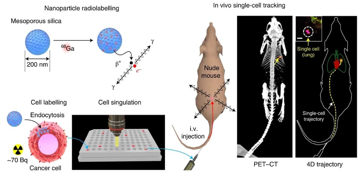
The CellGPS workflow: nanoparticles are used to concentrate 68Ga and then loaded into cells; isolated single cells are administered into mice; emitted gamma rays are captured using a small-animal PET scanner and processed to estimate the location of the moving cell in real time. (Courtesy: Nat. Biomed. Eng. 10.1038/s41551-020-0570-5)
The ability to accurately track the movement of cancer cells as they metastasize to other locations in the body could allow oncologists to better target therapies and start treatments earlier. To achieve this, researchers at Stanford University School of Medicine have developed CellGPS, an extremely sensitive and accurate method of single cell tracking using positron emission tomography (PET). They describe the technique and its feasibility to track single cell migration in laboratory mice in Nature Biomedical Engineering.
Applications for single cancer cell tracking using CellGPS include modelling and investigating the earliest stages of metastatic migration, including the circulation of tumour cell clusters in the blood, and the role of haemodynamic forces, cell adhesion, blood flow and biomechanical interactions. CellGPS could also be used to non-invasively measure the routes and kinetics of immune-cell mobilization in response to injury.
The CellGPS technique uses mesoporous silica nanoparticles (MSNs) to efficiently transfer radionuclides into cells for real-time cell tracking. The researchers labelled the nanoparticles with gallium 68 (68Ga), a radiometal with a 67-min half-life that’s commonly used for PET imaging of neuroendocrine tumours and metastatic prostate cancer. To enhance cell loading, they coated the nanoparticles with a lipid bilayer. MSNs with a diameter of 200 nm and a pore size of 4 nm carried 68Ga into live MDA-MB-231 breast cancer cells (a common model of breast cancer metastasis to the lungs), achieving a radioactivity level of nearly 100 Bq per cell.
The researchers initially assessed the metabolic activity of the cells after radiolabelling, noting that there was a small (5–15%) decrease in overall metabolic activity within 24 hours. They verified that DNA damage in cells, although detectable, did not result in significant biological differences in apoptosis, proliferation and migration.
After determining the optimal settings for acquiring PET images and data, the researchers injected approximately 100 breast cancer cells labelled with 68Ga-MSNs intravenously into mice. They used PET to confirm that the cancer cells arrested in the lungs. When they subsequently reduced the number of injected cells to just one, PET-CT showed a single focus of radiotracer accumulation in their lungs, appearing as a hot spot of approximately 1.7 mm diameter.
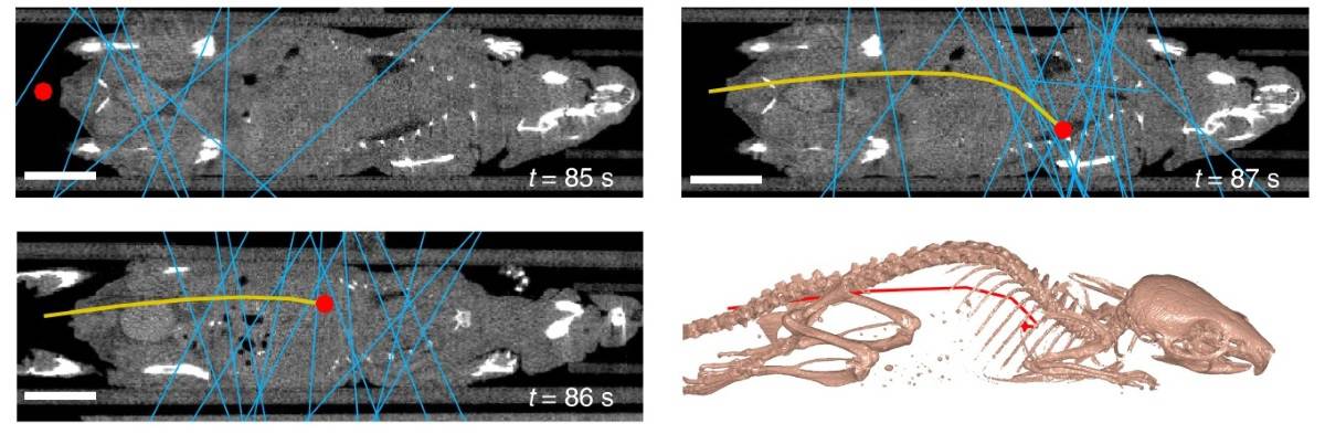
The researchers also used PET data to dynamically track the position of a moving cell in vivo. Using the list-mode data from the scanner, they computed the 3D position of a single cell injected into a tail vein through a butterfly catheter. The cell, traveling at a velocity of up to 50 mm/s, was tracked to the lungs within 3 s of intravenous injection. Its location matched the focal signal seen on conventional PET-CT imaging.
Dynamic tracking of a single cell in vivo at three time points. The blue lines represent detected coincidence events, the red dot is the estimated cell location, the yellow line is the reconstructed trajectory. Bottom-right: reconstructed trajectory with respect to the bony anatomy. (Courtesy: Nat. Biomed. Eng. 10.1038/s41551-020-0570-5)
To demonstrate that single cells can be imaged beyond the 67 min half-life of 68Ga, the researchers also tested the CellGPS workflow using zirconium-89 (89Zr, a positron-emitting radionuclide with a half-life of 78 hr). They intravenously injected single cells labelled with 89Zr-MSNs into two mice and imaged them over multiple days using PET. A single focus of uptake could be detected in the lungs for up to 48 hr and remained stable.
Guillem Pratx.
Principal investigator Guillem Pratx and colleagues state that the two most important findings of their research are that PET is sufficiently sensitive to image static single cells labelled with more than 20 Bq per cell of 68Ga, and that single migrating cells can be tracked dynamically directly from list-mode PET data.
“The CellGPS methodology could be extended to enable tracking of multiple cells simultaneously in the same individual,” they explain. “In the case of cellular therapies in which millions of cells are injected, it will be possible to label and track a small subset of cells using the CellGPS methodology.”
“We plan to push the technology to enable tracking of multiple single cells at the same time in the same individual by multiplexing the PET signals,” Pratx tells Physics World. “We also plan to investigate new applications where cell migration is key, primarily in the area of immune cell tracking for cancer immunotherapy applications, stem cell regenerative medicine, and more advanced models of cancer metastasis.”
A micron-scale “armour” that protects highly water-repellent nanostructures from damage has been developed by researchers in China and Finland. The new extra-durable coating could make it possible to employ these “superhydrophobic” surfaces on devices such as solar panels and vehicle windscreens that experience tough environmental conditions.
As their name suggests, superhydrophobic materials repel water extremely well. They owe this impressive ability to a thin layer of air that develops around nanometre-scale structures on their surface. By ensuring that droplets barely touch the solid part of the surface at all, the air layer effectively acts as a lubricant, allowing water droplets to roll off with near-zero friction.
These nanostructured surfaces are, however, mechanically fragile and can easily be wiped away. To address this drawback, a research team led by Xu Deng of the University of Electronic Science and Technology of China in Chengdu and Robin Ras of Finland’s Aalto University created a superhydrophobic surface containing structures at two different length scales: a nanoscale structure that is water repellent and a microscale one that provides durability.
The microstructure consists of an interconnected frame containing “pockets” of tiny inverted pyramids. Within these pyramids are the highly water-repellent and mechanically fragile nanostructures. The frame thus acts as a shield, preventing the nanostructure coating from being removed by abradants larger than the frame. “A finger, screwdriver or even sandpaper glides over these microstructures, leaving the nanostructures untouched, thereby preserving the surface’s attractive water-repellent feature,” Ras says.
A generic concept
Ras goes on to explain that the microscale armour could be made from almost any material. “It’s the interconnecting structure of the surface frame that makes it strong and rigid,” he says. “We made ours with pockets of different sizes, shapes and materials, including silicon, ceramics, metals and transparent glass. The beauty of this approach is that it is a generic concept that works for numerous materials, giving us the flexibility to design a wide range of durable waterproof surfaces.”
In one of their experiments, Ras, Deng and colleagues constructed a superhydrophobic surface from transparent glass and used it as a cover for solar cells. “One problem with solar cells is that when they are on rooftops or other difficult-to-reach places, they become gradually contaminated with sand and dust, which results in poorer efficiency over time,” Ras says. “For maintaining the high efficiency of current-generation solar panels, regular cleaning is needed, especially in places where there is a lot of dust in the air. Our durable water-repellent coating means that they get cleaned automatically by rain or dew.”
Withstanding difficult working environments
Other applications for the superhydrophobic surfaces include anti-icing and anti-fogging surfaces, he adds, or machines and vehicles that operate in harsh conditions. The surfaces may even have antimicrobial properties since bacteria, viruses and pathogens cannot cling to the nanostructures.
To prove that their surfaces could withstand difficult working environments, the researchers put them through a battery of tests: baking them at 100°C for several weeks; immersing them in highly corrosive liquids for hours; blasting them with high-pressure water jets; and exposing them to extreme humidity while bending them. They even rubbed them with sandpaper and cut them with a sharp steel blade. None of these treatments appeared to affect the surfaces and they continued to repel liquids as effectively as before.
The team members, who report their work in Nature, say they now plan to commercialize their coatings. “The benefits for the user are that they can keep surfaces spotless and clean, function in otherwise challenging conditions and reduce downtime by doing away with the need for constant cleaning,” Ras tells Physics World.
The Internet – our latest innovation in the history of written communication, which itself began in the fourth millennium BC – is now half a century old. It dates from the moment in October 1969 when a computer at the University of California, Los Angeles, accessed a computer at the Stanford Research Institute in Menlo Park. It spread slowly, funded by the US Department of Defense’s Advanced Research Projects Agency, as a project known as ARPANET. In 1970 it connected about 10 computers, and by 1977 there were around 100. Not until 1985 did the number reach 1000. From then, the numbers grew exponentially: from 10,000 by 1987, to 100,000 by 1990, to 1000,000 by 1993, following the invention of the World Wide Web by Tim Berners-Lee at CERN in 1989. Today, about half of the world’s population has access to the Internet, and the total number of Internet-connected devices is estimated at around 25–30 billion – that is, four devices for each person on the planet.
Thus, the Internet went from being “a closed community of US government staff and academic researchers, to a fringe community of nerds, to the bright shining hope of capitalism, to suddenly being critical infrastructure”, writes author and journalist James Ball. “And all of that happened with few of us paying attention and fewer still thinking even one step ahead.” His latest book, The System: Who Owns the Internet, and How It Owns Us, is a compelling historical analysis.
But it also serves as a clarion call to attention and action before the Internet degrades and becomes a source of division instead of its earlier source of unity – more like the profit-driven website Facebook than the non-profit Wikipedia, so to speak. “The joy and the wonder of the Internet is that everything is connected. Clearly, for anyone trying to look at how to fix its problems, the fact that everything is connected makes everything a lot harder, too.”
Ball is well qualified to write on this subject. He was a self-proclaimed teenage Internet nerd. Then, after graduating from a UK university in 2008, he became an investigative journalist, who has now spent more than a decade reporting on the Internet. In 2014 he was a member of the Guardian US team awarded a Pulitzer Prize for its coverage of US National Security Agency online surveillance, arising from the whistleblowing of Edward Snowden.
The System begins at the beginning in 1969 with a section, “The mechanics”, on the structure and operation of the Internet, divided into three chapters. “The architects” discusses the engineers and academics who invented the system. “The cable guys” covers the less glamorous fibre-optic companies that provide the physical links. “The custodians” profiles those who supervise its operation, notably ICANN, the Internet Corporation for Assigned Names and Numbers: until 2016 a US government agency, but now an international body that makes its income by taking a portion of the fees paid by people buying online addresses.
The second section of the book, “The money”, deals with the venture capitalists and chief executives responsible for starting and promoting Internet companies – some of whom have become billionaires – and the invasively inquisitive system of online advertising that earns these vast profits.
The third and final section, “The melee”, picks up on earlier discussions, covering topics such as government intelligence agencies and anonymous hackers (“The cyber warriors”), and the US Federal Communications Commission (“The rulemakers”). The final chapter, “The resistance”, focuses on activist groups, such as the San Francisco-based Electronic Frontier Foundation, trying to hold the Internet’s power in check.
Along the way, Ball includes many significant interviews with pioneers in the field, including Steve Crocker, a software engineer present at the first log-in in 1969; Gorän Marby, a Swedish regulator who took over the helm of ICANN in 2016; Brian O’Kelley, the former chief executive of huge online advertising company AppNexus; Cindy Cohn, a lawyer who directs the Electronic Frontier Foundation, and Wikipedia founder Jimmy Wales. But certain key players do not feature in interviews, notably the founders of Amazon, Facebook and Google, or, unsurprisingly, the intelligence chief in charge of the National Security Agency at the time of the Snowden revelations.
In discussing the need for future regulation of the Internet, Ball makes an illuminating historical comparison. The four best-known Internet companies – Amazon, Apple, Facebook and Google (now formally called Alphabet) – are “engines running along the railway lines set out by the Internet’s very structure”. And “just as railways could be used to create new monopolies in other industries – by charging different rates to different companies, or playing favourites” – so too can the Internet, when its structure has been harnessed by the big players.
Hard-fought competition laws introduced to regulate railway monopolies in the early decades of the 20th century, must now be reinvented to control the Internet for the good of the world, rather than just a group of billionaires
Following Tim Wu, a Columbia Law School professor and author of The Curse of Bigness: Antitrust in the New Gilded Age(2018), Ball argues that hard-fought competition laws introduced to regulate railway monopolies in the early decades of the 20th century, must now be reinvented to control the Internet for the good of the world, rather than just a group of billionaires. As Wu concludes: “If there is a sector more ripe for the reinvigoration of the big case tradition, I don’t know it.” Otherwise, the major Internet companies will continue to follow the example of Facebook’s 2014 purchase of WhatsApp for $19bn, as well as an analytics app that had allowed Facebook to spot and quash potential rivals. As Ball comments, “no-one was there to stop it, or even to question it. No-one ever is” – until now that is.
Maybe, he speculates in conclusion, the spur to democratic reform and regulation will be the current rise of authoritarian China’s technology giants and the consequent ebbing of the US dominance of the Internet after half a century. If so, ironically, a world-changing innovation originally triggered by military research may once again benefit from military considerations.
Blue skies, uncertain forecast: Karina Voggel at the Observatoire Astronomique de Strasbourg, France. (Courtesy: Karina Voggel)
Between March and May, France went through a two-month period of strict confinement. During this time, we were only allowed to leave our homes for essentials such as grocery shopping, and we needed to fill in a form every time explaining why we were out. Once home working became the default at my institute, I joined my partner (who is also an academic) in the south of France, as I did not want to be on my own for such a long period. Working from home was doable, although our small, open-plan apartment posed some challenges; for example, we had to share the kitchen table for our work and co-ordinate all our virtual meetings and talks so they wouldn’t interfere with each other.
Then, in early April, just before Easter, my partner came down with a heavy cough and fever. We immediately suspected COVID-19 and entered an even stricter quarantine, during which we did not set foot outside our apartment. At first, I had no symptoms, but within two days I was also not feeling well, and for the next week we were both severely ill. I am a young and healthy person, someone who runs marathons and has not a single medical condition or risk factor, yet COVID really wiped me out. It was scary how little energy we had left once the virus took over. We could not even find the energy to eat – a problem made worse by the fact that we entirely lost our senses of smell and taste.
After a week of symptoms, I began to improve. However, although I had cleared the acute illness, I was still severely weakened. It took me five or six weeks to get back to normal energy levels. Meanwhile, my partner continued to battle a fever and other severe symptoms for more than two weeks. This was a challenge, as it meant that I needed to nurse him back to health while I myself was far from well. Worse still, after an initial recovery, he developed a serious secondary bacterial infection and was hospitalized. This was an extremely hard time, as no visitors were allowed in the hospital and all I could do was hope he would get better. Luckily, he eventually did – albeit only after an ordeal that spanned more than two months.
Returning to a new normal
Now that our health permits it, we are back to working from home, and have been for more than a month. The Strasbourg Observatory has also implemented a limited in-person return to the office, as the number of active coronavirus cases in France has been low since May. Even so, there are many precautions. We are only allowed to have one person in an office at a time, and we organize this by filling in online sheets specifying the times when we are present. Upon entering the buildings, we are greeted with a hand-sanitizing station and provided with masks.
As astronomers, we don’t have labs, which makes social distancing easier. However, all our typical social gatherings and seminars are still prohibited. I think the return to in-person work has been managed very well, as it is possible to keep our distance from each other and people who want to continue working from home can do so. I think it is important to let individuals decide what they are comfortable with, depending on their own situation and risk assessment.
Lost opportunities
My biggest professional concern is how the crisis is going to impact my future in academia. I am in my second postdoc and I need to find a more permanent position soon. However, I lost several months of productivity to the virus and I am only on a two-year contract. I also had a busy March–May period planned, including several visits to the US and an invited conference talk. Obviously, none of that happened. It is really hard to showcase your science without conferences, especially in the critical early stage of your career. Although I have antibodies now and should be protected from the virus for a while, I do not plan to travel in the next few months or attend in-person meetings and conferences. Even later this year, organizing in-person events would put attendees at risk, especially since very few will have antibodies.
Another problem that is severely affecting astronomers is the cessation of operations at most telescopes, as support staff cannot get to and from the facilities. This means that anyone who was granted observing time during this period has not been able to do their observations – something that has impacted me personally. Because my partner is also an academic, my prospects in academia were already pretty bleak due to the “two-body problem”, and my impression is that COVID has worsened the outlook on all fronts. Many other junior scientists are in similar positions, and with universities in the US and UK issuing hiring freezes, I assume the pressure on the job market will only increase.
Words of caution
An even bigger concern, though, is that members of my family or other loved ones will get the virus, especially if there is a second wave in the autumn. When I see what it did to two healthy and fit people in their early 30s, I do not even want to imagine what it would do to people with medical conditions or other risk factors. So I urge everyone to be very careful. Someone else in our apartment building had the virus a bit earlier than we did, and it is likely that we caught it from them just by passing through the same hallway, as we never met them in person during that time. We observed all the strict confinement rules and took no risks, yet we were not able to avoid it – which shows that one can never be too careful.
But after surviving the virus, the silver lining is that it really aligns your perspective. In academia we can become obsessed with the workaholic mentality, trying to squeeze the maximum productivity out of our days. However, an experience like this teaches you that no matter what’s on your CV, all that really matters is that you and your loved ones are healthy and safe. My partner and I are alive, and now fully healthy, and so are all our family and friends. I have a newfound appreciation for that very simple fact.
Spider silk has been used by researchers in Taiwan to create tuneable lenses capable of focusing the shapes of incoming laser beams. The work was done by a team led by Cheng-Yang Liu at National Yang-Ming University, who developed the lenses by treating freshly extracted silk with drips of resin, under tightly controlled conditions. Their approach could offer significant improvements to existing biomedical imaging techniques.
Spider silk is well known for its useful mechanical properties including high values of elasticity, toughness and tensile strength. Yet the material also has fascinating optical properties. In recent studies, for example, researchers have noted its potential for photonics applications such as light guiding, imaging and sensing.
In their study, Liu’s team focussed on creating photonic nanojets (PNJs) using spider silk draglines – the strong spokes that form the basic structures of many spiderwebs. Typically, nanojets are formed when a transparent microscopic lens is illuminated on one side, creating an intense, focused point of light on the shadowed side through the effects of scattering, refraction, and diffraction.
Dripping resin
To create the lenses, the researchers first reeled stretches of dragline silk directly from long-legged cellar spiders. They carefully controlled the temperature, humidity, and reeling speed to ensure the fibres were smooth and uniform. They then dripped a resin onto the raw surface of the silk. Due to the wetting properties of the material, this caused it to distort into domes, whose shapes varied depending on how long they were left under the drip. Once the desired shapes were reached, the silk was placed in an ultraviolet oven for the resin to cure.
To produce PNJs from these lenses, Liu’s team first explored their optical properties through numerical simulations. These calculations revealed how the focal lengths, focusing sharpnesses, and peak focusing intensities of the beams each depended on the lens shapes. The calculations also showed that these properties could be finely tuned by controlling the time the domes spent under the resin drip. Through experiments, the researchers then showed that when visible laser beams were fired at their lenses, sharp PNJs could be focused far away from their flat, shadowed sides.
The mechanical properties of spider silk offer many advantages over the synthetic fibres currently used in tissue engineering. For example, the material is biocompatible and can be easily absorbed by the body. Because of this, Liu’s team believe the PNJs made possible with their lenses may lead to safe and reliable new ways to image biological tissues. If achieved, this could enable researchers to create large-area, high-resolution images of samples, and to scan tissues to a range of different depths.
In 2017 the US rapper B.o.B (real name Bobby Ray Simmons Jr) started a crowd-funding campaign to launch a satellite. The rapper, a vocal proponent of “flat-Earth theory”, wanted to seek evidence that our planet is a disc, not a globe. His aim was to raise $200,000 (later upped to $1m) on the GoFundMe website, with the aim of sending one or more craft into space to help him “find the curve” – the term that “flat-Earthers” use to describe the edge of our supposed disc-shaped planet.
The rapper’s quest may seem like a joke or publicity stunt. Indeed, there’s currently no evidence that B.o.B raised much money or got anywhere near his goal. However, in the last few years there has been an alarming rise in the number of people who, like B.o.B, believe in flat-Earth theories. There’s now an annual flat-Earth conference in the US – the most recent of which was attended by more than 600 people – while YouTube is full of videos purporting to provide evidence that the Earth is flat.
Physicists may mock the notion of a flat Earth, but the idea is gaining traction, particularly among people susceptible to other conspiracy theories. “They actually really do believe it,” says Lee McIntyre, a philosopher from Boston University and an expert in the phenomenon of science denial, whose books include Respecting Truth: Wilful Ignorance in the Internet Age (Routledge, 2015). McIntyre knows first-hand how sincerely flat-Earthers hold their views: he attended the 2018 Flat Earth International Conference in Denver, Colorado.
Asheley Landrum, a psychologist from Texas Tech University who was also at the Denver meeting, agrees that flat-Earthers are genuine, and not goofing around. “If they were [trolling], they are very good actors,” she says. “We talked to more than 90 members of the flat-Earth community and they’re all very sincere in their beliefs”. Lectures at the Denver event included “Talking to your family and friends about flat Earth”, “NASA and other space lies” and “14+ ways the Bible says flat Earth”.
Flat-Earth ideas are based on basic scientific misunderstandings that can be easily refuted. For most people, even those who have no physics background, the evidence for a spherical Earth is obvious. So we need to ask ourselves why these ideas still persist in the 21st century and, perhaps more importantly for the physics community: how exactly should we respond?
A circular history
The idea that the Earth is a sphere was all but settled by ancient Greek philosophers such as Aristotle (384–322 BC), who obtained empirical evidence after travelling to Egypt and seeing new constellations of stars. Eratosthenes, in the third century BC, became the first person to calculate the circumference of the Earth. Islamic scholars made further advanced measurements from about the 9th century AD onwards, while European navigators circled the Earth in the 16th century. Images from space were final proof, if any were needed.
Today’s flat-Earth believers are not, though, the first to doubt what seems unquestionable. The notion of a flat Earth initially resurfaced in the 1800s as a backlash to scientific progress, especially among those who wished to return to biblical literalism. Perhaps the most famous proponent was the British writer Samuel Rowbotham (1816–1884). He proposed the Earth is a flat immovable disc, centred at the North Pole, with Antarctica replaced by an ice wall at the disc’s outer boundary.
The International Flat Earth Research Society, which was set up in 1956 by Samuel Shenton, a signwriter living in Dover, UK, was regarded by many people as merely a symbol of British eccentricity – amusing and of little consequence. But in the early 2000s, with the Internet now a well-established vehicle for off-beat views, the idea began to bubble up again, mostly in the US. Discussions sprouted in online forums, the Flat Earth Society was relaunched in October 2009 and the annual flat-Earth conference began in earnest.
Round impact One manifestation of the Earth being spherical is that low-pressure weather systems rotate anti-clockwise in the northern hemisphere due to the Coriolis effect. (Courtesy: Jacques Descloitres, MODIS Rapid Response Team, NASA/GSFC)
As with any fringe movement there are disagreements and several different flat-Earth models exist to choose from. Some models propose that the Earth’s edges are surrounded by a wall of ice holding in the oceans. Others suggest our flat planet and its atmosphere are encased in a huge, hemispherical snow globe from which nothing can fall off the edges. To account for night and day, most flat-Earthers think the Sun moves in circles around the North Pole, with its light acting like a spotlight. The most recent “US model”, for example, suggests that the Sun and Moon are 50 km in diameter and circle the disc-shaped Earth at a height of 5500 km, with the stars above this on a rotating dome. Many flat-Earthers also reject gravity, with the “UK model” suggesting that the disc is itself accelerating up at 9.8 m/s2 to give the illusion of gravity.
Physicists will scoff at these ideas, but the worrying thing is that they are spreading rapidly and gaining proponents outside America too. “While they may not be as many [in Europe], they are as loud as their colleagues in the US,” says Jan Slegr, a physicist from the University of Hradec Králové in the Czech Republic, who in 2018 co-authored a paper outlining ways for teachers and others to confront outlandish flat-Earth ideas with physics (Phys. Educ.53 045014).
Such efforts are important. Alarming polling data by the firm Datafolha, for example, indicate that 7% of the Brazilian population – some 11 million people – believe that the Earth is flat. This shocking number has been attributed to a resurgent evangelical Christian church, but there are also signs that religious fundamentalism is spreading these ideas in Islamic countries too. In 2017 the website Jeune-Afrique reported that a geology student in Tunisia was intending to submit a PhD defending her work on a flat-Earth model.
Conspiracy mentality
It would be easy to dismiss flat-Earthers as simply being misguided due to a lack of education. While there are indications that those susceptible to such views have low levels of scientific literacy, Landrum at Texas Tech says that flat-Earthers aren’t necessarily people who don’t believe in science. “It’s not really an education thing,” she says. “It really is about distrusting authorities and institutions. [It] seems to be based on both a conspiracy mentality and a deeply held belief that looks a lot like religiosity but isn’t necessarily specifically tied to a religion”.
Landrum thinks this conspiracy mentality is linked to science denial and a susceptibility to believing deceptive claims on social media (Politics and the Life Sciences38 193). No longer the domain of a “foil-hat-wearing fringe”, she believes those with a conspiracy mentality have lost the ability to judge when to trust and when to be a sceptic. Their lack of trust in authority includes not just scientists but scientific bodies such as NASA, all of whom (they think) are part of a massive conspiracy to prevent the flat-Earth truth being revealed. “[They] view the world through this really dark filter where [they] assume that all authorities and institutions and corporations are just there to exploit you.”
McIntyre adds that the flat-Earthers he interacted with each believed a selection of conspiracy theories, including that governments control the weather and that chem-trails from aeroplanes consist of chemical or biological agents. “The only one I found that they all believed,” he says, “was that we hadn’t gone to the Moon. If you offer them back evidence, like the view of the Earth from the Moon, they say it’s fake.” Indeed, many flat-Earthers are more invested in the idea of a conspiracy than in providing a workable model of a flat Earth.
Nikk Effingham, a philosopher at the University of Birmingham in the UK who has met flat-Earthers at a London meet-up, says that we often don’t recognize the extent to which confidence in authority shapes our beliefs. “When we try and prove something like the Earth being round, because it’s a belief that we are so sure of, we under-play the justified role of authority in that,” he says. Most people are therefore comfortable accepting the world is a globe, even if they can’t immediately recount the scientific evidence.
Flat-Earthers seem to have a very low standard of evidence for what they want to believe but an impossibly high standard of evidence for what they don’t want to believe
Lee McIntyre, Boston University
But that’s not the case for those mired in a conspiracy mentality. What’s also clear is that the rise in flat-Earth beliefs has been fuelled by the Internet and YouTube videos in particular. “Almost everybody that we spoke to said that either they were directly exposed to flat Earth on YouTube or they were exposed to it via a family member who was exposed to it on YouTube,” says Landrum. Flat-Earth videos often present numerous arguments in rapid succession with what Landrum dubs “an illusion of fluency”.
Key to the videos’ success has also been the algorithms that serve them up to viewers of other conspiracy-related content. “The algorithms facilitate the normalizing of conspiracies and the feeling of a consensus within your community,” explains Landrum. “Flat Earth is just another example of that.” In 2019 YouTube did acknowledge the problem and said it would be tweaking its algorithm to reduce its recommendations of conspiracy-theory videos. But the fact remains that the videos are still on its platform.
Proving the Earth isn’t flat
It was McIntyre’s work on science denial that led him to the 2018 flat-Earth conference in Denver, where delegates spent time discussing the “evidence” and finer details of their theory as well as the supposed conspiracy that flat-Earthers believe is shielding their ideas from the wider public. “I thought that if I could understand how to push back against flat-Earthers, I could use the same techniques to fight back against climate-change deniers and anti-vaxxers,” he says. After all, their ideas are all generally based on fallacies and misunderstanding of science. “Some of the flat-Earthers know enough physics to throw around the vocabulary, but they don’t actually understand enough physics to be compelled by the truth.”
Time curve The plane of a free-swinging Foucault’s pendulum shifts with time, proving that the Earth is round. (Courtesy: iStock/Meinzahn)
But even without the visual confirmation of pictures taken from space, many of the arguments used by flat-Earth proponents can be easily dismissed with trigonometry or basic physical laws. A good place to start is with a Foucault’s pendulum, the device named after the French physicist Léon Foucault, who in 1851 famously hung a heavy 28 kg brass bob from a 67 m chain in the Panthéon in Paris. Such a pendulum, which can swing in any plane, changes direction during the course of a day, yielding direct evidence of the Earth’s rotation. (Though as Slegr points out, that hasn’t stopped some flat-Earthers claiming that all Foucault pendulums are fraudulent and that museums use magnetic coils to turn the plane of the pendulum’s rotation to make the Earth seem to rotate.)
Another phenomenon that proves the Earth is a spinning globe is the Coriolis force, which acts perpendicular to the direction of motion of a spinning mass. This force leads to cylones swirling clockwise in the southern hemisphere and counterclockwise in the northern hemisphere; through the direction of winds, it also impacts ocean currents. Long-range military snipers even have to make allowance for deflections caused by the Coriolis effect. Indeed, as Slegr points out, getting physics students to explain the evidence for a spherical spinning Earth is a great critical-thinking exercise.
But that deep critical thinking is what’s often missing among flat-Earthers. Consider photos of distant skylines, which are often wheeled out as “proof” that the Earth is flat. In his interactions with flat-Earth theorists, McIntyre was commonly shown a picture of Chicago, taken from Lake Michigan, in which the city’s skyscrapers are clearly visible despite being viewed from a distance of 100 km out. “Given the curvature of the Earth, you shouldn’t [in principle] be able to see the skyline of the city from that far out,” he says.
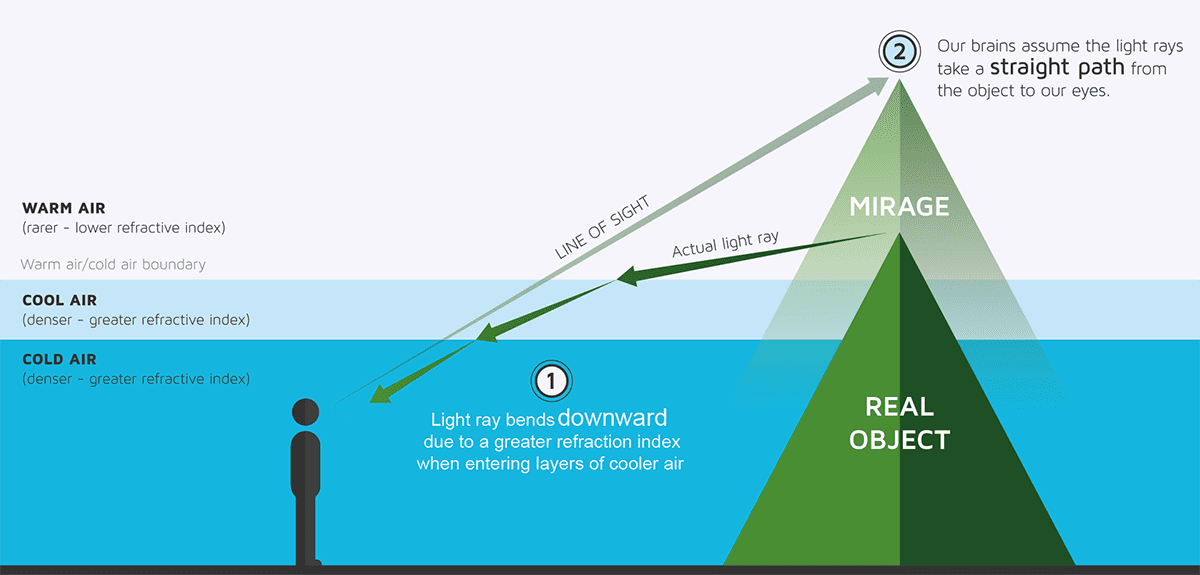
The reason the buildings are visible, as McIntyre knows, lies in the fact that air directly above the water’s surface is colder than the air higher up. This inverse temperature gradient means that light rays refract toward the colder, denser air, allowing an image of the reflected skyline, formed on the water below the horizon, to appear almost hovering above the horizon (figure 1). This notion can easily be verified by taking a photo even further away, where the “superior mirage” will disappear.
1 Why distant skyscrapers are visible despite the curvature of the Earth
This photo was taken from Mount Baldy in Indiana Dunes National Park on the south-east coast of Lake Michigan, roughly 60 km across the water from the city of Chicago, which lies on the opposite bank. At that distance, Chicago’s skyline should not be visible as the curvature of the Earth takes it beyond the horizon. The fact that the buildings are visible is in fact simply a mirage. Mirages are usually created when a cold, dense layer of air sits above a layer of warmer, less dense air, for example when the Sun beats down on a black road on a hot summer’s day. The warm ground heats the bottom few centimetres of air, refracting sunlight up to your eyes to create an “inferior mirage”. But if a layer of warm air sits above your line of sight, with a cool layer beneath, you get a “superior mirage”. Light bends down towards the denser air, but because our eyes assume the light has travelled in a straight line, the object appears higher than it is. The effect also explains why a far-off ship can be seen even though it might have dipped below the horizon. It can even make distant boats appear to float in the air.
(CC-BY-SA / Ludovica Lorenzelli, DensityDesign Research Lab)
But, as McIntyre found, this type of reasoning is unlikely to convince flat-Earthers. “They seem to have a very low standard of evidence for what they want to believe but an impossibly high standard of evidence for what they don’t want to believe.” One of their key experimental tools is a Nikon P900 camera with a ×83 optical zoom, in which flat-Earthers place an almost religious faith. Able to capture details not visible to the naked eye, they hope to use it to show that objects don’t disappear over the horizon but come back into view when examined at high enough resolution.
McIntyre described his frustrations with flat-Earthers in a paper last year in the American Journal of Physics (87 694), in which he challenged physicists to come up with simple, straightforward answers to refute the “evidence” for a flat Earth that could be understood by a general audience. Someone who rose to the bait was retired physicist Bruce Sherwood, who realized that “just citing the scientific facts is not going to convince anybody”. Instead, given that flat-Earthers place so much emphasis on naked-eye observations, he and colleague Derek Roff decided to create a navigable 3D computer simulation of a flat Earth to see how well it could replicate what we see.
Based on the US version of the flat-Earth model, it allows anyone to virtually roam a flat world. “Walking round in it, there were many things that show tremendous discrepancies,” says Sherwood. One of the major problems is the size and brightness of the Sun. In the flat-Earth model this varies by more than a factor of two from sunrise to midday, something we obviously do not see. The night sky also differs. In the northern hemisphere we see constellations rising in the east and arcing across the sky but in the flat-Earth model they would just circle at a constant height. “What [Sherwood] has created is something that’s much harder for [flat-Earth proponents] to laugh off, because it takes their own views seriously, [and] traces out the consequences,” says McIntyre. “I think that on this basis, other physicists can go out and help to push.”
Dangerous liaisons
From McIntyre’s perspective, flat-Earth conspiracies are a danger and need confronting. “Maybe 10 or 20 years ago, I would have said, just laugh at them, how much traction are they going to get? I no longer feel that way.” If these ideas are not challenged, he fears that as with supporters of “intelligent design”, proponents of a flat Earth will start running for US school boards, looking to push their ideas into the US education system. “The sort of reasoning that they use is infectious and if you don’t push back against them, it just gets worse and they’re able to recruit new members,” he warns.
But Effingham, who has also interacted with flat-Earthers on Facebook, wonders if physics is the place to start combating these conspiracy-based ideas. “I’m not saying that the perfect formula doesn’t have some kind of physics argument in it, but just turning on a YouTube video of physics lectures is not going to do it.” Instead, Effingham has tried to get flat-Earthers to understand that, by watching YouTube videos, they too are slavishly following an authority – not a scientific authority, but the authority of whoever is proposing the conspiracy theories they subscribe to.
Effingham also tries to point out their inconsistencies. “Every position they took required a different view of the conspiracy, and required the conspiracy to be bigger or smaller, and it was impossible to get a consistent conspiracy going that explained everything.” McIntyre, for example, recalls asking one flat-Earther why planes flying over Antarctica from, say, Chile to New Zealand don’t have to refuel, which they’d have to if the continent were (as they believe) an ice wall tens of thousands of kilometres long. He was simply told that planes can fly on one tank of fuel and refuelling planes could just be a giant hoax to stop us realizing that the Earth is flat.
Joining forces The rise in flat-Earth beliefs is partly due to the erosion of trust in science, which will require action on the part of scientists to rebuild. (Courtesy: iStock/prawny)
Landrum agrees the underlying problem is one of trust rather than physics. “We really should figure out as a scientific community, and as a society as a whole, how we can start building back trust in our organizations and institutions.” And she feels we need to do this face-to-face. “I don’t mean go yell at them on Twitter – that’s not engaging.” It’s also vital, she says, for scientists not to patronize flat-Earthers but to take questions seriously. That may seem like an excruciatingly painful process, but a necessary one, for people to gain trust in science as an institution again.
A level of sustained personal engagement can change minds. “It does work to push back against science deniers,” argues McIntyre, pointing to current NASA boss Jim Bridenstine. He was appointed by Donald Trump in 2018 and was known to have disputed climate change. “[But] once [he] became head of NASA, within a matter of two months or so, he changed his mind on climate change, and publicly said, ‘I was wrong’,” says McIntyre. The difference was that the evidence was presented to him by scientists he had grown to trust.
Oddly, Landrum says that many flat-Earthers may distrust scientists, but they are not against the scientific method. “The majority of them put a lot of faith, for lack of a better word, in science. There’s a lot of curiosity and a lot of scepticism and a lot of the really good qualities that make scientists.” But while the spirit of experimentation may be there, flat-Earthers are not always prepared to change their minds when their experiments fail. And that’s why McIntrye hopes some physicists might go with him to future flat-Earth conferences.
“I think that physicists need to be more involved,” he says. “There’s really no excuse for us to just sit back and laugh at them. Because while we’re laughing, they are recruiting people to believe these crazy things.”
Biomedical electronic implants, such as cardiac pacemakers, deep brain stimulators or spinal cord stimulators, enhance quality-of-life by providing diagnostics and treatments within the human body. Most of these devices, however, are powered by batteries. And once these batteries run down, patients must undergo invasive surgery to replace them.
To address this obstacle, researchers at Gwangju Institute of Science and Technology (GIST) propose a new way to provide sustainable electrical power within the body without the risks of surgical complications – via a concept called active photonic power transfer. They have developed a power transfer system comprising a skin-attachable light-source patch and a photovoltaic array integrated into a flexible medical implant.
The thin, flexible micro-LED patch emits photons that penetrate through tissue and are captured by the photovoltaic array, which then generates electrical energy to power the implanted device. The authors note that unlike other implanted photovoltaics that rely on ambient light, this system can generate power indoors or outdoors, day or night and regardless of covering by clothes.
“One of the greatest demands in biomedical electronic implants is to provide sustainable electrical power for extended healthy life without battery replacement surgeries,” explains lead author Jongho Lee. “Currently, a lack of a reliable source of power limits the functionality and performance of implant devices. If we can secure enough electrical power in our body, new types of medical implants with diverse functions and high performance can be developed.”
Power patch
Lee and colleagues created the light-source patch from an array of AlGaInP micro-LEDs powered by a conventional 3 V battery. The micro-LEDs emit light at 670 nm, which penetrates roughly 2.5 mm through tissue and can be converted into electricity by photovoltaic materials such as GaAs (which has a bandgap lower than the photon energy).
The skin-attachable light-emitting patch over mouse skin. (Courtesy: Juho Kim)
The researchers first demonstrated the patch’s thermal and mechanical compatibility with skin samples. Then to investigate its potential for power transfer, they tested the system in live mice. They fabricated a tiny (1 × 0.65 cm) flexible stimulator comprising a 4 × 8 array of GaAs photovoltaic devices, with a total active area of 11.1 mm2, and a small rechargeable microbattery. They implanted the stimulator through an incision in the animal’s skin and placed the light-source patch on the skin surface.
When the researchers switched on the light-source patch, the emitted photons successfully penetrated the animal’s tissues and wirelessly transferred power to the implanted stimulator. The power transmitted through the skin was 8.2 μW – greater than the required power consumption of the custom-built stimulator (roughly 2.3 μW). This power was enough to generate periodic stimulating pulses, as well as to charge the built-in microbattery. They note that even after turning off the light-source patch (at 106 min), the stimulator operated for an extra 90 min, powered by the charged battery.
The team also demonstrated that the implanted stimulator could regulate the heartrate of a mouse in bradycardia (a slower than normal heart rate) whilst wirelessly powered by the skin-attached patch. The stimulator generates pulses with a frequency of 3.3 Hz. As soon as the stimulator’s output lead wires were brought into contact with the right atrium and left ventricle, the animal’s heart started to beat regularly at around 3.3 Hz.
Commercial cardiac pacemakers require a power of between 1 and 10 μW, depending on operating mode. The researchers point out that that the power transferred by their custom-fabricated optoelectronic device (with an active photovoltaic area of 11.1 mm2) is already within this range, and that a pacemaker requiring 10 μW would need a minimum active area of 13.5 mm2.
“Currently, we are planning to develop the technology further to apply it for use in humans,” Lee tells Physics World. “We have to check or improve the mechanical reliability, long term biocompatibility and power density.”