Breakthrough Listen is an organization that searches for evidence of technological life by surveying one million nearby stars in the Milky Way as well as 100 nearby galaxies. The search has been going for five years and uses a wide range of radio and optical bands.
Needless to say, no definitive evidence of life has been found. In a bid to expand the scope of the search, Breakthrough Listen has come up with a catalogue of “Exotica”, which includes 700 distinct objects that will targeted in the search. The idea is to go beyond the usual targets of stars that could have Earth-like planets and look at “one of everything” in the observable universe.
The targets range from comets to galaxies as well as some of the most rare and violent celestial phenomena. Examples include Tabby’s Star, which has a bizarre dimming behaviour; and the interstellar object ’Oumuamua, which passed near Earth in 2017.
Cubestat scientist
Julie Sage runs experiments in space on cubesats and hosts a science news channel on YouTube – not bad for a 16-year-old. In this Via Satellite podcast, Sage talks about the challenges of being a young scientist, space exploration and communicating science to young people.
In the video below, Sage talks about Physics World‘s favourite crustacean: the mighty mantis shrimp.
Ocean Insight, a US manufacturer of specialist spectral systems, software and optical solutions, has high hopes for its Liquid Transmission Measurement System (LTMS), a spectroscopy-based unit for real-time, in-line optical monitoring of liquid concentration levels in a range of industrial plating, colouring and coating applications.
The modular system comprises a rugged optical sensing platform – spectrometer, control unit and machine-learning software – within a compact benchtop configuration that’s suitable for day-to-day deployment into harsh industrial environments. As such, the LTMS is able to generate high-precision colour and concentration measurements from the factory floor on a 24/7 basis, with flow-cell set-up and automatic fluid handling or built-in cuvette holder.
Right now, the LTMS is being sold directly into industry end-users and, once tailored to the customer’s requirements, comes ready to use “out of the box” in the manufacturing facility. “Alternatively, if the end-user has a preferred OEM integrator, the LTMS can be incorporated as part of their existing ‘smart factory system’, with or without fluid-handling capability,” explains Scott Scargle, director of strategic markets at Ocean Insight.
Getting the priorities right
Despite a myriad of potential LTMS applications, Ocean Insight’s near-term focus for the product is in the consumer electronics and food processing industries. Colour consistency of external parts – for Bluetooth speakers, MP3 players and the like – is a major pain-point in the consumer electronics supply chain, with batch-to-batch consistency of anodized aluminium being notoriously difficult to control.
Anodization is an electrolytic process to create a protective, anti-corrosion layer on the aluminium surface – a coating that can subsequently be dyed and decorated in a range of colours. Trouble is, anodization chemistry remains something of a black art, with colour materials and finishing (CMF) designers and contract manufacturers forced to work with wide colour tolerances and complex production processes.
Scott Scargle: “Our scientists and engineers create value in all sorts of ways.” (Courtesy: Ocean Insight)
If that’s the problem, the LTMS looks a lot like the answer. With its ability to measure solution concentrations in the anodization tank out on the factory floor, those CMF tolerances can actually get a lot tighter – which translates into enhanced repeatability, less scrap and streamlined product development cycles. “In short,” notes Scargle, “the LTMS gives the CMF designers a wider colour palette to choose from, while the contract manufacturers can make their parts with better consistency.”
In the food-processing industry, meanwhile, the LTMS is being lined up to support specialist coating applications on a range of foodstuffs. A case in point is the wax coatings that packing-houses apply to citrus fruits – typically to prolong the shelf-life and enhance the visual appeal of the fruit, also to act as a carrier for antifungal agents. “It’s a balancing act,” says Scargle. “The LTMS can be used to make sure concentration levels are within a certain tolerance to meet health and safety requirements, while high enough to maintain a level of effectiveness.”
Another use-case is the application of pesticides to food crops. Before a pesticide mix is applied in the field, for example, the LTMS is able to verify that the concentration falls within a window of acceptance to meet statutory safety guidelines. “Equally important,” adds Scargle, “the LTMS can be used to check the rinse water after the pesticide storage tank is cleaned, ensuring there are no unwanted residuals left behind that may end up being sprayed on crops later.”
Benefits and efficiencies
When it comes to customer upside, one of the big advantages of the LTMS is in-line deployment – a feature that makes it possible to more readily address quality issues as they occur. Consider an aluminium anodization coating tank, the electrolytes of which need to be analysed versus pass/fail criteria on a regular basis. Traditionally that testing has relied on expensive analytical equipment – for example, high-performance liquid chromatography (HPLC) – in a central laboratory (or more often an offsite testing facility for smaller contract manufacturers).
All of which is time-consuming, manually intensive and can mean significant process downtime while the results are pending. “The LTMS essentially eliminates that whole walk-to-lab workflow,” says Lu Luo, sales director at Ocean Insight and head of the company’s development facility in Rochester, New York. “It’s about the benefits of real-time, in-line diagnostics – the workflow and resource efficiencies plus greater process uptime – versus manually intensive and centralized lab testing.” What’s more, the LTMS workflow is such that scientific staff are no longer tied up on routine quality-control tasks. “It’s a portable unit and can be operated from a user-friendly interface by a technician,” Luo adds.
Lu Luo: “The LTMS essentially eliminates that whole walk-to-lab workflow.” (Courtesy: Ocean Insight)
Another notable feature of the LTMS is the innovative use of machine-learning technology. Each customer gets a tailored system that’s “trained” for their specific optical sensing applications versus a number of chemical solutions. “The machine-learning models provide a direct answer on solution concentrations for pass–fail manufacturing decisions – either on the technician’s display or sent digitally to a central manufacturing execution system,” says Luo.
It’s a neat innovation that comes with in-built scalability. “Customers can step-and-repeat for different liquids and coating solutions, training new machine-learning models as they go and growing their database of models,” Luo adds.
Ultimately, the LTMS represents a further signal of intent from Ocean Insight as it seeks to reinforce its credentials as the “Applied Spectral Knowledge Company” across the photonics value chain – optical components, systems, software and industrial solutions. “We are a global optical solutions company – helping research customers to unlock new scientific insights and industry customers to deliver new commercial opportunities,” notes Scargle.
“Our scientists and engineers create value in all sorts of ways,” he concludes, “whether that’s feasibility testing through our lab services group; machine-learning advances from our Ocean Intelligence group; or custom product development in our solutions engineering team.”
Computer simulations and experiments with algae provided the team with information about the jamming and unjamming of cells at high densities. This research could lead to a better understanding of the differences between healthy and cancerous cells in human tissue.
The self-assembly of cells into tissues sits firmly at the interface of physics and biology. Cells are complex biological systems that sense changes in their environment and communicate with other cells, but they can also exhibit self-organization that is driven purely by thermodynamics.
Out of equilibrium
The motion of a cell (its motility) requires a constant input of energy and therefore motile cells exist out of equilibrium. Such systems of motile elements belong to the category of active matter. Startling dynamic behaviours have been observed in active systems, including motility induced phase separation and accumulation at solid boundaries. Most active matter research focuses on self-propulsion, but collective dynamics are also observed in systems of self-rotating particles.
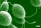
C. reinhardtii is a single celled alga with a light sensitive eyespot, which scans its environment by rotating as it swims. Ravazzano and colleagues investigated the extent to which the collective behaviour of the algae can be reproduced by computer simulations of rotating disks. They then showed in simulation that rotation can induce jamming–unjamming transitions at high volume fractions.
Self- assembly of active particles is generally observed at high densities where interactions between the particles become significant. However, algae tend to aggregate in response to stress and so may behave collectively in dilute suspensions. The addition of sodium chloride to the medium induces the algae to form rotating clusters. This was also observed in simulations of disks subject to active torque, but only in the presence of adhesion between the disks. This indicates the significance of the stress induced aggregation and the active torque in the motion of the algae.
Active torque and jamming
The jamming transition occurs when, without crystallizing, a system of particles becomes so closely packed that it behaves as a solid. This phenomenon, which has been observed in living cells, generally occurs when the density of the system is increased.
C. reinhardtii cells do not exhibit jamming at high density, because when they become crowded, their motility increases. Ravazzano and colleagues suggest that the active rotation of the algae increases in response to crowding, which opposes jamming.
This hypothesis was tested in simulation by preparing the disks with zero propulsion at the passive jamming volume fraction and increasing the rotation. A transition from a jammed to an unjammed state at a threshold torque was observed. Though more research on the response of the algae is needed, it is evidently possible for active torque to trigger unjamming.
The addition of self-propulsion to the model complicates the self-assembly behaviour. As the torque is increased, the system first jams and then unjams. The explanation offered by the Milan team is that a crossover between propulsion and rotation determines the behaviour of the algae.
At low torques, the self-propelled particles avoid jamming because they move coherently, but the rotation randomizes their motion and they undergo jamming as it is increased. At higher torques, the rotation dominates over self- propulsion and the unjamming transition is observed as before.
The outlook
In their paper describing the study, the researchers highlight the similarities between the jamming transition of the disks and the change from a solid to liquid like state observed in healthy versus cancerous cells. They also remark on the “possible role for rotations in collective cell migration,” and give the observed formation of vortices in confined epithelial cells as an example.
Laboratory sessions that are designed to emphasize and teach experimentation skills lead to gender imbalances among students, with men and women taking on different roles. That is according to a study by researchers in the US, which found no significant differences in role division in more traditional labs that attempt to reinforce lecture content (Phys. Rev. Phys. Educ. Res.16 010129).
In their study, the researchers observed students enrolled in a mechanics class as part of a calculus-based physics course. Students attended the same lectures, but two different lab courses: one set up to reinforce knowledge introduced elsewhere on the course, and the other designed to teach experimentation skills. In the traditional lab sessions students were given instructions of experimental procedures and worksheets to complete. However, the experimentation lab students were expected to make decisions about the design and analysis of their experiments. The study included 143 students: 109 men, 32 women and two students who did not disclose their gender.
To assess the roles students took, the researchers observed the lab sessions at five-minute intervals, noting what each student was handling. The choice was a desktop computer, lab equipment, a personal laptop, a notebook or “other” – a broad category designed to ensure all student time was accounted for. In the inquiry-led labs men systematically handled the equipment more, while video analysis showed that women spent about twice as much time as men on laptops.
The study also revealed that students rarely discussed the roles they would assume, and there were no instances of students being explicitly allocated roles by other group members or lab instructors. The researchers suggest that the differences they saw are driven by subtle verbal and non-verbal interactions at the individual level that accumulate to create class-level patterns.
Lead author Natasha Holmes from Cornell University says that it is unclear if there are negative implications from this finding, but it needs to be explored. It could, she explains, reinforce gender stereotypes, which is concerning in a field like physics that has significant diversity issues. “If looking around the room you see that all the male students are handling the equipment what sort of message is that sending?” Holmes told Physics World. “There is a risk that there are implications down the line for students’ feelings of whether they belong.”
Holmes and her colleagues have previously highlighted the benefits of inquiry-led labs, showing that they boost student engagement. Indeed, she says there is “overwhelming evidence that traditional labs are unproductive”. The team says that this latest work does not distract from this, but shows that active steps need to be taken when redesigning lab courses to ensure gendered roles are not reinforced. In particular, inquiry-led labs, with the work structure removed, attention needs to be paid to the group dynamics.
Reporting their findings in Nanoletters, the research teams have jointly demonstrated a unique approach that might stop SARS-CoV-2, the COVID-19 virus, from infecting human cells.
Cellular decoys
The researchers have developed two types of cellular nanosponge that bind to SARS-CoV-2 before it can enter human cells. These nanosponges consist of polymeric nanoparticles covered with membranes from one of the two human cell types that are known to be invaded by the virus, namely lung epithelial cells and macrophages. This means that the surfaces of the nanoparticles feature the exact same receptors and proteins that the virus would normally bind to. In a sense, the nanosponges act as cellular decoys by mimicking human cells and therefore offering alternative binding sites to SARS-CoV-2.
Schematic of the working principle of the nanosponges. Polymeric nanoparticles are cloaked with human cell membranes so that SARS-CoV-2 is surrounded by the nanosponges before it can enter any host cell. (Courtesy: ACS Nanoletters 10.1021/acs.nanolett.0c02278)
The key idea is to deploy many of these nanosponges, so that they can surround the virus, effectively soaking it up before it can enter any host cell. And if the virus cannot enter a cell, it cannot replicate and is neutralized, therefore preventing the spread of the infection to the rest of the body.
The researchers first tested the preliminary safety of the fabricated nanosponges in mouse models. They found that a 3-day exposure to both types of nanosponge did not damage the animals’ lung tissue. In addition, blood markers as well as platelets, red and white blood cell counts were not negatively affected, therefore ruling out any short-term toxic effects.
Next, the team tested the efficacy of the nanosponges in vitro, by exposing monkey cell cultures to the virus in the presence of the nanosponges. Both types of nanosponge reduced the infectivity of SARS-CoV-2 in a dose-dependent manner, with the best result leading to a reduction of up to 90% compared with non-treated cultures.
A treatment for COVID-19?
These nanosponges might represent a unique solution to the race for a treatment of COVID-19. They are relatively easy to fabricate, and come with the added benefit of being insensitive to viral mutations and potentially applicable to other types of coronavirus.
“Because the cellular nanosponges are not targeting a specific virus, as other drugs normally do, these sponges are potentially agnostic to viral mutations and viral species. As long as the virus still attacks human lung epithelial cells or human macrophages, these nanosponges can bind to the virus and neutralize it,” says senior author Liangfang Zhang.
Zhang adds that the team is now focused on testing the efficacy of these nanosponges in appropriate animal models of COVID-19. If the data are positive, they will advance the nanosponges towards human clinical trials.
This episode of the Physics World Weekly podcast features three physicists at McMaster University in Canada. They responded to COVID-19 restrictions on in-person learning by mailing out simple equipment so their students could do undergraduate lab experiments at home. Instead of just getting by with the new arrangements, Sara Cormier, Adam Fortais and Kari Dalnoki-Veress were delighted to find that their students learned new skills working at home and often did experiments with family members – giving physics a wider audience in the community.
Dalnoki-Veress also explains how he and his co-organizers of the Soft Matter Canada Symposium scrambled to put the event online after it was cancelled earlier this month. Again, much to the organizers’ delight, the Zoom-based event grew from a symposium into the much larger Soft Matter Canada Conference as more and more people signed up to participate. Indeed, the organizers now hope to hold the online event several times a year.
You can contact Dalnoki-Veress at kdalnokiveress@gmail.com to find out about the next Soft Matter event.
Optical fluorescence scans of excised cervical tissue, with the redox ratio of coenzymes shown in distinctive colours. Sections of healthy tissue, low-grade cancer and high-grade cancer show distinctive patterns that can be automatically evaluated for a rapid diagnostic result. (Courtesy: Dimitra Pouli, Tufts University)
A label-free fluorescence microscopy technique can detect the metabolic and structural signatures of cancer in epithelial tissue even before it develops. A team in the US and Spain used two-photon excitation fluorescence (TPEF) microscopy to map the presence of two metabolism-related coenzymes in cervical biopsy samples. They found that the distribution and ratio of the coenzymes vary with depth in a way that picks out changes in morphology associated with precancerous lesions. The researchers say that the method may ultimately be incorporated into routine screening to identify cancers early on, when treatment is most effective.
When a cancer has grown to the point at which it can be diagnosed from the symptoms that it causes, it might already be too late for successful treatment. Far better is to catch the cancer before it develops, but this means spotting the subtle biochemical changes that indicate that a cell’s metabolism has ramped up ready for proliferation.
While it is possible to measure these changes non-invasively, current techniques – nuclear medicine or MRI, for example – require dedicated imaging facilities and the injection of tracers. The alternative is to take biopsies for laboratory analysis, but tissue sampling sites are still typically chosen using low-specificity and sub-optimal-sensitivity visual means, and the procedure can cause pain and other side effects.
In conventional fluorescence microscopy, specific molecules emit visible light when they are excited by higher-frequency photons. In TPEF microscopy, the target molecules emit after absorbing two relatively low-energy photons that, individually, could not trigger such fluorescence. This technique has an advantage for physiological applications because the two near-infrared (NIR) photons that excite the molecules are scattered less by tissue than higher-frequency light, allowing cells beneath the tissue surface to be imaged. The depth at which the NIR beam is focused can also be varied, giving a depth-resolved map of fluorophore distribution.
Georgakoudi and her colleagues exploited these advantages to quantify in samples of cervical epithelium the presence of two molecules: the reduced form of nicotinamide adenine dinucleotide (NAD(P)H) and flavin adenine dinucleotide (FAD).
“These enzymes play an important role in several of the pathways involved in producing energy and synthesizing molecules that the cell needs to survive,” explains Georgakoudi. “The balance of the pathways that the cell utilizes to do this often changes as it becomes cancerous.”
The relative quantities of FAD and NAD(P)H therefore give a window onto cancer-related metabolic changes, but they also provide a picture of how cells are structured. Because these molecules are concentrated in mitochondria (subcellular structures found in the cells’ cytoplasm but not in their nuclei and borders), their presence can be used to infer the cytoplasmic-to-nuclear ratio – the ratio of the size of the cell nucleus to the overall cell size – and the degree to which mitochondria cluster together. In healthy epithelia, both of these properties vary significantly with depth. “That is one of the markers that the cells are differentiating (maturing) as they are normally expected to do, as we move from the deeper cell layers of the epithelium to the surface,” says Georgakoudi.
In precancerous lesions, in contrast, this normal differentiation process is disrupted, and the epithelial cells display no such depth-dependent variation. The researchers found that this lack of differentiation was detectable by TPEF microscopy. Moreover, they found that the process of tissue classification could be automated by combining cell morphology and mitochondrial organization measurements with the FAD:(FAD+NAD(P)H) ratio, which also displayed less variability with depth in precancerous tissues.
Although Georgakoudi and colleagues studied cervical epithelial tissues specifically, in which cancer is usually caused by a particular strain of human papillomavirus, they say that the same cell-morphological and biochemical patterns should be present in many epithelial cancers. To apply the technique in the clinic, however, will require advances in the delivery of high-energy pulses and improvements in image acquisition speed.
“We are starting this summer a project to develop an instrument that will enable us to test this technique in humans in the clinic within two years,” says Georgakoudi. “It will of course take a couple of years at least to go through initial testing and optimization, but there is no question that the ability to assess subtle metabolic changes in human tissues in vivo will enable new insights into the process of cancer development so that we can detect and treat it more effectively.”
There’s much ado about next to nothing, it seems, in the rarefied world of ultrahigh-vacuum (UHV) systems. Operating at pressures of 10–7 Pa and lower, UHV provides a core enabling technology for all manner of surface-science studies that rely on the interaction of photon, electron or ion beams to probe the physical and chemical properties of sample surfaces – among them X-ray photoelectron spectroscopy (XPS), low-energy electron diffraction (LEED) and secondary-ion mass spectroscopy (SIMS). At the same time, UHV conditions ensure that researchers are able to study a chemically clean sample surface free from unwanted adsorbates – also a must-have requirement for thin-film growth and preparation techniques such as molecular beam epitaxy (MBE) and UHV physical vapour deposition (PVD).
Within the UHV environment, the mechanical manipulation, positioning and preparation of the sample represent a complex engineering challenge, typically requiring analytical stages that can deliver a combination of precise linear motion along three axes (xyz) as well as rotation around one or two of those axes (polar and azimuthal). A case in point is the MultiCentre family of analytical stages from UHV Design , a specialist UK developer of UHV motion and heating products. These configurable stages offer scientific users up to five axes of motion alongside options for additional control and testing of the sample, including motorization, temperature measurement, the ability to apply a voltage (sample biasing), heating to 1200 °C, and liquid-nitrogen or liquid-helium cooling.
MultiCentre applications span fundamental research in materials science, particularly at surfaces, as well as thin-film process development and quality control.
Nick Clark, UHV Design
As such, the MultiCentres are an essential building block of analytical experimental techniques for chemical and structural analysis in the fields of thin-film fabrication, semiconductor science, catalysis and nanotechnology, amongst others. “MultiCentre applications span fundamental research in materials science, particularly at surfaces, as well as thin-film process development and quality control,” explains Nick Clark, chief scientist at UHV Design. “The stages and associated accessories are a complete solution for sample manipulation and transfer as well as preparation ahead of analysis – including removal of surface contaminants, crystallization and thin-film deposition.”
Centre stage
The MultiCentre range comprises two main product lines: the general-purpose XL-T series, a compact, single-bellows stage that’s designed specifically for surface-science chambers where space is at a premium; and the XL-R series, a dual-bellows stage with a secondary shaft support and z-axis travel up to 1000 mm – a higher-end specification for surface analytical and synchrotron end-station applications that require longer travel and enhanced stability.
One of the key features of the MultiCentre stages is the motorization of any or all axes of motion, with the emphasis on user-friendly motor assembly and disassembly. “Our motors are neat, compact and easy to remove – a big advantage when it comes to bake-out and maintenance of the UHV chamber and subsystems,” Clark explains. The approach to motorization also plays out in terms of vacuum integrity and mechanical reliability.
Nick Clark: the focus on user-friendly product design underpins the MultiCentre family of analytical stages. (Courtesy: UHV Design)
Take the four- and five-axis stages, in which the polar and azimuthal axes of rotation exploit magnetically coupled drives. “The use of magnetically coupled drives is fundamentally more reliable because you’re not twisting a bellows around to give you the rotation,” Clark adds. “Ultimately, that means less chance of a vacuum leak, while the drive components are less prone to mechanical damage.”
That focus on user-friendly product design underpins the MultiCentre offering. Many traditional stage designs, for example, require the services to be coiled around the shaft. This increases the swept radius of the stage, creates potential snagging areas and, after multiple cycles, the cooling pipes can fatigue to the point of failure. In contrast, the XL-T series uses the 65 mm internal-diameter bellows bore to route all services – including liquid-nitrogen cooling coils – to yield an uncluttered, compact design at the sample stage.
“The XL-T configuration significantly reduces the swept radius and eliminates the cycling stress on the cooling system whilst freeing up space for sources and detectors to get in close to the sample on multitechnique chambers,” notes Lukasz Rybacki, senior mechanical design engineer at UHV Design. “What’s more, the option to extend functionality when required – such as the addition of extra sample parking stages – provides an economic route to future-proofing your purchase.”
Hot stuff
For the end-user, this “scalability by design” yields significant upside. Customers can choose a four-axis MultiCentre configuration for polar rotation only, or the five-axis system if azimuthal rotation is also required. The same goes for heating and cooling services. If sample heating is needed, options for resistive heating (to 900 °C) and e-beam heating (to 1200 °C) are available.
The former employs a self-supporting tantalum foil heater (for minimum outgassing and a large ratio of heated to open surface area to ensure heater longevity). The foil is also yttria-coated to provide additional robustness in oxidizing atmospheres and for protection in the event of an accidental vent. To upgrade to e-beam heating, which gives users another 300 °C of heating, simply requires a change of power supply unit. An innovative liquid-nitrogen cryomodule provides sample cooling down to –165 °C.
MultiCentres provide continuous azimuthal rotation and temperature measurement even when cooling with liquid nitrogen and when heating to 1200 °C.
Lukasz Rybacki, UHV Design
“In addition,” says Rybacki, “the MultiCentres are unique in their ability to provide continuous azimuthal rotation and temperature measurement even when cooling with liquid nitrogen and when heating to 1200 °C. That functionality can help in the uniform growth and crystallization of thin films and the uniform removal of material during depth-profiling experiments.”
Finally, all XL-T and XL-R MultiCentre stages can be configured to accept the most common surface analysis sample holders, including pucks, flags and ESCA stubs. A lot of attention has also gone into the design of the sample holder to make sure it is virtually nonmagnetic and therefore compatible with low-energy analysis techniques – such as angle-resolved photoelectron spectroscopy (ARPES) – which are very sensitive to magnetism.
MultiCentre stages: versatile by design
UHV Design’s MultiCentre analysis stages are “a complete solution” for surface-science studies, combining sample manipulation, transfer and preparation – including removal of surface contaminants, crystallization and thin-film deposition. A selection of leading-edge applications is highlighted below.
A four-axis MultiCentre XL-T stage with heating, liquid-nitrogen cooling and deposition shielding supports an advanced UHV system capability at the Centre for Designer Quantum Materials, University of St Andrews, UK. Phil King and colleagues are investigating the electronic structure and many-body interactions of quantum materials using electron spectroscopy as well as creating novel designer quantum materials via atomic layer-by-layer growth. Using the XL-T, the researchers are able to prepare spin targets for investigation by their spin- and angle-resolved photoelectron spectroscopy (SARPES) system.
A Chinese research collaboration, headed up by Qing Huan and Kui Jin at the Institute of Physics, Chinese Academy of Sciences, Beijing, has developed a custom UHV facility to accelerate advanced materials discovery. Comprising a combinatorial laser MBE system and an in-situ scanning tunnelling microscope (STM), the six-chamber UHV system provides high-throughput film synthesis techniques and subsequent rapid characterization of surface morphology and electronic states of the resulting combinatorial thin films. The preparation chamber is installed with a customized MultiCentre analytical stage and an ion gun, allowing cycles of ion bombardment and annealing of the sample (up to 1200 °C).
Two highly customized MultiCentre stages are being put to use by the Nanoscale Processes and Measurements Group at the US National Institute of Standards and Technology (NIST) in Gaithersburg, Maryland. A high-temperature XL-T stage (operating at up to 1200 °C and tilting along a non-standard axis) supports crystalline thin-film growth analysis using reflection high-energy diffraction (RHEED), while a high-voltage (20 kV) XL-T stage is being applied in field-ion microscopy (FIM) imaging and preparation of scanning tips for UHV, cryogenic, high-magnetic-field STM studies.
A distant object that could be smallest known black hole, or the largest known neutron star, has been spotted by the LIGO–Virgo gravitational-wave detectors. The 2.6 solar-mass object appears to have merged with a 23 solar-mass black hole, creating gravitational waves that were detected here on Earth in August 2019. Unlike a previously observed merger between two neutron stars, no electromagnetic signal was observed. At nearly 9:1, the ratio of masses of the two objects is the greatest ever detected by LIGO–Virgo.
LIGO and Virgo are three huge interferometers – two in the US and one in Italy – that have detected gravitational waves from the mergers of black holes and neutron stars for nearly five years.
Neutron stars and stellar black holes are the final stages of evolution for large stars – with black holes being more massive than neutron stars. In theory, the maximum mass of a neutron star is about 2.1 solar masses. However, there is some indirect evidence that more massive neutron stars could exist. There is little evidence for the existence of black holes smaller than about 5 solar masses, leading to a mass gap in our observations of these compact objects.
Record breaking
What is intriguing about the August 2019 merger – dubbed GW190814 – is the mass of the smaller object, which appears to fall within this gap. “Whether any objects exist in the mass gap has been an ongoing mystery in astrophysics for decades,” says Charlie Hoy of the UK’s Cardiff University, who played a key role in analysing data from the detection and writing the paper that describes the observation, which has been published in The Astrophysical Journal Letters. “What we still don’t know is whether this object is the heaviest known neutron star or the lightest known black hole, but we do know that either way it breaks a record.”
LIGO Scientific Collaboration spokesperson Patrick Brady at the University of Wisconsin, Milwaukee adds, “This is going to change how scientists talk about neutron stars and black holes. The mass gap may in fact not exist at all but may have been due to limitations in observational capabilities. Time and more observations will tell.”
According to LIGO team member Vicky Kalogera of Northwestern University in the US, the large mass ratio will encourage astrophysicists to rethink models of how such binary compact objects form. “It’s a challenge for current theoretical models to form merging pairs of compact objects with such a large mass ratio in which the low-mass partner resides in the mass gap,” she says.
I think of Pac-Man eating a little dot
Vicky Kalogera
Unlike the merger of two neutron stars that was observed by LIGO–Virgo in 2017, no electromagnetic radiation was detected with the gravitational waves of GW190814. According to LIGO-Virgo scientists there are three possible explanations for this. One is the great distance to where the merger occurred – 800 million light-years – which is about six times the distance as the 2017 neutron-star merger. Another possibility is that both objects were black holes, and no electromagnetic radiation has been detected from any black-hole mergers spotted by LIGO–Virgo. A third possible explanation is that the neutron star was “swallowed whole” by the black hole in such a way that no radiation was emitted.
“I think of Pac-Man eating a little dot,” quips Kalogera, adding “When the masses are highly asymmetric, the smaller neutron star can be eaten in one bite.”
The above video is a visualization of the period leading up to the GW190814 merger, showing the two objects and the gravitational waves they emitted.
The COVID-19 pandemic has led to a sudden increase in data sharing, multicentre image data collection, online data annotation, deep learning and the building of large repositories, according to informatics expert Peter van Ooijen, who calls for more consideration of these topics in radiology training.
“It is not ‘just’ about the deep learning itself, but also about infrastructure, legal issues, standardization, etc,” he told AuntMinnieEurope.com, noting that a multitude of initiatives from the imaging informatics side have occurred since March, although they are not always communicated as such.
“At EuSoMII [European Society of Medical Imaging Informatics], we are involved in the imagingcovid19ai.eu initiative,” added van Ooijen, who is coordinator of the Machine Learning Lab at the Data Science Center in Health (DASH) of the University Medical Center Groningen, the Netherlands.
In the radiology learning curricula, there’s a strong need to include more training sessions about imaging informatics, particularly artificial intelligence (AI), and medical students and radiologists in training are requiring a more extensive knowledge on imaging informatics issues, he said.
Peter van Ooijen.
“In the Netherlands, we are covering imaging informatics in the formal training of our residents, but also residents start their own initiatives to organize meetings on these topics,” Van Ooijen explained. “In my institution, medical students came to me asking if we could help them to learn more about AI, so they formed their own team, and at DASH, we joined up with them to increase the data science training for medical students.”
EuSoMII proposed a plan to the European Society of Radiology (ESR) that is now part of the European Diploma in Radiology (EDiR) curriculum, although no formal examination is currently available. This curriculum has a wide spectrum of topics, ranging from the standards used, such as DICOM/HL7, to the ethical issues surrounding the implementation of decision-support systems and more in-depth knowledge of deep learning.
For the ESR curriculum, EuSoMII proposed different knowledge and skill levels on imaging informatics in the formal training of radiologists – from the first year of training all the way up to a specialization in medical imaging informatics.
The impact of developments in imaging informatics on the day-to-day work of the radiology department is significant, and given the development of informatics and the implementation of an increasing number of automated software tools, the way radiologists are trained is becoming even more important, Van Ooijen and colleagues wrote in an editorial posted on 19 May by European Radiology.
“Currently, most radiologists lack knowledge and skills in the area of imaging informatics, although there is a clear will to learn about these topics,” they noted. “Studies have shown that most radiologists and residents agree that academic training in imaging informatics should be implemented, although it is also recognized that time constraints during radiology training hampers the inclusion of imaging informatics.”
Growth of radiomics
A major research trend is radiomics and texture analysis, the popularity of which is due to its symbiosis between high-throughput data and clinical decision-making, the authors continued.
Defined as a data-mining approach aiming to extract high-dimensional data in the form of a multitude of features from clinical images for building machine learning or statistical models, radiomics can be applied to various imaging modalities to answer relevant clinical questions in, for instance, head-and-neck masses, pancreatic fistulas, hip osteoporosis, lymph nodes and lung disease.
Illustration of radiomics workflow for various applications involving image acquisition, radiomic feature extraction, and model evaluation for diagnosis and prediction. (Courtesy: Yeshaswini Nagaraj and European Radiology)
“The successful application of radiomics depends on the different stages in image analysis such as image acquisition, feature extraction and model validation. Each stage needs to be carefully evaluated to achieve reliable construction of a model that can be transferred into clinical practice for the purposes of prognosis, disease prediction and evaluation of disease response to treatment,” they pointed out.
As part of the radiomics approach, machine-learning techniques can be employed to learn from given examples and detect hard-to-discern patterns from large and complex datasets. This approach leads to the selection of quantitative features that may not be straightforward for a human observer.
“The performance of radiomics models are fluctuating due to high-dimensionality features, some studies report performance that exceeds that of radiologists,” the authors observed. “One of the additional advantages of radiomics is that the outcome is shown to be less susceptible for changes in the acquisition protocol.”