Neutrino symmetry violation could help explain why matter dominates antimatter

Physicists operating a huge detector in Japan have confirmed previous hints that neutrinos behave differently to their antimatter counterparts. The researchers, who make up the T2K collaboration, point out that it is still too early to claim discovery of the long-sought leptonic charge-parity (CP) violation. But they say that the latest results impose the strongest constraints yet on the type of matter-antimatter imbalance that neutrinos can experience.

CP violation was introduced by Andrei Sakharov in 1967 as a way of explaining why the universe (or at least the part visible to us) appears to consist almost entirely of matter even though equal quantities of matter and antimatter ought to have been produced in the Big Bang. Previously it had been thought that particles and antiparticles would always behave in exactly the same way if their spatial coordinates were reversed. But Sakharov posited that this symmetry was broken in the early universe.

Such an asymmetry has been observed in the decays of certain quark-containing particles, namely kaons and B-mesons. However, the amount of CP violation observed in these processes is far too small to explain the huge imbalance that exists between matter and antimatter.

Very heavy neutrinos

The nearly 500-strong international T2K collaboration is investigating whether the shortfall might in fact be made up by neutrinos, which are leptons. The idea is that the decay of very heavy neutrinos shortly after the Big Bang would have yielded the CP violation needed to generate a matter-dominated universe.

Neutrinos come in three flavours – electron, muon and tau – and oscillate from one flavour to another as they travel through space as a result of their tiny but finite mass. CP-violation would mean muon neutrinos transform into electron neutrinos more readily than muon antineutrinos morph into electron antineutrinos, or vice-versa.

T2K uses a neutrino beam generated at the Japan Proton Accelerator Research Complex (J-PARC) in Tokai on the east coast of Japan. Energetic protons strike a graphite target to create pions and kaons, which decay to create muon neutrinos and muon antineutrons. The source can be adjusted to create beams that are mostly muon neutrinos or mostly muon antineutrinos.

Extremely pure water

The neutrinos are sent through the ground (which offers little resistance to neutrinos) towards the Super-Kamiokande detector underneath a mountain in Kamioka, nearly 300 km to the west. The detector comprises 50,000 tonnes of extremely pure water, within which a small fraction of neutrinos will interact with neutrons to produce Cherenkov light that is picked up by an array of 13,000 photomultiplier tubes.

Because electron neutrinos and antineutrinos cannot be measured simultaneously, the experiment is first run in “neutrino mode” and then switched to “antineutrino mode”. However, in both cases the detector and beam lines consist entirely of matter, as opposed to antimatter, which tends to bias the results in favour of enhanced neutrino, rather than antineutrino, oscillation. As such, the collaboration has to correct its measured results to identify any CP violation, which it does using data from magnetized detectors positioned near the graphite target at the source in Tokai.

Analysing events recorded between 2009 and 2018, the researchers report in Nature that they observed 90 electron neutrinos and just 15 electron antineutrinos. Even allowing for the effect of the purely matter-based components, this major imbalance points to CP-violation in neutrinos, they say. Indeed, the results suggest an almost maximum bias towards matter and comes close to excluding any favouring of antimatter. Zero asymmetry between the two is ruled out with a confidence of 95%, as was the case when the collaboration last published an analysis in 2017.

“Undeniably exciting”

In a commentary piece to accompany the paper in Nature, Silvia Pascoli of Durham University in the UK and Jessica Turner of Fermilab in the US write that the “undeniably exciting” results could represent “the first indications” of a solution to the universe’s matter–antimatter asymmetry. But they argue that a true discovery of leptonic CP violation will require “more intense beams, larger detectors and better-understood experimental features”.

The T2K collaboration plans next to reduce systematic uncertainties by upgrading the magnetized detectors as well as collecting more data, while J-PARC intends to upgrade the accelerator and beamline in order to raise the intensity of the beam. T2K spokesperson, Atsuko Ichikawa of Kyoto University in Japan, says that the collaboration plans to carry out the upgrade within the next two years, although she adds that the schedule might slip due to the coronavirus pandemic.

Super-Kamiokande should then be superseded by the much bigger Hyper-Kamiokande. More than 70 m deep and nearly 70 m wide, Hyper-K will contain 260,000 tonnes of ultrapure water – allowing researchers to make more precise probes of CP-violation as well as look for proton decay. The project was approved officially by Japan in February and is due to start operating in 2027, but here too, says Ichikawa, the virus could lead to delays.

Also this decade researchers hope to switch on the Deep Underground Neutrino Experiment at the Sanford Lab in South Dakota, US. This will use several thousand tonnes of liquid argon to intercept a neutrino beam that has travelled 1300 km from Fermilab in Illinois.
Peeking inside our brains: can MRI quantify axonal features?

Confocal microscopy and dMRI

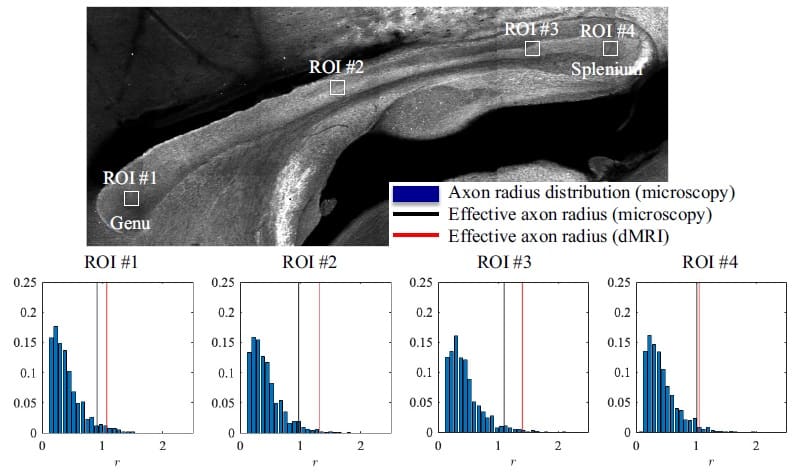
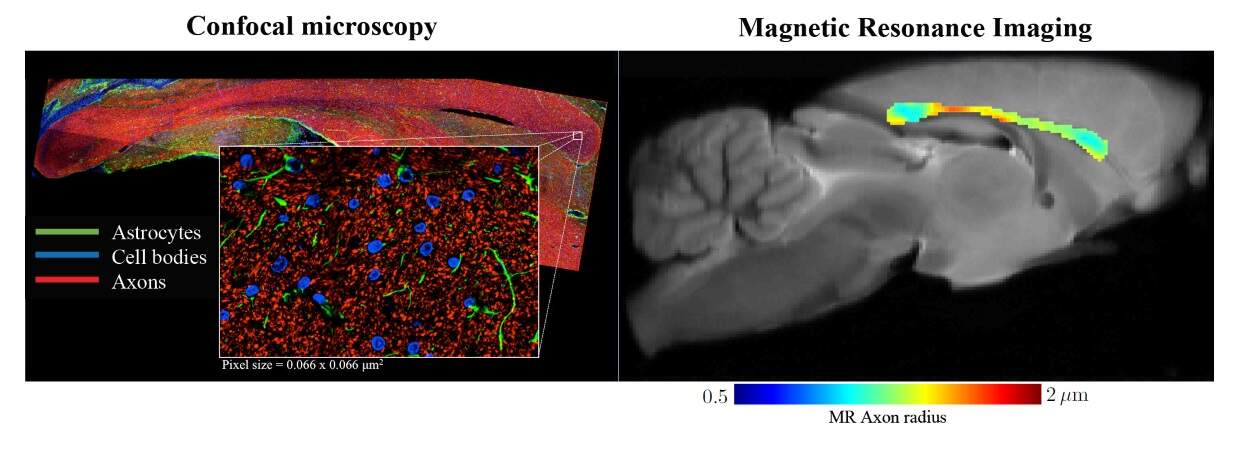
An international team of researchers has established a way to non-invasively measure the radii of axons, fine nerve fibres in the brain, using diffusion MRI (dMRI). Their proposed method showed good agreement with histological studies in both rodents and humans, and outperformed previous techniques in which reported measurements were an order of magnitude larger than histologically-derived axonal sizes (eLife 10.7554/eLife.49855).

Axons are the wire-like protrusions of a neuronal cell, involved in conducting electrical impulses away from the cell body. They are microscopic in diameter and, as bundles, comprise the primary form of communication of the nervous system. Clinical and histological studies have shown that axon radii can range from 0.1 µm to more than 3 µm in the human brain, and that this size, along with myelination, is responsible for the speed of neuronal communication.

In addition, clinical studies of neurodegenerative diseases, such as multiple sclerosis, have revealed preferential damage to smaller axons, while an electron microscopy study involving subjects with autism spectrum disorder showed a significant difference in axon size distribution compared with healthy controls.

Non-invasive axon radii quantification…

Clearly, accurate quantification of axon radius is an important neuroimaging biomarker. This, however, has proven to be a highly challenging task to perform non-invasively. Nevertheless, a team of researchers from the Champalimaud Centre for the Unknown in Portugal, NYU Grossman School of Medicine in the USA and the Cardiff University Brain Research Imaging Centre (CUBRIC) in the UK, set out to achieve just that.

The team used dMRI, a non-invasive and non-ionizing imaging modality that measures the random motion of water molecules in tissues, revealing details of the tissue microarchitecture. The key novelty of the approach is the researchers’ proposed method to separate MRI signals originating from different compartments of the probed tissue. More specifically, they modelled how the water signal behaves in different tissue types, and thereby managed to suppress signal arising from surrounding tissue outside the axons. In doing so, they could more accurately quantify the properties of the axons in the probed brain tissue samples.

…shows unprecedented agreement with histology

To validate their work, the researchers first tested their model on rodents using high-resolution (100 x 100 x 850 µm) dMRI images acquired on a 16.4T MR scanner (Bruker BioSpin). After scanning, they collected 50 µm-thick slices from two rat brains, corresponding to the imaged volume, and stained them to highlight the axons. Finally, they used confocal microscopy to visualize and analyse the tissue slices. Their results showed that the median MR-derived effective axon radius was 3–13% larger than the median radius derived from histology, an error that could be due to shrinkage of the filaments through staining.

Histological validation

The second part of the study focused on human subjects, who were imaged using a lower resolution (3 x 3 x 3 mm) Siemens Connectom 3T MR scanner. Here, the team used histological values reported in the literature for comparison. Despite this, the researchers showed that their method was able to accurately estimate known axonal sizes with errors an order of magnitude smaller than those in previous MRI studies.

The researchers conclude that their study revealed “a realistic perspective on MR axon radius mapping by showing MR-derived effective radii that have good quantitative agreement with histology”. Thinking about the next steps, first author Jelle Veraart, adds: “The non-invasive quantification of axon diameters using MRI allows clinicians and researchers to identify problems and developmental pathways that arise in the depths of the brain, driving forward treatment and understanding of development and disease progression.”

Healing spinal cord injuries with graphene

Spinal cord injuries often result from impacts such as car accidents, but they can also occur due to tumour growth and other non traumatic causes. They can leave patients fully or partially paralysed and can affect the functioning or organs leading to a myriad of health complications. Now, a group of researchers at the Materials Science Institute of Madrid (ICMM) in Spain is developing a novel treatment using graphene – a single layer of carbon with unique mechanical and electrical properties.

Taking a biomaterials approach, the group is developing graphene-based foams that could be implanted in the spinal cord. These foams can act like a scaffold or trellis, assisting with the regeneration of neurological tissue within the healthy region surrounding the injury site. Research group leader María Concepción Serrano López-Terradas describes the project in this interview with Physics World, recorded before Spain’s nationwide lockdown began in March due to the COVID-19 pandemic.

Find out more about the innovative materials research taking place at the ICMM in this video profile we shared last week. Also take a look at the Physics World Nanotechnology Briefing, published in April 2020. This free-to-read collection celebrates how nanotechnology is playing an increasingly important role in applications as diverse as medicine, fire safety and quantum information.

Hubble’s best shots: a cheeky gravitational grin

Gravitational lensing – the ability of a large mass to bend the path that light takes through the space around it – is surely one of Albert Einstein’s most dramatic predictions. Nowhere is this phenomenon demonstrated better than in Hubble’s many images of galaxies (and clusters of galaxies) that act as cosmic gravitational lenses, magnifying and bending the light of far more distant objects.

In this image, the smiling face is a happy coincidence of line-of-sight alignments and gravitational lensing. A galaxy cluster catalogued as SDSS J1038+4849 includes the two orange elliptical galaxies that make up the face’s eyes, which bend and magnify the light of a more distant background galaxy. In fact, the lensing alignment is so perfect that the path the light takes through space is bent enough to form a partial Einstein ring, which is the arc of light forming the face’s beaming grin.

Zoom backgrounds from Perimeter Institute, physics infographic from Alan’s Factory Outlet, clever magnetic clock

Now that video meeting services like Zoom are all the rage, you might want to impress your friends and colleagues with a background image from one of the world’s most prestigious centres for physics research. The Perimeter Institute for Theoretical Physics (PI) in Canada has released a series of images including an artist’s impression of two colliding black holes and a dark matter map of the cosmos. But my favourite is an exterior shot of the PI building in Waterloo, Ontario – which I had been planning to visit in June, but have sadly had to cancel.

As well as running Alan’s Factory Outlet in Luray, Virginia, Alan Bernau has a passion for creating science infographics. Back in August 2019, we featured an infographic created by Bernau and his work colleagues about the abundance of matter and energy in the universe. He has just been in touch to let us know about his team’s latest creation, which is called “38 radioactive elements and what they are used for”. The elements are grouped according to their half-lives, with the shortest-lived entries being elements like flerovium and tennessine, which are created in tiny amounts in nuclear physics facilities.

The above video is of Chris Kalogroulis and the beautiful magnetic clock that he built – which bagged him the GSK UK Young Engineer of the Year 2020. The clock’s hidden mechanism has a Heath Robinson/Rube Goldberg feel to it, which contrasts to the simplicity of the clock face.

And finally, we couldn’t leave you without revealing the answers to last week’s Red Folder physics-trivia quiz, which was designed to keep you amused during the global lockdown.

You can hear us discussing the quiz on the latest episode of the Physics World Weekly podcast, but here, for the record, are the answers.

We should add that, since creating the quiz, we’ve started to harbour doubts about the final question, which asked which US institution once offered a professorship to Galileo. We had thought that it was Harvard, but this blog from 2013 questions that “fact”, which just goes to show that crafting quizzes isn’t as easy as it looks.

  1. C – keyboards
  2. B – swimming
  3. A – in a brewery
  4. D – telescope
  5. C – dog
  6. D – shower curtain
  7. A – Caltech
  8. C – REM
  9. D – professional footballer
  10. B – Harvard University

Wide-band-gap semiconductors could harvest sunlight underwater

Solar cells based on wide-band-gap semiconductors work better under water than the narrow-band-gap ones used in conventional silicon photovoltaic devices. This finding by researchers at New York University (NYU), US, could aid the development of more efficient solar cells to power autonomous submersible vehicles and sensors.

Underwater vehicles cannot operate for long periods of time due to the lack of durable power sources. At present, they typically rely on batteries, connections to shore-based grid power, or power derived from solar cells situated above water – for example on a surface ship.

Onboard solar panels could be an attractive alternative for vehicles that need long-term power far from shore or support vessels, but conventional solar cells based on silicon or amorphous silicon are far from ideal for underwater operation. Thanks to their narrow band gaps of around 1.1 and 1.8 eV respectively, cells of this type absorb a large amount of red and infrared light. Unfortunately, water is also very good at absorbing these wavelengths, even at shallow depths, explains study lead author Jason A Röhr.

Blue and yellow light (400–600 nm), in contrast, is less easily absorbed by water, which means that it can penetrate further below the surface. To Röhr and his colleagues in André Taylor’s Transformative Materials and Devices Lab at NYU’s Tandon School of Engineering, this suggested that semiconductors with wider band gaps could be a better “fit” for supplying energy underwater, he says.

Detailed-balance model

To investigate these types of semiconductors further, the team used a detailed-balance model to measure the efficiency limits of different light-absorbing materials in various oceans and lakes around the world. These included parts of the Atlantic and Pacific oceans that are relatively clear and absorb light well, and a lake in Finland, which is more turbid and has poorer light absorption characteristics.

The model calculates the power-conversion efficiency of an underwater solar cell, which is given by the ratio of the power density output, pout, to the power density input from the solar spectrum, pin. It then measures the output power from the maximum power point on the solar cell current density-voltage (J-V) curve, pout = JmaxVmax, and measures pin by integrating the photon flux density – that is, the number of photons hitting the cell’s surface – over time. By combining a given absorption spectrum together with known values for the band-gap energy of a semiconductor, the depth below sea level, and water temperature, the researchers then obtained the detailed-balance efficiency limit of a semiconductor material in different locations.

For their input data, the researchers sifted through more than 400 published articles to unearth light-absorption spectra that not only covered the relevant spectral region, but was also representative of many areas on Earth. “We wanted to make sure that we didn’t just consider the clearest waters,” Röhr explains.

The calculations, which are detailed in Joule, reveal that at a depth of two metres, solar-cell absorbers would function best with a band gap of 1.8 eV, while at 50 metres a band gap of 2.4 eV was optimal. The devices’ balance efficiency ranged from around 55% in shallow waters to more than 65% at greater depths, while generating power greater than 5 mW/cm2. These efficiencies increase in cold water, and the researchers found that both balance efficiency and power output were independent of geographic location. This is good news because it means that the solar cells could be tailored to specific operating depths rather than locations.

The best underwater semiconductors

So which types of semiconductors might best fit these requirements? According to Röhr, good candidates include solar cells made from organic materials, which are lightweight, cheap to make and work well under low light conditions. Poly(3-hex- ylthiophene-2,5-diyl) (P3HT), for example, has a band gap of around 2.1 eV, can easily be produced in large quantities, and could be perfect for operation in shallow waters. Materials such as rubrene (2.2 eV band gap) and pentacene (2.2 eV) would work well at large depths, as would poly(p-phenylene vinylene) derivatives (2.3–2.4 eV).

Alternatively, alloys made with elements from groups III and V of the periodic table, such as the ternary and quaternary cadmium zinc telluride (CZT), copper zinc antimony sulphide (CZAS), AlGaAs, InGaP and GaAsP could also be good because they have tuneable band gaps, and so could be tailored to more efficiently absorb light at various depths.

While the composition of underwater sun-harvesting materials would be different to conventional solar cells, their general design would not need to change all that much, says Röhr. Obviously, they would need to be waterproof and stable for long periods in marine environments, but researchers have already shown that silicon solar panels can be encapsulated and remain submerged underwater for months without suffering any significant loss in power conversion. Similar encapsulation techniques might thus be employed to stabilize solar cells made from wide-band-gap semiconductors.

“Alternatively, we recently demonstrated that organic solar cells can be made resilient to water by selectively removing the electron acceptor from the top surface of the material,” Röhr tells Physics World. “However, we still need to show that this type of cell can be made more efficient than traditional silicon-based cells.”

This, he concedes, will be no easy task, as silicon’s cost and efficiency are both so competitive. However, he adds that new materials are constantly being developed, and he hopes that these results will inspire other research groups to look for new materials for underwater cells, too.

AI checks CT scans for COVID-19

Artificial intelligence (AI) can diagnose COVID-19 from CT scans, researchers in China claim. At least two teams have released studies that they say demonstrate that deep learning can analyse radiological features for accurate COVID-19 diagnosis faster than current blood tests, saving critical time for disease control.

COVID-19 first appeared in Wuhan in China at the end of last year and has since spread across the globe. By early March, the World Health Organization had declared the outbreak a pandemic, and there have now been more than 130,000 confirmed deaths worldwide.

Presentation of the viral disease ranges from asymptomatic to severe pneumonia with acute respiratory distress and multiple organ failure. COVID-19 is typically diagnosed by a reverse-transcription polymerase chain reaction (RT-PCR) test on a blood sample, but there are concerns about the sensitivity and availability of tests, and turnaround times for results.

CT scans are reported to be able to detect characteristic manifestations of COVID-19 in the lungs, with claims that this can lead to faster diagnosis than with current RT-PCR tests. But COVID-19 also shares similar imaging features with other types of pneumonia, making it difficult to differentiate. Could deep-learning rapidly analyse images and identify features of COVID-19?

To develop an AI tool to detect COVID-19, researchers led by Bo Xu of the Tianjin Medical University Cancer Institute and Hospital took CT images from 180 individuals who were diagnosed with typical viral pneumonia before the COVID-19 outbreak and 79 patients with confirmed COVID-19. They randomly assigned images from the patients to train or test the deep-learning algorithm.

In results published on medRxiv, the researchers claim that their model identified COVID-19 from CT images with an accuracy of 89.5%. Two radiologists who also assessed the images achieved an accuracy of around 55%. The team say that the results demonstrate that AI can offer accurate diagnosis from a CT scan (medRxiv 10.1101/2020.02.14.20023028).

In other work, published in Radiology, another team from China, led by Jun Xia of the Wuhan Huangpi People’s Hospital, trained a deep-learning model to detect COVID-19 using chest CT scans from 400 patients with COVID-19, almost 1400 people with community-acquired pneumonia and more than 1000 people without pneumonia (Radiology 10.1148/radiol.2020200905).

When they tested their AI on CT images from 450 patients, 20% with COVID-19, it achieved an accuracy of around 90%. Again, the researchers say that this shows that deep learning can differentiate COVID-19 from community-acquired pneumonia and other lung diseases.

Not everyone agrees, however. Michael Lu, a radiologist and expert in AI for imaging at Massachusetts General Hospital, tells Physics World that you first need to consider whether CT is good for diagnosing COVID-19. “The current US stance is not to do CT as the primary diagnostic test,” he explains.

“There was some initial data out of China saying that CT had high sensitivity, was accurate for detection of COVID-19,” Lu explains. “Subsequent to that, there’s been a lot of discussion of that paper, concern that there may have been selection bias involved. With any of these tests, the pre-test probability, sort of the prevalence or what stage of the COVID-19 pandemic you’re in, can actually have a major impact on the apparent performance of the test.”

Although he is keen to stress that the researchers are working rapidly with available data, Lu is also concerned that the mix of COVID-19 cases and historic lung images used to train and test the AI models does not reflect what a doctor dealing with suspected COVID-19 patients would see. “It’s an artificial distribution of patients,” he explains.

There are also issues around the practicalities and expense of mass testing using CT scans. “One of the issues is that, at least currently in the US, the scanners have to be cleaned after a patient comes in,” Lu says. In China, they have reportedly started covering people with plastic bags to prevent virus transmission between patients.

Lu believes that PCR tests will improve and will remain the gold standard for COVID-19 testing. He adds, however, that we should still pay attention to work on CT scans coming out of China, as they are ahead of the rest of the world in dealing with the pandemic, and things could change.

Physicists close in on a simpler route to quantum degenerate molecules

Cooling atoms to ultracold temperatures is a routine task in atomic physics labs, but molecules are a trickier proposition. Researchers in the US have now used a widely-applicable combination of methods to make molecules colder than ever before – a feat that could pave the way for applications in areas as diverse as high-temperature superconductivity and quantum computing.

In everyday life, we do not see the bizarre effects of quantum mechanics because the quantum states of the particles around us are constantly collapsing, or decohering, as they interact. At temperatures near absolute zero, however, some identical particles will simultaneously occupy the lowest energy quantum state available. This phenomenon is known as quantum degeneracy, and it was experimentally demonstrated in 1995, when groups led by Eric Cornell and Carl Wieman (then at the University of Colorado, Boulder) and Wolfgang Ketterle of the Massachusetts Institute of Technology (MIT) created the first Bose-Einstein condensates (BECs) with rubidium and sodium atoms, respectively.

Other groups have subsequently made condensates using other atomic species, and various techniques have been developed to cool atoms to quantum degeneracy. In one of the simplest methods, a sample of atoms is confined in a magnetic or optical trap. Hotter atoms with more kinetic energy are more readily able to escape, or evaporate, from this trap, so the remaining atoms become cooler. In another method, known as sympathetic cooling, one type of atom is cooled directly and allowed to thermalize with atoms of another type, thereby cooling them by extracting their kinetic energy.

More degrees of freedom, more problems

Physicists have also sought to cool molecules to quantum degeneracy, since their physics is richer and more complex than that of unpaired atoms. “Molecules have rotations and vibrations, and if you choose your molecule wisely, it can have a very strong electric dipole moment,” explains Florian Schreck, a physicist at the University of Amsterdam in the Netherlands. “That allows molecules to have very strong dipole-dipole interactions, which gives them huge potential for quantum simulation of spin-lattice systems, high-temperature superconductors, topological systems…an endless list of theory proposals.”

Unfortunately, this complexity also makes molecules more difficult to cool. The sympathetic cooling process, for example, relies on elastic collisions to transfer energy between particles. Molecules, however, can easily undergo inelastic collisions that release energy from one of their multiple rotational, vibrational and other degrees of freedom. This release of internal energy leads to a large increase of kinetic energy, and usually ends with the molecule being lost from the trap.

Although molecules have been created in BECs from paired atoms of the same atomic species since the early 2000s, it was not until 2019 that an equivalent milestone was reached for polar molecules. In that work, Jun Ye and colleagues at JILA in Boulder, Colorado cooled potassium and rubidium atoms to quantum degeneracy, then brought them together and tuned the magnetic interaction between them using a so-called Feshbach resonance. This tuning made it energetically favourable for the atoms to form a covalent bond.

Colder and colder

In the new work, Ketterle and colleagues at MIT adopted a more general – and thus potentially more widely applicable – approach. They began by using a Feshbach resonance to create sodium-lithium molecules in an optical trap formed at the focus of a laser beam. The molecules in this optical trap “feel” the trapping potential much more strongly than the atoms from which they are formed. They therefore exchange their potential energy for kinetic energy, becoming hotter than the unpaired sodium atoms in the process. However, by placing the molecules in stretched spin states (in which the electronic and nuclear spins are both aligned with the applied magnetic field) the researchers strongly suppress the inelastic collisions that would otherwise have ejected the molecules from the trap. This allows them to let the molecules thermalize with the colder atoms until the system reaches thermal equilibrium at a temperature of 2.23 μK.

To get colder still, the researchers utilize the weaker trapping potential felt by the atoms. “We decrease the power of the optical trap so that the most energetic atoms leave it,” explains Hyungmok Son, a PhD student in Ketterle’s group at the Harvard-MIT Center for Ultracold Atoms and the paper’s lead author. “The atoms left in the trap collide among themselves and get colder. These colder atoms collide with the molecules to do further cooling, and this is an ongoing process.”

Upgrades required

The researchers eventually cooled their sample to 220 nK, and Son says that they should be able to cool it to quantum degeneracy if they can trap more sodium atoms with their molecules. “That will require some technical upgrades to our experimental setup,” he adds. “We’re trying to get more powerful lasers.” In the meantime, the researchers intend to study how the cooling power changes as they vary the magnetic field to ascertain whether it matches theoretical predictions.

Schreck, who was not involved in the work, calls the technique “very powerful” and says it should work for other molecules, too. “There is no real limit to it,” he says. “With further improvements on the initial atom numbers they should be able to push this all the way to quantum degeneracy.”

The research is published in Nature. 

Hubble’s best shots: The Helix Nebula

Star death is a puzzle. We understand the broad strokes of what it entails – the most massive stars explode in an incandescent supernova, while smaller stars on the scale of our Sun expand into red giants and shed their outer layers to form a planetary nebula around their inert white dwarf core. What we are missing is some of the details. Planetary nebulae can adopt a variety of wonderful shapes, but what produces those shapes?

Take this Hubble image of the Helix Nebula, which is a planetary nebula about 650 light years away. From our orientation, it appears doughnut-shaped with the white dwarf in the hole at its centre. However, Hubble helped show that this shape is an illusion. What we’re really looking at is two discs of gas and dust almost perpendicular to one another. One popular theory is that complex planetary nebulae like these are the product of binary star systems, where a companion star ends up sculpting the material coming off its dying partner.

One more thing: look closely and you can see strange objects around the circumference of the illusory doughnut shape. They look a bit like comets, and are known as “cometary knots”, but they are far too large to be globs of ice and dust. Instead, they are dense globules of gas in the nebula.

Mirror-symmetry violation discovered in ground states of strontium and bromine isotopes

Physicists in the US have made the surprising discovery that two nuclear isotopes with exactly mirrored numbers of protons and neutrons have different ground states. The researchers, led by Daniel Hoff and Andrew Rogers at the University of Massachusetts Lowell, uncovered this mirror symmetry violation by accident, while investigating the nuclear physics of astronomical X-ray bursts. Their discovery could have profound implications for our knowledge of the physics that govern atomic nuclei.

Many processes in particle physics have properties rooted in conservation laws, which describe how certain quantities must remain the same throughout nuclear reactions and decays. One effect that emerges from these laws is called “mirror symmetry” and relates to isotope “partners” whose numbers of protons and neutrons are exactly switched with each other. Such partners are known to have identical ground states in terms of angular momentum and parity. This reflects the similarity of the behaviors of protons and neutrons in the nucleus – something that underlies theories of nuclear physics.

In their study, Hoff and colleagues looked at strontium-73 (38 protons and 35 neutrons) and bromine-73 (35 protons and 38 neutrons). However, their original plan was to study the rapid proton capture exhibited by another isotope, rubidium-73. This process is thought to be involved in the production of some astrophysical X-ray bursts. Rubidium-73 is very short lived and the team were producing it via the beta-decay of strontium-73.

Stack of silicon detectors

Using the National Superconducting Cyclotron Laboratory facility at Michigan State University, the researchers first fired a beam of nuclei containing strontium-73 into a stack of silicon detectors. After being slowed to a halt, the nuclei decayed into rubidium-73 by emitting a single positron, followed by a proton, which are detected.

Looking at how the two particles are emitted from the isotope provided information about how rubidium-73 captures protons. However, their analysis led them to a wholly unexpected discovery: that the ground state of strontium-73 must be identical to that of rubidium-73. The result came as a surprise because strontium-73 is a mirror partner of another isotope, bromine-73, which is known to have a different ground state from the strontium-73 ground state determined by Hoff and colleagues. This means that strontium-73 and bromine-73 appear to violate mirror symmetry.

The researchers suggest that the violation could be the result of several factors including differences in shape between the two partners, but more experiments will be required to confirm their influences. These include direct measurements of strontium-73’s mass and also use beta nuclear magnetic resonance (β-NMR) to determine the ground-state spin of the isotope.

The team already has further experiments planned. The research is described in Nature.

Copyright © 2026 by IOP Publishing Ltd and individual contributors