In 2011 parched soils in Texas caused agricultural losses of around $8 billion in one of the worst droughts the state has ever seen. 2017 saw the opposite extreme when Hurricane Harvey dumped 150 cm of rain over south-eastern Texas in six days, resulting in extensive flooding, 68 fatalities and economic losses of over $100 billion. Whilst we can’t control the rain, we can control the water. A new study demonstrates the potential of the “managed aquifer recharge” technique for capturing flood water in Texas and injecting it into aquifers.
This technique, which already helps Texas manage its water supply, diverts floodwater, reducing its impact. It temporarily stores the water at the surface, before treating it and gradually injecting it into depleted aquifers beneath. This tops up groundwater supplies ready for the next drought.
To investigate this approach in Texas, Qian Yang and Bridget Scanlon from the University of Texas at Austin, quantified how much water could be captured and stored from the ten major rivers that discharge into the Gulf of Mexico.
Between 2015 and 2017 around 37 cubic km of water could have been captured from these ten rivers during high magnitude flow events, the scientists found. The excess water from these three wet years is equivalent to around twice the annual water demand in Texas in 2016 and would have been enough to replenish the depleted Texas Gulf Coast Aquifer system.
“Compared to traditional surface reservoirs such as lakes, depleted aquifers provide much greater storage capacity,” says Yang, whose findings are published in Environmental Research Letters (ERL).
By studying rainfall and river flow records over the last 50 years, Yang and Scanlon also show that small numbers of long duration events – longer than a week – contribute most to the large flow volumes.
Such schemes are more cost-effective than expanding surface water storage. Texas already has three operational managed aquifer recharge systems in place — at El Paso, Kerrville and San Antonio. Other parts of the world, particularly areas where aquifers have been depleted and flash floods are common, are also taking advantage of this way of balancing water supply and demand. To date there are over 1000 schemes in operation worldwide.
Climate change is projected to increase weather extremes, with floods and droughts anticipated to become more frequent and severe in many regions. Yang and Scanlon believe that managed aquifer recharge is likely to become a serious mitigation tool.
“I think managed aquifer recharge will become very prevalent in the future as a way of better managing water resources, especially in those regions that suffer from floods, droughts, and groundwater depletion,” says Yang.
Nanopore sensors can identify a wide variety of single molecules and biomolecules, but all such devices made so far rely on electrodes to measure the current change through the nanopore as a molecule passes through it. Researchers at Nanjing University in China have now made the first nanopore sensor that works optically and does not require any electrical connections. The new technique, dubbed DiffusiOptoPhysiology (DOP), could find applications in drug screening, clinical diagnosis and DNA sequencing.
“Since the first single-molecule nanopore sensors were made a few decades ago, their mode of measurement – which requires a patch clamp amplifier for single-channel recording – has not changed,” explains Shuo Huang, who led this research effort. “This has set a high technical hurdle that has limited nanopore research to a few academic laboratories and specialists around the world.”
Much-simplified technology
“In our work, we were inspired by recent advances in calcium fluorescence imaging,” he says. “The result is a much-simplified technology, the DOP, which is adapted from optical single-channel recording (oSCR).”
oSCR optically monitors calcium ion (Ca2+) flux through individual nanopores embedded in a droplet interface bilayer (DIB). Although very efficient for high-throughput measurements, a pair of electrodes is still required to electrically drive Ca2+ions through the pores. Such a setup is not without risk since manually inserting electrodes into aqueous droplets requires much dexterity and delicate micro-manipulation skills to avoid inadvertently rupturing the DIB.
DOP does not require any electrical connections, explains Huang. It works by optically monitoring (the strong) fluorescence emission that occurs when Ca2+ ions bind with the indicator dye, Fluo-8 (a chemical reagent routinely employed by biologists and chemists) as they diffuse through the channels of a nanopore sensor.
Reporting single molecule activities and parallel measurements
“The fluorescence intensity is proportional to the ionic flux through the channels, which in turn can report single molecule activities, such as binding of small molecules to a nanopore or translocation of nucleic acids through it.”
DOP allows for parallel measurements from thousands of nanopores on a centimetre-sized chip and is cost effective – at less than $1 in consumables for a single measurement. This low cost, as well as its small size makes it highly suitable as a disposable device for use in clinical diagnosis, says Huang.
To test their device, the researchers detected small molecules such as cyclodextrin, PEG1500 and dsDNA as well as various macromolecules and biomacromolecules.
“The DOP chip may find applications in screening for new drug molecules,” says Huang. “Its low cost and the fact that it is easy to use may allow research teams around the world to now exploit the power of nanopores for potentially routine clinical diagnoses.” Indeed, the Nanjing team says that it has decided to share its technique for preparing biological nanopores via a public gene repository service.
Looking ahead, the researchers say they are now busy working on a highly portable imaging hardware that is compatible with DOP. “This hardware would resemble a mobile cell phone with integrated optical and laser diode-based illumination modules so that anyone anywhere could carry out DOP,” Huang tells Physics World.
A brain computer interface device – an electroencephalography cap. (Courtesy: Wyss Center)
Stroke is the second leading cause of death worldwide and the third cause of induced disability, according to estimates from the Global Burden of Diseases, Injuries, and Risk Factors Study. Treatments based on constraint-induced movement therapy, occupational practice, virtual reality and brain stimulation can work well for patients with mild impairment of upper limb movement, but they are not as effective for those burdened by severe disability. Therefore, novel individualized approaches are needed for this patient group.
Martina Coscia from the Wyss Center for Bio and Neuroengineering in Geneva, and colleagues from several other Swiss institutes, have published a review paper summarizing the most advanced techniques in use today for treatment of severe, chronic stroke patients. The researchers describe techniques being developed for upper limb motor rehabilitation: from robotics and muscular electrical stimulation, to brain stimulation and brain–computer/machine interfaces (Brain 10.1093/brain/awz181).
Robot-aided rehabilitation approaches include movement-assisting exoskeletons and end-effector devices, which enable upper arm movement by stimulating the peripheral nervous system. These techniques can also trigger reorganization of the impaired peripheral nervous system and encourage rehabilitation of the damaged somatosensory system. Several studies have reported the efficiency of robot-aided rehabilitation, alone or in combination with other techniques, in the treatment of upper limb motor impairment. One study that included severely impaired individuals also demonstrated encouraging results.
Muscular electrical stimulation can help improve the connection of motor neurons to the spinal cord and the motor cortex. Researchers have also demonstrated that application of electrical stimuli to the muscles provides positive effects on the neurons responsible for sensory signal transduction to the brain, thereby improving the motion control loop function. By modulating motor neurons’ sensitivity, muscular electrical stimulation inhibits the muscle spasms observed in other treatments.
More recently, therapies have moved on from the simple use of currents to harnessing coordinated stimuli to orchestrate more complex, task-related movements. Although this particular set of techniques didn’t show a particular advantage over physiotherapy in long-term studies of patients with mild upper limb impairment, it did seem to have a stronger effect for chronic severe patients.
Stimulating the brain
Brain stimulation, meanwhile, stimulates cortical neurons in order to improve their ability to form new connections within the affected neural network. Brain stimulation techniques can be divided into two branches – electrical and magnetic – both of which can activate or inhibit neural activity, depending on the polarity and intensity of the stimulus.
Personalized neurotechnology-aided rehabilitation of the arm could improve recovery in severe chronic stroke patients. (Courtesy: Wyss Center)
Researchers have achieved encouraging results using both techniques. In particular, magnetic field-triggered inhibition of the contralesional hemisphere (the hemisphere that was not affected by the stroke) activity yielded positive results. Magnetic, low-frequency stimulation of the contralesional hemisphere also proved encouraging – improving the reach to grasp ability of patients, although only for small objects. Excitingly, some studies suggest that coupling contralesional cortex inhibition with magnetic stimulation of the chronically affected area could achieve effective results.
Within these techniques, one promising approach is invasive brain stimulation, in which a device is surgically implanted in a superficial region of the brain. Such techniques allow for more sustained and spatially-oriented stimulation of the desired brain regions. The Everest trial used such methods and showed significant improvement for a larger percentage of patients after 24 weeks, compared with standard rehabilitation protocols.
Another promising recent development is non-invasive deep-brain stimulation, achieved by temporally interfering electric fields. The authors envision that a deeper understanding of the complex mechanisms involved in the brain’s reactions to magnetic and electrical stimulation will provide an important assistance in clinical application of these techniques.
The final category, brain–computer or brain–machine interfaces (BCIs or BMIs), exploit electroencephalogram (EEG) patterns to trigger feedback or an action output from an external device. Devices that produce feedback are used to train the patient to recruit the correct zone of the brain and help reorganize its interconnections. These techniques have only recently transitioned to the clinic; however, early results and observations are promising. For example, a BCI technique coupled with muscular electrical stimulation restored patients’ ability to extend their fingers.
In recent years, researchers have also tested combinations of the techniques described above. For example, combinations of robotics and muscular electrical stimulation have shown encouraging results, especially when more than one articulation was targeted by the treatment. Combining brain stimulation with muscular electrical stimulation and robotics has proved more effective in severe than in moderate cases. Also, coupling of muscular electrical stimulation with magnetic inhibitory brain stimulation provided better results than either individual technique. Interestingly, addition of electrical brain stimulation to a BCI system coupled with a robotic motor feedback enhanced the outcome, helping to achieve adaptive brain remodelling at the expense of inappropriate reorganization.
Coscia and co-authors highlight that all the techniques studied share a range of limitations that should be addressed, such as small sample size, limited understanding of the underlying mechanisms, lack of treatment personalization and minimal attention to the training task, which they note is often of limited importance for daily life. Addressing these limitations might be key to improving the clinical outcome for patients with severe stroke-induced upper limb paralysis treated with neurotechnology-aided interventions. Moreover, the authors plan to begin a clinical trial to test the use of a novel personalized therapy approach that will include a combination of the described techniques.
Epilepsy is the world’s most common chronic neurological disorder, affecting one in 100 people. It is characterized by recurrent seizures that often have no apparent external trigger but are the result of neurons in the brain behaving irregularly. The affliction was in fact one of the first medical conditions to be recognized in the ancient world. There is a 2600-year-old Babylonian cuneiform tablet that distinguishes between various type of seizures, while Roman emperor Julius Caesar was said to have “the falling sickness”.
Despite the Greek physician Hippocrates proposing in the fifth century BC that epilepsy was a medical condition that originated in the brain, for a long time many people mistakenly thought it was caused by spirits. This superstition continued to be believed until the 17th century when it was finally accepted that the brain was indeed the site of the problem. However, no-one really knew how to treat epilepsy, with remedies including everything from prescribed diets and special living conditions, to medicinal herbs. Some people with epilepsy even had holes drilled in their skulls or were subject to bloodletting – but these surgical techniques were far from effective.
There are millions of people who live with unpredictable seizures that are – for reasons we don’t completely understand – not well controlled by medication or even surgery
Modern medicine has thankfully made huge progress since then, but there are still millions of people who live with unpredictable seizures that are – for reasons we don’t completely understand – not well controlled by medication or even surgery. Indeed, for the 30% of people with epilepsy who have symptoms that cannot be treated, the unpredictable onset of seizures can greatly hamper their daily lives, as they are, for example, prohibited from driving, swimming or operating machines – activities we usually think are commonplace. Some people have been seriously injured – or even died – as a direct result of sudden, unexpected seizures. An early warning system that would allow these patients to take the necessary precautions to prepare for or even avert the seizures entirely, would obviously be highly desirable.
So, what can physics say about this challenge?
It’s all a phase
1 Firing neurons Neurons are cells in our nervous system that receive and send information via electrochemical signals. (Adapted from Noba textbook series: Psychology 2019 DEF publishers “Neurons” chapter.)
The brain is an incredibly complex network consisting of tens of billions of neurons forming hundreds of trillions of connections, which makes the task of prediction seem daunting. However, each neuron is still a physical object that obeys the laws of physics.
The fundamental equations that describe the electrical voltages inside neurons were discovered by Alan Lloyd Hodgkin and Andrew Fielding Huxley in 1952, for which they received the Nobel Prize for Physiology or Medicine in 1963. The Hodgkin–Huxley equations are considered the gold standard for understanding the intricate dance of sodium and potassium ions as they rush in and out of neurons. Their movement depends on changes in voltage that control specialized ion gates – proteins that open and close pores in the membrane to allow the passage of ions. Solving this system of coupled mathematical equations allows researchers to keep track of the voltage of each neuron over time.
One of the key processes that can be modelled is the rapid rise and fall of the voltage each time a neuron fires. When a neuron has been sufficiently stimulated by its neighbours, ion gates that were locking sodium ions out suddenly open. A massive influx of these charged particles ensues, causing the interior voltage to spike. In this way, a signal can quickly propagate along the length of the neuron, and then to other connected brain cells.
We have carried out computer simulations of the behaviour of small networks of neurons, which has let us monitor the effects of slight changes to individual parameters, such as the threshold for each neuron to fire
As part of our research at Nova Southeastern University in Fort Lauderdale, US, we have carried out computer simulations of the behaviour of small networks of neurons using the Hodgkin–Huxley equations. This work has let us monitor the effects of slight changes to individual parameters, such as the threshold for each neuron to fire, or the “stickiness” of ion gates, which makes them stay open longer than they should. All of these may have a significant impact on the excitability of each neuron.
2 Communication network Simulation of a small network of neurons, with the nodes that are “firing” highlighted. (Courtesy: Louis Nemzer)
Understanding the behaviour of individual neurons is only a starting point. Complex systems like the brain show collective or emergent behaviour, in which the whole acts very differently from the sum of its parts. This can include sudden, marked changes caused by smooth alterations in external conditions, rather like the phase transition that occurs when you pull an ice cube out of your freezer. It remains frozen solid right until you raise its temperature to the critical point, 0 °C, whereupon it promptly melts into liquid water. This transition is due to complicated interactions between water molecules that you’d never guess no matter how long you studied a single water molecule in isolation.
Similarly, an epileptic seizure occurs when the normal functioning of the brain is interrupted by neurons locked into a single rhythm, and it’s likely that this is also a kind of phase transition. In this case, a group of hyperexcitable neurons at a specific location – the seizure focus – start firing in unison. This recruits other neurons to synchronize with them, which in turn recruits other neurons, setting off a synchronization avalanche. In this picture, the seizure focus acts much like a seed crystal that, when dropped into a pot of sugar dissolved in hot water, causes the whole thing to crystallize into rock candy.
Edge of chaos
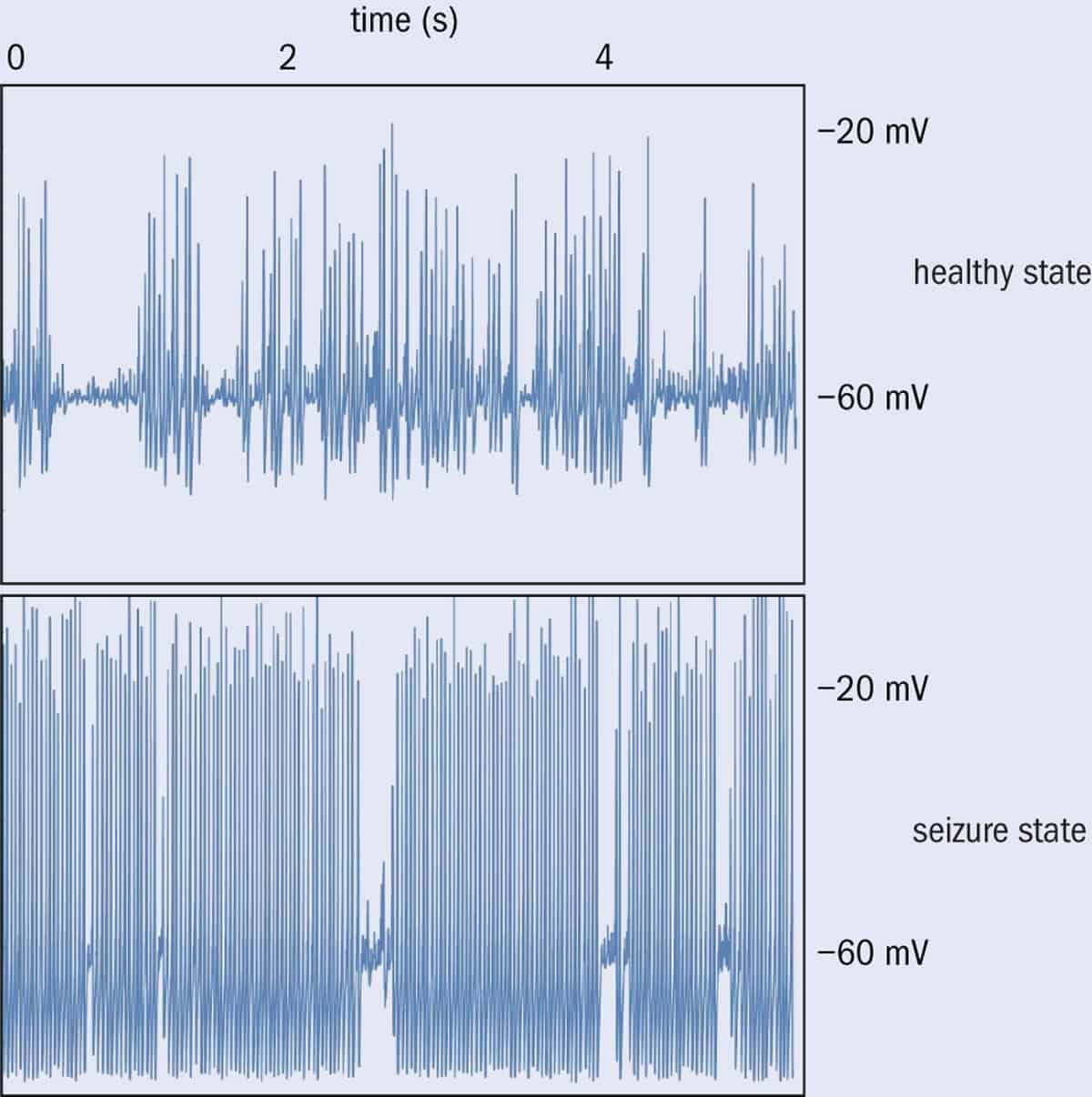
Clearly, the brain must be finely tuned between too much excitation, which can lead to chaos or runaway synchronization, and too much inhibition, which leads to stasis and inactivity. In fact, many scientists believe that healthy brain function is perched right on the edge of chaos. In our simulations using small networks of neurons, we can identify the “healthy” state because it shows bursts of activity of all sizes. That is, we see many small bursts, fewer medium bursts, and a small number of large bursts. Since there is no characteristic burst size, this distribution is called “scale-free”.
In contrast, if the sodium ion gates become sticky, each neuron is now slightly hyperexcitable, leading to all-or-nothing synchronization in which many neurons fire in unison, as in an epileptic seizure. The effect of a tiny change therefore cascades until the network is pushed “over the cliff” into pathological synchronization.
3 Simulating seizures Comparing the “healthy” and “seizure” states of the simulated network. The healthy state shows bursts of various sizes, while the seizure state is usually locked into a repeating, synchronized pattern.
Our computer simulations help us understand the hallmarks of the seizure state. But predicting if a seizure is likely to start within the next 15 minutes is a much bigger task. It’s the difference between trying to tell if it’s raining right now (which you can do by just sticking your hand outside) and predicting whether it’ll rain tomorrow (better call in your best team of meteorologists).
For this task, we need actual patient data. Monitoring the activity of the brain is conventionally done via an electroencephalogram (EEG), in which electrodes are placed directly on to a patient’s scalp to detect the brain’s electrical signals. However, EEG data are imperfect because high-frequency signals cannot penetrate the skull, and there may be spurious blips from eyeblinks and other muscle movements. A more accurate method is electrocorticography (ECoG), which uses an electrode grid surgically implanted inside the cranium, just on top of the brain. Obviously, this technique involves a very invasive operation and is not something patients undergo lightly. Only people with severe seizures that continue to occur even after repeated attempts at treatment will elect to have the electrodes implanted.
But even after scouring ECoG data for identifiable signs that indicate when a seizure is about to start, nothing stands out. What’s the point of generating high-quality data if you don’t even know what patterns to look for? This is where machine learning, the burgeoning new field of computer science, can help.
The je ne sais quoi of Jane
You may not realize it, but machine learning is already hard at work in your daily life, tagging people in photos on Facebook, recommending your next movie on Netflix, and alerting your bank to questionable transactions (see “A learning revolution”).
To understand the power of machine learning, you first need to think about how conventional programming takes advantage of what computers are very good at: namely, following the rules. Computers, after all, can run through a flowchart much faster than any human. However, there are some tasks that we mortals are still better at, like identifying your friend Jane. Think how long it would take to list the “rules” for telling a picture of Jane apart from anyone else, especially if you include photos with different lighting conditions and angles.
In fact, it might seem hopeless to try and train a computer using a set of rigid rules. But this is where machine learning comes in. Instead of attempting to programme the computer manually, we train it with many labelled examples. We can take a hard drive full of snaps and tell the algorithm, “this is Jane” or “this is not Jane”. Over time, the computer learns, via reinforcement, the features that distinguish Jane from everyone else.
To classify the risk of seizures, our group in Florida is adapting machine-learning algorithms that are already used in medicine to diagnose radiology images
So to classify the risk of seizures, our group in Florida is adapting machine-learning algorithms that are already used in medicine to diagnose radiology images. We are currently training the machine-learning models – and the simulations we created before are helping by generating simulated data that the algorithms can learn from. Next, as long as we have enough labelled ECoG data from patients, we hope to be able to build a highly accurate warning program even if we do not know which features in the signal the algorithm is using. While this “black-box” approach may seem disconcerting at first, the primary test will be the usefulness, if not the explainability, of the resulting system.
Ultimately, we hope to use the algorithm to develop an accurate, straightforward smartphone app that would use data transmitted from wireless scalp electrodes to provide real-time information about seizure risk to patients and healthcare providers – just as people with diabetes can now receive automated alerts from a blood-sugar monitor. This information would allow patients to take medication or at least get to a safe space. Some people could even have an electric shock administered to avert the seizure entirely.
Detect and treat
If a seizure has been predicted to be imminent – or has already begun – ideally you want to be able to prevent it escalating. One possible method is to directly administer drugs to the seizure focus inside the brain – an approach that may work even for patients who do not usually respond to oral medications.
George Malliaras and colleagues at the University of Cambridge in the UK have even been able to fabricate such a method. Their hybrid device can be implanted into the brain, and not only detects when a seizure is starting, but also automatically delivers GABA – a neural inhibitor – to quickly calm it (Science Advances 4 eaau1291). Improvements in this kind of “on-demand” drug release complement seizure prediction methods, since knowing when a seizure is coming is much more valuable if there are effective, rapid means for preventing it entirely, especially if the warning comes just seconds before the seizure is about to start.
Humanity has been dealing with epilepsy for millennia, and its effects on patients range from mildly inconvenient to devastating. However, modern physics is providing new optimism that real improvements in quality of life are possible. People who do not receive relief with drugs or surgery may still be able to lead normal lives by monitoring their condition as a manageable chronic disorder. My hope is that an increased understanding of the physics of the brain will have a real positive impact on people’s lives.
Sounding out the focus
Some epilepsy patients who do not find relief using medications are candidates for more invasive treatments, including surgery to remove the seizure focus. However, while operations can often be effective, there is a big risk that they could damage normal brain structures. One promising new method involves using targeted ultrasonic waves to eliminate the seizure focus, without having to open the skull at all – just as sound waves can be focused on a kidney stone inside a patient to destroy it non-invasively. An early test of this technology was led by Vibhor Krishna, a surgeon at Ohio State University in the US. His patient was awake inside a magnetic resonance imaging (MRI) machine, when focused ultrasound waves were used to safely ablate the seizure focus while minimizing side effects. According to the Focused Ultrasound Foundation – which is dedicated to the development and application of focused-ultrasound technologies in medicine – the process minimizes damage to healthy brain by eliminating the need for incisions, holes in the skull, or electrodes in the brain.
Stepping down: Joi Ito, head of the Massachusetts Institute of Technology’s Media Lab, has stepped down following his and the lab’s connections with the financier Jeffrey Epstein. (CC BY 2.0 Joi Ito/Mizuka)
The head of the renowned Media Lab, based at Massachusetts Institute of Technology (MIT) in the US, has stepped down following his and the lab’s connections with the disgraced financier and child-sex trafficker, Jeffrey Epstein, who died by suicide in a New York City jail in August. Joi Ito, an entrepreneur who has served as director of the MIT Media Lab since 2011, resigned from his position over the weekend following an investigation by The New Yorker. The article charges that he and Peter Cohen, the lab’s former director of development and strategy, had worked to keep Epstein’s contributions to the lab as anonymous despite MIT’s donor database listing Epstein as “disqualified” from providing funds.
Epstein’s actions have reverberated throughout the US scientific community over the past month after investigations revealed that he supported many top research universities and labs by offering their scientists significant financial support. His targets included several prominent physicists and physical scientists including George Church, Lawrence Krauss, Kip Thorne, Frank Wilczek, as well as the late physicists Murray Gell-Mann and Stephen Hawking. Although there is no indication that the scientists were involved in any dubious activity, Epstein’s connections – apparently intended in part to support his ideas about eugenics and cryogenics – have embarrassed scientists and their institutions.
We recognize with shame and distress that we allowed MIT to contribute to the elevation of [Epstein’s] reputation, which in turn served to distract from his horrifying acts
Rafael Reif
Initial evidence of Epstein’s behaviour first emerged in a Florida court case in 2005 when a 14-year-old girl and her parents reported that Epstein had molested her at his Florida home. The report led to a police investigation and multiple charges that Epstein sexually assaulted underage girls. However, the local district attorney at the time – Alexander Acosta – agreed to a plea of guilty to two counts of soliciting prostitution, one of them involving a girl under the age of 18. Epstein was sentenced to 18 months in jail – during which he was allowed to travel to work in his office six days a week – and placed on a sex offender list.
In November 2018 the Miami Herald then published a full account of 60 women who said that Epstein had abused them. In July this year, he was arrested on sex trafficking charges in New York state. A week later, the situation led to Acosta’s resignation as Secretary of Labor in the Trump administration, owing to his agreement that the sentence was too light. Epstein killed himself in his Manhattan jail cell on 10 August.
‘A mistake of judgement’
While some contributions to research institutions predated Epstein’s original conviction and registration as a sex offender, others continued well after that event, until at least 2017. But once the latest case against him became public, the scientific community began to voice its regrets for their connections. On 22 August MIT president Rafael Reif apologized in a letter to the institute for accepting about $800,000 from Epstein for the Media Lab and individual researchers.
“We recognize with shame and distress that we allowed MIT to contribute to the elevation of his reputation, which in turn served to distract from his horrifying acts,” Reif stated in the letter. Faculty members’ decisions about accepting financial gifts “are always subject to longstanding Institute processes and principles,” Reif went on. “To my great regret, despite following the processes that have served MIT well for many years, in this instance we made a mistake of judgment.”
The Media Lab’s links to Epstein led to two prominent members – Ethan Zuckerman and Nathan Matias – cutting ties with the lab in late August. But The New Yorkerrevealed that the connections between the lab and Epstein ran much deeper,reporting that Epstein “appeared to serve as an intermediary between the lab and other wealthy donors…securing at least $7.5m in donations for the lab” from the philanthropist Bill Gates and investor Leon Black. Its report also reveals how lab leaders attempted to put Epstein’s investments as anonymous, despite MIT’s donor database listing Epstein as “disqualified” from providing funds. In an open letter on 7 September announcing that Ito has offered his resignation, Reif says that he has asked MIT’s General Counsel to carry out an independent investigation into the situation. Ito had also previously admitted receiving $1.2m from Epstein for investment funds that he controlled.
‘Moral failings’
Other scientists at MIT have also become embroiled in the scandal, including the MIT physicist Seth Lloyd, who apologized publicly for taking funds from Epstein. In a blog post on 22 August he wrote that he was “deeply disturbed” at Epstein’s original conviction but agreed to visit him during his first prison term. He also received grants from Epstein’s foundation in 2012 and 2015. “These were professional as well as moral failings,” he writes, adding that he has committed financial resources to aid women harmed by Epstein’s abuse.
Yet Epstein’s links with the scientific community extend well beyond MIT. Although Epstein had no background in science, he formed links with high-profile scientists in the early 1990s when he made donations to the Santa Fe Institute, which had been co-founded by Gell-Mann in 1984. In 1998 Epstein also provided research funding for science historian Anne Harrington from Harvard University. “He offered me modest funding to pull together an interdisciplinary group,” she told the Harvard Crimson. “Had I known even a hint of what we all have subsequently learned about him, I never would have accepted it.”
Later, in 2003, Epstein donated $30m to Harvard to fund its programme for evolutionary dynamics, headed by mathematical biologist Martin Nowak. Epstein used his wealth to gather scientists into his orbit beyond Harvard, hosting glittering lunches and dinners and flying them to his private island in the US Virgin Islands. According to the New York Times, he frequently discussed his idea of eugenics in which he planned to inseminate several women to give birth to his offspring.
George Church, who is a chemist and geneticist at Harvard, apologized for his “poor awareness and judgment” in accepting research funds from Epstein in 2007 and meeting him frequently since then, despite admitting to Stat News that he had read articles about Epstein’s conviction in 2008. “But they weren’t clear enough for me to know there was a serious problem,” he claimed. Harvard itself reports that it has received no financial support from Epstein since 2007, although the university says it has no plan to return $6.5m (of the $30m promised) that he had donated before then.
As for Krauss, he received financial support from Epstein for Arizona State University’s Origin Project, which he headed before it closed in 2018. Subject himself to charges of sexual misconduct that led to his departure from Arizona State in May, Krauss had also organized a conference about gravity on Epstein’s island. Other physicists whose names have appeared in the news in connection with Epstein appear merely to have accepted his invitations to conferences and meals.
A new generation of neutron sources is providing scientists with unprecedented power to probe the structure and properties of materials ranging from biomolecules through to superconductors. Spallation sources recently built in the US, Japan and China are now the brightest neutron sources in the world, while the European Spallation Source (ESS) now being built in Lund, Sweden, will set the bar even higher when it comes online for users in 2023.
But these powerful new instruments present a challenge for the engineering systems needed to support them. High neutron fluxes generate large amounts of radiation, which creates issues for critical infrastructure such as the vacuum systems needed to maintain ultrahigh vacuum conditions within the experimental facility. “We need to build reliable vacuum equipment that won’t be damaged by radiation,” says Andreas Schopphoff of Pfeiffer Vacuum, who has worked closely with users to devise custom vacuum solutions for this new breed of spallation sources. “It can cause problems both for the electronic systems used to control vacuum pumps and gauges, and the materials we use to build them.”
Neutrons reveal material secrets
Neutrons are widely used to study material properties because they penetrate deep into the sample, scattering off atomic nuclei to generate data that researchers can analyse to determine the detailed structure within the material. Neutrons also have the right wavelength to transfer their momentum and energy to the particles inside the sample, which provides valuable information about fundamental vibrations that control chemical bonding and magnetic interactions. What’s more, neutrons have a nuclear spin that makes them very sensitive to the location and orientation of magnetic moments in materials.
While neutron sources have been available for many years, this new generation of spallation sources can deliver neutron beams that are orders of magnitude more powerful than previous designs based on nuclear reactors – which can speed up experiments and enable scientists to probe materials and their interactions with more detail than ever before.
The most intense reactor-based source available today, the Institut Laue-Langevin in Grenoble, France, achieves a power of 58 kV, while the ESS will reach 5000 kV by smashing a high-energy proton beam into a solid target made of tungsten. The force of the impact creates free or “spalled” neutrons, which are then slowed and collimated into an intense neutron beam that can easily be pulsed for neutron scattering experiments. Other spallation sources operate in a similar way, but can use different acceleration methods and different types of target – although heavy metals are chosen for their ability to absorb radioactive species produced during the spallation process.
The tungsten target at the China Neutron Spallation Source. (Courtesy: IHEP)
But radiation is still a major concern for these high-power neutron sources. At the Japanese Proton Accelerator Research Complex (J-PARC) – the most intense neutron source in operation today – radiation levels can reach millions of grays in the vicinity of the neutron beam.
According to Schopphoff, such high levels of ionizing radiation creates two major problems for vacuum pumps and gauges stationed close to the neutron source. “First, we cannot connect electronic systems directly to vacuum pumps or gauges because the radiation would cause them to fail and stop working,” he says. “We also need to think about eliminating certain materials from the pump design, such as Teflon, that would also be affected by high levels of radiation.”
Finding radiation-resistant solutions
The solution for the electronics, says Schopphoff, is to position them a long way from the pumps. At the China Spallation Neutron Source, for example – which has just completed its first round of operations – some of the vacuum gauges were connected to their controllers via cables up to 220 m long. Vacuum generation is equally important, and both the turbopumps and the backing pumps must be designed to be radiation resistant.
“For some applications it’s important to bring enough electrical power to the pump,” he explains. “So we have to install large cables, particularly for the backing pumps. At J-PARC we worked with the project team to develop an electronic system specifically for their needs.”
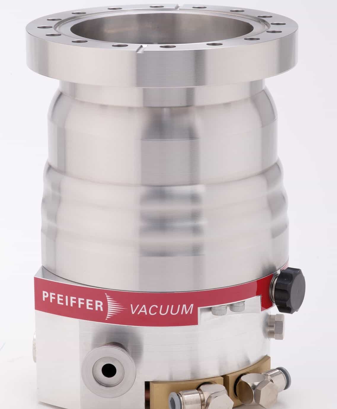
The HiPace turbomolecular pump from Pfeiffer Vacuum.
Meanwhile, the effect of ionizing radiation on materials can demand a whole new approach to pump design. Most large-scale experimental facilities exploit high-speed turbopumps to maintain UHV conditions, but to be effective they need a backing pump to reduce atmospheric pressure to a “rough” vacuum. In the case of J-PARC, the project team were concerned that the Teflon used within the two most common types of backing pump – oil-sealed or dry backing pumps – would become more brittle when exposed to radiation.
The solution, says Schopphoff, was to introduce an oil-free backing pump design called the multistage roots pump, which reduces the air pressure via a series of rotors spinning at speeds of up to 6000 rpm. “A multistage roots pump does not have any kind of sealing material, so there is no need to use Teflon,” explains Schopphoff. “It is a combination of up to six stages of roots blowers, which can compress the gas from medium vacuum to atmospheric pressure.” The frictionless operation of the pump also prevents wear and reduces the need for maintenance, which is particularly useful when the pump does not include any Teflon.
According to Schopphoff, J-PARC was the first neutron facility to exploit a multistage roots pump for the initial stage of the pumpdown. “Since then we have developed a new model that is attracting more interest from other spallation sources, particularly the ESS, as well from other customers who need vacuum systems that are exposed to high levels of radiation.”
New challenges at the ESS
For the ESS, the higher neutron flux means that radiation will be an even greater problem. As with the backing pump, the ESS project team is also concerned with the tiny amounts of Teflon that are used inside the turbopumps. As an example, explains Schopphoff, a turbopump incorporates an electrical motor that is insulated with Teflon. Any damage to the insulation could cause an electrical short circuit that stops the pump from working and interrupts the experimental run.
“We are currently discussing the options with the ESS project team,” says Schopphoff. “It would be pretty much impossible to remove all the Teflon from a turbopump, but it might be possible in some places. We have to look at the amount of effort it would take to change all these materials, or whether it might be better to leave them in.”
One of the problems both for Pfeiffer Vacuum and the ESS project team is that there is no easy way to test the long-term behaviour of turbopumps when exposed to the radiation levels expected at the facility. “This will be the first time that a vacuum system will be used in these conditions, and so we have to rely on the users and their experience of what types of materials can be used,” says Schopphoff. ”That’s the reason we always need to have close communication with the users.”
Each year an estimated half a million people slip illegally over the Mexico-US border. Trying to stop them are over 20,000 Border Patrol agents, guarding a more than 3000 km stretch of arid, semi-arid and desert landscape. The human activity along this narrow strip of land changes the environment, according to a recent study.
Shrubs and woody plants have encroached landscapes in the US Southwest border region for the last 150 years. In some areas brushy and woody plants have taken over and it is unlikely that the original grassland will ever regenerate — once mesquite shrubs march in there is no tempting the yucca or ocotillo cactus back. Without a doubt climate change and over-grazing have played a role. The dry climate and limited water resources make the region sensitive to drought and warmth. However, anecdotal evidence suggested that illegal border crossings had also had a substantial impact.
To test this theory Haoying Wang from New Mexico Tech, US, integrated large-scale remote sensing data with socio-economic data and modelled the trends in vegetation cover change and impact of human disturbance from 2008 to 2017. The remote sensing data reveal a large increase in shrubland and barren areas during this short period, and a significant decrease in grassland and pasture.
Gathering accurate data on illegal crossings is difficult but border patrol staffing figures and apprehensions act as a reasonable proxy. By combining these two datasets Wang confirmed that illegal crossings do significantly damage the vegetation and ecosystem in the border area, and that increased border patrol activity also causes serious damage.
Wang’s model showed that within a 3-mile buffer of the border a 10% increase in illegal border crossings would lead the vegetation cover to decline by an additional 13% above the expected annual background decline. And a 10% increase in border patrol staffing would bring an extra 135% vegetation cover decline relative to the annual background.
However, Wang cautions that these two scenarios aren’t directly comparable as they are not equally likely and therefore follow different statistical distributions.
Sometimes policy change, such as tightening up security at border crossings, can result in a displacement effect and lead to more illegal crossings in remote regions.
“Those remote routes usually go through areas with rich wilderness or protected wilderness areas like the Big Bend National Park and the Organ Pipe Cactus National Monument,” says Wang, who published his findings in Environmental Research Letters (ERL).
One of the reasons that increased border patrol activity is so damaging is the use of heavy-duty vehicles and the necessity to travel off-road.
“In a semi-arid/arid environment, the ecosystem is very weak,” says Wang. “When you drive on a route often enough, it becomes a new road. If the road does not get proper maintenance the erosion will expand around. Eventually, it is hard for vegetation cover to come back.”
Losing the native vegetation has a host of unwelcome consequences. For example, native grasslands absorb more carbon dioxide than barren areas or shrubland. In addition, native vegetation helps to bind the soil, absorb rainwater into underground aquifers, prevent flooding and soil erosion, support a diverse ecosystem and, if managed properly, provide sustainable livestock feed.
By understanding the impacts of illegal crossing and border security activity, and identifying the hotspots for vegetation change, Wang believes it will be possible to stem some of the damage. He is currently in discussion with the US National Park Service to see what kind of policymaking and public communication measures might be used to protect these fragile lands.
The colour-changing properties of living cells that some animals use to camouflage their bodies have been mimicked in a material created by Andrew Salmon and colleagues at the University of Cambridge in the UK. The team made their material by loading tiny water droplets with specialized gold nanoparticles. Through a series of experiments, they showed that these droplets can change colour reversibly when heated by light – and that the movement of the droplets can also be controlled using light.
Animals including cuttlefish, zebrafish and chameleons can actively camouflage themselves in varied environments by changing the colour and patterns on their skin. This is done using chromatophore skin cells, in which pigments are continually shifted around by proteins that expand and contract.
Researchers are keen on mimicking this behaviour using artificial chromatophores, in which the proteins are replaced by light-powered mechanisms. In their study, Salmon’s team created such a structure using tiny water droplets in a thin film of oil. The water droplets contain gold nanoparticles that are coated in a specialized polymer shell.
Hydrophobic transition
Above a critical temperature of 32 °C, the polymer shell undergoes a transition that makes it become hydrophobic. This causes the nanoparticles to cluster together in the droplet as they try to minimize their contact with water. In this configuration, the nanoparticles appear bluer in colour. The transition is reversible so if the temperature dips below 32 °C, the particles are no longer hydrophobic and will move apart, which makes them appear redder (see video).
The process is controlled by illuminating the droplets with focussed beams of light. The nanoparticles are much better at absorbing light than water, which means that the nanoparticles can be heated rapidly by light without unduly heating the water.
Salmon’s team also discovered that the nanoparticles tend to cluster at the bases of the droplets and realized that this can be used to generate locomotion through two separate mechanisms. Firstly, with strong, off-centre laser illumination, they found that high-pressure microbubbles will rapidly form at the bases of the droplets, propelling them forward as they expand. Alternatively, with weaker illumination, local surface tension can be increased towards the base of the droplet. This introduces a surface tension gradient, and a subsequent shear force across the droplet, causing it to move.
The researchers demonstrated these effects both in individual droplets, and in larger, centimetre-scale areas of closely packed droplets in oil films. They now hope to recreate their results on metre-scale sheets with multiple layers. They will also look at different nanoparticle materials and shapes try to use light-triggered swimming to “herd” droplets. With further improvements, their technology could soon become suitable for applications including active camouflage and large-scale dynamic displays.
Narasimhan Rajaram, a biomedical engineer at the University of Arkansas, has received a $2.03 million grant to develop optical imaging technologies that identify therapy-resistant tumours early in the treatment process. The five-year grant, from the National Cancer Institute, will support the creation of a device to monitor the response of head-and-neck cancer patients to radiotherapy and chemotherapy during treatment.
The current standard-of-care for head-and-neck tumours involves a seven-week regimen of radiation and chemotherapy, followed by MRI and X-ray CT eight weeks after treatment to determine whether the tumour has responded.
“The long treatment duration makes it imperative to find out right away if changes are required to the treatment regimen for non-responding tumours,” says Rajaram. “Exceptional responders could also benefit by allowing potential de-escalation of the radiation dose. Unfortunately, there are currently no methods that can identify treatment response in the clinic during therapy, which causes patients – both responsive and resistant – to lose critical time when alternative approaches could be considered.”
To remedy this situation, Rajaram’s team has partnered with researchers from Johns Hopkins University and the University of Arkansas for Medical Sciences (UAMS). Technology development and pre-clinical studies will be conducted at the University of Arkansas and Johns Hopkins, while the clinical trials will be conducted at UAMS.
The researchers aim to develop an endoscope-compatible fibre-optic probe that combines diffuse reflectance spectroscopy and Raman spectroscopy. Diffuse reflectance spectroscopy uses optical fibres to deliver low-power, non-ionizing visible light onto tissue and collect the diffusely reflected light. The Rajaram lab has developed models of light–tissue interaction to extract quantitative information, such as tissue oxygenation, from this reflected light.
Raman spectroscopy uses inelastic scattering of near-infrared laser light to provide a highly specific fingerprint of molecules in tissue. Since every molecule has unique Raman features, mathematical models can be used to identify and quantify the contributions of individual molecules.
“These complementary tools can provide information about tumour oxygenation levels, which is critical for radiation therapy to work, as well as the contributions of key biomolecules in the tumour microenvironment that contribute to the development of radiation resistance,” Rajaram explains.
He adds that the technologies developed in this project could also be used to evaluate other treatments, such as new drugs being developed to treat different cancers.
Researchers have succeeded in stabilizing antiferromagnetic skyrmions in an ordinary material system at room temperature for the first time. The new result will be important for future real-world applications that make use of these tiny magnetic particle objects.
Magnetic skyrmions are quasiparticle magnetic spin configurations with a swirling vortex-like structure. They can be thought of as 2D knots (or “spin textures”) in which the magnetic moments rotate about 360° within a plane. They were first discovered about ten years ago in non-centrosymmetric manganese-silicon and cobalt-iron-silicon crystals, but they are now known to occur in a wide range of materials, including ultra-thin magnetic multilayers, which are much more compatible with potential future applications.
Magnetic skyrmions could be used as storage bits in next-generation memories that have a much higher density than today’s disk drives thanks to their small size and the fact that they can be efficiently controlled with spin currents. They are also robust to external perturbations.
In recent years, researchers have made skyrmions in low-dimensional magnetic materials and in nanoscale thin-film multilayers. They have also isolated skyrmions as room-temperature metastable states in ferromagnets by applying an external magnetic field. However, measuring around 100 nm across, the structures made so far are still too big for competitive real-world applications. Ideally, they need to be reduced in size to the 10-nm range or smaller.
One of the main difficulties in hosting skyrmions in ferromagnetic thin films or multilayers, however, is that dipolar interactions in these materials do not allow for such small skyrmions. They also make it extremely difficult to stabilize skyrmions without applying external magnetic fields.
Researchers at the CNRS/Thales/Paris-Sud University in France say they have now overcome this problem and have stabilized antiferromagnetic skyrmions in synthetic antiferromagnets (SAFs) – at room temperature. “The SAF systems we employed in our experiments are widely employed in industry,” explains study lead author William Legrand. “This means that we now have easy access to antiferromagnetic skyrmions, which were before restrained to more complex alloys called ferrimagnets under specific conditions at one particular working temperature only.”
The strategy works because antiferromagnets contain two coupled equivalent magnetic subsystems aligned antiparallel to each other, he explains. This arrangement has no net magnetic moment and thus does not generate a dipolar field.
Antiferromagnetic coupling
The researchers made their SAF by stacking several layers of different ferromagnetic and non-ferromagnetic metals (platinum, cobalt and ruthenium in this case) with individual layer thicknesses of around a nanometre (or just three to seven atoms-thick) in a periodic fashion. These ferromagnetic layers are coupled antiferromagnetically through a non-magnetic spacer layer by Ruderman-Kittel-Kasuya-Yoshida (RKKY)-type interlayer electronic coupling.
Just as in antiferromagnets, SAFs do not have any dipolar interactions. However, there may be a small but measurable local dipolar field because of the presence of the non-magnetic spacer. Far from being a nuisance, this field could actually help the researchers to image the antiferromagnetic skyrmions hosted in the SAFs using local probe techniques such as Magnetic Force Microscopy (MFM).
Stabilizing spin textures
The researchers stabilized a particular kind of spin texture in their SAF, which is a “spin-spiral”, by adjusting the individual layer thicknesses in the material. These spin textures can then be turned into antiferromagnetic skyrmions by coupling the SAF electronically to a third magnetic layer, itself robust and adjacent to the SAF. “This layer biases only one of the layers of the two that make up the SAF,” explains Legrand. “It thus defines the exterior of the skyrmions and allows them to be stabilized.”
We believe that this platform for experimentally obtaining antiferromagnetic skyrmions without the need for an external magnetic field could be used to miniaturize skyrmion-based devices to much smaller sizes, he tells Physics World.
The researchers, led by Nicolas Reyren, Vincent Cros and Albert Fert, say that they now plan to study the dynamics of these antiferromagnetic skyrmions in more detail. “We would also like to build more robust SAF assemblies to further reduce the minimal size of the skyrmions we have stabilized. We shall then be truly on the way to atomic-sized skyrmions at ambient conditions.”