Top: the injectable hydrogel promptly recovers its stiffness after injection through a catheter; the centre panel shows the gel regaining its stiffness; on the right, the gel also sticks to the glass in a vertical position. Below: schematic representation of the differential release rate from the two phases of the hydrogel. (Courtesy: Advanced Healthcare Materials 10.1002/adhm.201801147)
Hydrogels represent one of the current frontiers in tissue engineering and regenerative medicine, due to their biocompatibility and physiologically-relevant properties. During the last few decades, numerous advances in the field led to the development of a variety of hydrogel formulations capable of delivering drugs and cells directly to the site of injury in various tissue types.
The application of hydrogel scaffolds to the clinic is currently hindered by two major hurdles: scalability and the invasive nature of delivery techniques. Injectable hydrogels represent an innovative way to reduce the invasiveness of in vivo delivery. However, currently available injectable hydrogels fail to combine functionality, biocompatibility and scalability, mainly due to extensive synthesis protocols and/or complex formulations.
The hydrogel developed by the Stanford team overcomes such limitations, being both biocompatible and easy and quick to produce. The major breakthrough, however, is the technique through which the hydrogel is delivered: it can be quickly and effortlessly injected through a long and thin clinical catheter. Most importantly, the transiently “broken” gel readily recovers its original structure immediately after injection, overcoming the limitations of current hydrogels.
The researchers achieved these characteristics through the combined use of high molecular weight hyaluronic acid (HA), a polymer that is widely distributed in the human body, and poly(ethylene glycol)‐block‐poly(lactic acid) nanoparticles. The functionalization of HA with long hydrophobic moieties allowed for a strong, but reversible, interaction between the two components of the hydrogel. This enabled the transient loss of stiffness upon pressure during injection, and the subsequent quick gelation once the viscous solution reached the other side of the catheter.
Moreover, the authors demonstrated that the characteristic, beneficial effects of HA (cell survival, proliferation and migration) were maintained by the polymer in the formulation. They also showed the hydrogel’s robust haemocompatibility and biocompatibility in vivo in rat models.
Incorporation of a blood vessel growth-promoting molecule, ESA, into the hydrogel resulted in the sustained release of this functional molecule over time. Interestingly, the researchers found that incorporating ESA into the nanoparticles markedly prolonged its release rate, suggesting the possibility for the hydrogel to be employed as a tool for administering complex therapies.
“In future studies, we plan to investigate this hydrogel as a minimally-invasive delivery vehicle of growth factors and cells to treat myocardial infarction,” says first author Amanda Steele. The authors add that their future work will also focus on the loading of more than one therapeutic molecule into the gel. Considering the different release rates of the two phases of the hydrogel, their efforts could provide a valuable tool for in vivo delivery of different molecules at programmed times.
Consider an electric car. Whether your image is of a Nissan Leaf, a Tesla Model S or a BMW i3, such vehicles essentially consist of two main parts. There is an electrical part (the battery and the motor) and a structural part (the body of the car). The battery’s only job is to store and distribute electrical energy. Its structural function, as far as it has one, depends solely on the properties of its casing and is generally limited to protecting the battery itself. The car body, meanwhile, provides structural integrity but stores no electrical energy. In some circumstances, however, it may be possible (and indeed desirable) to combine these two aspects into a single material – one that can perform both structural and energy-storage functions.
As their name suggests, such multifunctional structural materials simultaneously carry out two or more functions that would normally have to be addressed separately. For example, a structural role might combine with optical, electrical, magnetic or thermal properties. In some cases, entire complex devices can be built either within or from the primary structural material.
This versatility means that such materials can be used in many different areas, from energy storage to packaging. The potential to save weight and volume makes them especially attractive for mobile electronic devices, aircraft and electric or hybrid vehicles.
With the growing popularity of electric vehicles, multifunctional devices that combine structural performance with energy storage are of particular interest. Broadly speaking, there are two approaches to creating these devices. One strategy is to save volume by incorporating a conventional energy-storage device, such as a battery, into the primary structural material, thereby creating a multifunctional structure. Indeed, such systems were already being discussed in the mid-2000s, for example by James Thomas from the US Naval Research Laboratory and Muhammad Qidwai from the defence firm Geo-Centers in 2005 (J. Minerals, Metals and Materials Soc.57 18). However, while creating these multifunctional structures is relatively straightforward, they provide only limited weight saving.
The second, more sophisticated – but also more challenging – approach is to ensure that each component of the multifunctional device can perform more than one role, and is itself formed of a multifunctional material, as my colleagues and I described in 2013 (Composites: Part A46 96). Our team, led by Milo Shaffer, Alexander Bismarck, Anthony Kucernak, Emile Greenhalgh and myself, is now working to develop multifunctional energy-storage materials as part of a project funded by the UK’s Engineering and Physical Sciences Research Council. The goal is to develop the knowledge we need to create a range of materials with properties we can control and tailor. But we aren’t alone: other researchers around the world are also developing multifunctional electrical energy-storage materials and electrochemical devices, based on different principles of energy storage. Some researchers are doing this through electrochemical reactions (as in batteries and fuel cells), while others are focusing on electrostatic accumulation of charges (as in capacitors and supercapacitors). Ultimately, work in this area could lead to fully electric aircraft, credit-card-sized mobile phones and even the demise of “monofunctional” batteries that serve only one purpose.
Making trade-offs
In general, a single multifunctional material will not achieve the full performance of each monofunctional material it replaces. However, since it replaces two materials or devices, its performance only needs to be good enough to reduce the overall system weight. To understand what constitutes “good enough”, it can be helpful to define efficiency indices, as suggested by Eric Wetzel in 2004 (AMPTIAC Quarterly8 91). As long as the sum of the structural and electrical efficiencies is greater than 1 (assuming that each individual property in monofunctional material has an efficiency equal to 1) then it is possible to save on net weight and volume.
Another important consideration is the energy-storage device’s overall performance. The key factors here are its energy density, which measures how much energy can be stored in the device, and its power density, which governs how quickly the energy can be transferred. Batteries and fuel cells, for example, are characterized by high energy density but low power density, while capacitors offer a limited energy density with a high power density.
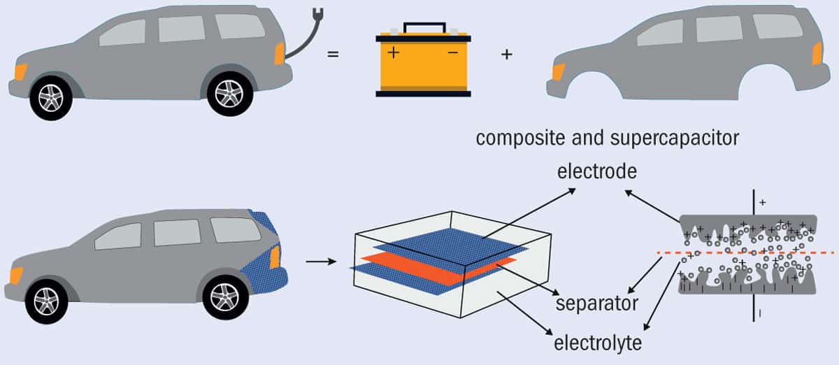
1 Multifunctionality concept (a) In conceptual terms, an electric car consists of a body (which provides structure) and a battery (which stores electrical energy – the motor was omitted as it is not included in the scope of the research). (b) Multifunctional electrical storage materials like the car boot lid shown here combine both aspects into a single structure. (Adapted from an original by Polina S Stefanova)
Supercapacitors lie somewhere between batteries and capacitors in terms of their energy and power densities, and their fast charge/discharge rates are particularly relevant to electric vehicles. A typical supercapacitor consists of an electrolyte, a separator, and two high-surface-area electrodes made from the same material. After adding a secondary, structural, function, it is essential that all these components still do what they did in their original, monofunctional form. For example, the surface area of the electrodes is very important, because the stored electrical energy is based on separating the charged species in an electrical double layer at the electrode/electrolyte interface after a voltage is applied.
Despite these limiting requirements, supercapacitors are attractive for multifunctional material developers. One reason is that their electrodes do not change dimensions when they are being electrically charged or discharged, and the material can last for an extremely long time (surviving for up to a million charging cycles). Battery systems, in contrast, offer higher energy densities, but their electrodes typically change shape as current passes through them, leading to stresses and degradation. Another attraction of supercapacitors is their layered architecture, which strongly resembles that of conventional composite laminates (figure 1).
There are other similarities between conventional supercapacitors and composite materials too. For example, carbon is used in both, albeit usually in different forms. Conventional supercapacitors use carbon as an electrode material because it has a high surface area and conducts electricity well; in contrast, carbon in composite materials is typically used as a reinforcement due to its excellent mechanical performance. So to achieve multifunctionality, these two sets of properties must be combined, which opens a range of possibilities for experimenting with modifying carbon.
Polymers are another component common to both energy-storage devices and composite materials. In their case, the overlap in function is less straightforward. In composites, the polymers must be rigid, as they have to transfer high shear loads; in electrochemical devices, in combination with electrolytes and solvents, the polymers must be mobile enough to allow ion conduction. Achieving the required combination of high ionic conductivity and good mechanical performance is challenging, as the intrinsic material characteristics conflict with each other.
One promising way around this conflict is to design a material that has two bi-continuous, interconnected phases: one responsible for structural performance and another that provides ionic conductivity. This route has shown potential not only in the preparation of structural electrolytes but also in electrode development (J. Comp. Mater.49 1823). In the latter, a bi-continuous structure can be achieved by impregnating unmodified carbon, for example carbon fibres, with a highly porous carbon material such as carbon aerogel (figure 2).
2 Versatile element Carbon is attractive for developers of structural superconductors because different forms of the material offer good electrical conductivity and excellent mechanical performance. This schematic illustration shows carbon fibres impregnated with carbon aerogel.
These amorphous carbonaceous materials are made of a 3D network of interconnected nanometre-sized particles with low density, large surface area, and high electrical conductivity. Integrating the properties of structural carbon fibres and carbon aerogel may make it possible to form an electrode material that not only has a high capacity for energy storage (due to its high surface area) but can also reinforce the structure of the composite.
Future challenges
Despite much progress in designing and developing individual multifunctional materials, this is only the first part of the challenge. Before they can perform well, such materials have to be integrated into effective systems that combine multifunctional materials. This process naturally creates further multifunctional demands – for example, the need for multifunctional current collectors, separators or encapsulation materials.
In addition, the behaviour and use of multifunctional materials does not always fit into traditional views of either composite materials or energy-storage devices. At times, even the language of the two communities can be inconsistent; for example, does the standard symbol σ denote stress, or electrical conductivity? New interdisciplinary collaborations will be needed to find practical solutions to these challenges, and thus to bring these unique systems closer to real-life applications.
Almost 60 societies in science, technology, engineering, mathematics and medicine (STEMM) have formed a consortium to tackle sexual harassment. Announced at the annual meeting of the American Association for the Advancement of Sciences (AAAS), in Washington, DC, the consortium has begun to collect model policies, law guidance as well as tools to prevent and respond to sexual and gender harassment in STEMM.
The importance of the consortium is that they can fill in where academic institutions cannot, by setting professional standards and raising the bar
Vicki Magley
The initial idea for a consortium was initiated in 2016 when the AAAS and the American Geophysical Union (AGU) recognized the need to tackle sexual harassment in STEMM. There then followed a report published in June 2018 by the National Academies of Sciences, Engineering, and Medicine (NASEM), which noted that current efforts to tackle sexual harassment in the sciences at US universities are failing and need urgent reform. The new consortium will be project managed by EducationCounsel, an education-consulting firm. Jamie Lewis Keith, a partner at EducationCounsel, notes that the consortium could also include international members or affiliations.
“A lot of STEMM disciplines are very male dominated, and we know from research that sexual harassment is more likely to occur in a male dominated context,” says psychologist Vicki Magley from the University of Connecticut, who sat on the NASEM report’s committee and is working with the consortium. “The importance of the consortium is that they can fill in where academic institutions cannot, by setting professional standards and raising the bar.”
Common principles
The hope is for societies and institutions to create a set of common principles and offer their experiences and resources to other members of the consortium, for example, the AGU already has the Ethics and Equity Resource Center that will be available to other STEMM societies. The AAAS, meanwhile, hosts SEA Change which is a voluntary certification, metrics and self-assessment voluntary programme for “barrier removal for women, blacks, Hispanics, Native Americans, and people with disabilities, as well as others who are marginalized”. Shirley Malcom, AAAS senior advisor and director of SEA Change, says that many of the consortium members are working to become SEA Change providers.
Beth Cunningham, executive officer of the American Association of Physics Teachers (AAPT), says she filed her organization’s consortium membership two days before the public launch. Joining the consortium will make sure the AAPT’s meetings are “inclusive and a welcoming space and safe” and help the association apply it to its members’ work environments. Cunningham adds that joining the consortium “shows our support for making science a safe space”, which is vital for her as too many women in physics have experienced harassment. “We have persisted, but it shouldn’t be that way,” says Cunningham.
Data from 27 net-importers of maize across Africa, Asia and Latin America since the turn of the millennium indicate that the variability in maize price could rise by 10% by mid-century due to climate-induced supply shocks. But this rise could be offset by a 10% boost in those nations’ ratio of imports to total consumption.
“International markets act as a source of stability rather than a source of risk,” says Bowen Chen of Kansas State University, US. “This is at least the case for maize.”
It’s well known that importing food products has the potential to alleviate domestic price instabilities brought about, for example, by local weather changes. But imports also expose countries to supply shocks that originate overseas – a problem made more acute because the export of many staple food products comes from just a handful of countries, such as the US.
Several studies have explored the stability of food prices for countries with different approaches to imports and market intervention. But none, says Chen, has specifically explored the effect of imports on domestic price stability while keeping other variables constant.
By performing a linear-regression analysis on data taken from 76 maize markets in 27 countries from 2000–2015, Chen and his Kansas colleague Nelson Villoria found that a 1% rise in the import ratio resulted in a 0.29% reduction in an intra-annual coefficient of variability of maize prices. In comparison, a 1% rise in stored maize for future consumption reduced the variability coefficient by 0.22%.
“At least in these data, imports and buffer stocks are substitutes to achieve similar targets of price instability,” says Chen.
Chen and Villoria point out that storing food brings additional costs, as well as the risk of spoilage. Nevertheless, says Chen, the variability coefficient is a summary statistic that can be easily understood and can “therefore help to facilitate policy debates regarding the role of imports in mitigating the price effects of domestic supply shocks”.
The researchers predicted future price variability using projections of maize yields under climate change. Calculations showed that a 10% boost in the import ratio would be enough to offset the greater future variability. It’s a target that would be easier for some countries than others. In some central African countries, for example, the maize import ratio is already less than 10%, so an additional 10% would, in relative terms, be roughly a doubling.
Chen and Villoria, who reported their findings in Environmental Research Letters (ERL), are now exploring how foreign yield shocks could affect their analysis. “The possibility that crop yield shocks in the exporting countries translate into food price instability in the importing countries has been a major concern of food importing and developing countries,” says Chen.
The 2019 Tyler Prize has been awarded to climate scientists Warren Washington of the US National Center for Atmospheric Research and Michael Mann of Penn State University, US.
“One of the things that makes this award special for me is sharing it with a personal hero of mine, Warren Washington…who has contributed fundamentally to the field of climate modelling,” says Mann.
Washington helped build one of the first computer models of Earth’s climate. “Dr Washington has been a pioneering climate scientist for over 40 years,” says John Shepherd, former deputy director of the Tyndall Centre for Climate Change Research. “Much of what is known about the Earth’s climate system and climate modeling is directly traceable to [his] lifelong work.”
Mann is perhaps most famous for his work on reconstructing past climate and his “hockey-stick” graph. He received “intense public scrutiny”, as the Tyler Prize press release puts it, then “chose not to retreat to the lab, but instead doubled-down on his efforts to make climate change science accessible to the public”.
“Professor Mann did not choose the easy way out,” says Naomi Oreskes of Harvard University, US, author of the book Merchants of Doubt. “For his courage in the face of this challenge, Mike Mann is not just a great scientist, but also a hero.”
“This award means a lot to me because it recognizes the two things that are most near and dear when it comes to my work and that’s contributing both to the advancement of our science and the effort to communicate that science to the public and policymakers,” says Mann.
Some refer to the $200,000 Tyler Prize, which was founded in 1973 by the late John and Alice Tyler, as the “Nobel Prize for the environment”. The prize covers environmental science, environmental health and energy. Previous winners include climate scientists Richard Alley, Veerabhadran Ramanathan and Roger Revelle, biologist Edward O. Wilson, primatologist Jane Goodall and conservation biologists Anne Ehrlich and Paul Ehrlich.
Washington and Mann will give a public lecture on their work in San Francisco on May 2nd and receive their prize at a ceremony the following day.
The MoS2 FET-based biosensor and response-concentration graph. Reprinted with permission from Nano Lett. 10.1021/acs.nanolett.8b03818 Copyright 2019 American Chemical Society
A new, sensitive field-effect transistor-based biosensor made from 2D molybdenum disulphide could be used in non-invasive prenatal testing for Down syndrome. The device, which is functionalized with gold nanoparticles containing DNA probes that specifically target DNA fragments of chromosome 21, is sensitive to this DNA at concentrations as low as 0.1 femtomoles/litre.
Down, or trisomy 21, syndrome is caused by the presence of an extra copy of chromosome 21 within the genome and is the most common birth defect, occurring in roughly one in 800 births. Current screening techniques include ultrasound scans or indirect biomarkers tests – for example, testing for α-fetoprotein, chorionic gonadotropin and free estriol. However, these have limited accuracy and high misdiagnosis rates. Amniocentesis, which does provide a definitive diagnosis, is not without risk to both the future mother and foetus, and whole-genome sequencing – a highly accurate technique – is slow and expensive because it needs to amplify all genomic segments and sequences owing to the extremely low, sub-femtomole, concentrations of foetal DNA.
The researchers functionalized the MoS2 with gold nanoparticles and then topped these with probe DNA sequences. These DNAs specifically capture target DNA fragments of chromosome 21 in maternal blood samples. When bound to the probes, these fragments cause the electric current measured across the FET sensor to drop.
The researchers believe that the main mechanism behind this drop is the p-doping of the MoS2 channel by the probe DNA or target DNA, which should lead to a decrease in the number of electrons within local regions of the MoS2 channel.
Ultrahigh detection limit
Compared with previously reported MoS2 FET biosensors, the new sensor has an ultrahigh detection limit of 100 attomoles/litre, a high response of 240% and excellent selectivity. A real-time test also showed that the biosensor responds to the target DNA at concentrations as low as 1 fM/L. All these properties satisfy the essential requirement for Down syndrome screening, says the team.
According to Zhang and colleagues, the high sensitivity and large response of the FET biosensor come mainly thanks to two factors. The first is the high-quality and uniform monolayer MoS2 used as the channel in the FET that is extremely sensitive to surface dopants. Second, the gold nanoparticles employed, which are of the right size and right density, functionalized on the MoS2 channel serve as excellent linkers for the probe DNA.
And that is not all: as well as detecting chromosome 21 for Down syndrome, the sensor might also be used as a universal platform to detect receptors like proteins, viruses, antibodies, and nucleotides simply by replacing the probe molecules, say the researchers.
Communities in remote rural locations around the world often lack access to medical equipment such as imaging devices. Ultrasound scanners, for example, could bring numerous benefits to such low-resource populations, both for obstetrics and more general medical applications. And as smaller, more portable ultrasound units are developed, it should become easier to bring such devices into remote areas. The use of ultrasound in austere environments, however, is limited by battery life.
In the Zanskar Mountain Range in India, for example, most people live in small villages that are inaccessible for over half of the year, due to extreme cold and heavy snow. To help such remote communities, the Himalayan Health Exchange organizes a trek each August to provide medical and dental services to these under-served villages. And in 2017 and 2018, four emergency medicine residents from Palmetto Health Richland joined the trek.
The Palmetto physicians brought along a portable Lumify ultrasound system and demonstrated that it could be powered using only a solar panel for the entire month-long trip. This was the first time that an ultrasound unit had been brought on the remote medical mission and represented the first use of medical imaging in this remote Himalayan region (J. Ultrasound Med. 10.1002/jum.14923).
First author Laura Nolting, ultrasound director at Palmetto, described how this came about: “I was working in the paediatric emergency department one evening when I noticed Dr Daniel Baker was wearing hiking boots. I inquired about his unorthodox shoe selection and he informed me he was breaking them in for an upcoming global health trip to the Himalayas,” she explains. “He told me about the trek and using a solar pad as the primary source of power and I asked if he thought the portable Lumify would work; and we just went from there.”
Team members on the Kargaikh High Altitude Trek. (Courtesy: Marc Kushinka)
The trek supplied medical care to eight clinics, all located between 12,000 and 16,400 feet above sea level and accessible only by foot. All the required medical and personal equipment was transported by the team — 20–30 medical students, residents, support personnel and physician instructors — with limited use of donkeys and horses.
With no central power grid or infrastructure in this region, the team relied exclusively on solar power. They used a low-cost, lightweight Anker solar panel, which has three ports that provide 2.4 A each with enough direct sunlight. They brought along two handheld ultrasound Lumify probes (curved linear and linear), along with a compatible Galaxy tablet to display the images. In total, the tablet, probes and solar pad added only about 1.1 kg of weight.
Case studies
On a typical clinic day, the team saw 20 to 50 patients, approximately half of whom underwent scanning. Patients that received ultrasound exams included, for example, a woman with ankle pain following a stumble. The high-frequency linear probe clearly showed that she had a fractured ankle and she was placed in a splint and crutches.
The team also saw several children with high blood pressure and heart rate. The children underwent physical examination with additional ultrasound exams that revealed normal-appearing cardiac activity with no evidence of pulmonary hypertension. Another patient was a pregnant woman with occasional abdominal pain. The woman, who lived a 2–3-day walk from the closest hospital, received foetal sonography and was reassured that the foetus was developing as expected.
The solar panel was charged on the roof of the tent used for medical care. The authors note that the time required to completely charge the ultrasound probe was dependent upon the available direct sunlight on each particular day, making charging times unpredictable. However, they were still able to scan as and when needed and did not encounter a time when the machine was inadequately charged for use.
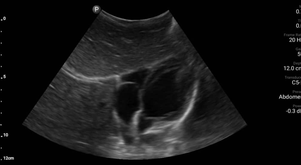
Ultrasound scan of a paediatric male’s heart demonstrating normal-appearing cardiac activity without evidence of pulmonary hypertension. (Courtesy: L Nolting et al J. Ultrasound Med. 10.1002/jum.14923).
Other problems faced included low ambient light inside the tent making the tablet screen difficult to see at times, and software updates for the probes, which had to be done before the trip began. But even with its limitations, the team concluded that solar-powered sonography is a feasible option to enhance medical care in some of the most isolated and inaccessible regions of our planet.
“The Himalayan Health Exchange has been offering this trek for the past 17 years. I have had residents participate for the past several years. I am not sure if anyone is planning on going this upcoming year but a few residents have voiced interest,” Nolting tells Physics World.
In a twist on a classic physics experiment, researchers in the Netherlands and France have worked out why room-temperature alcohol droplets will levitate and propel themselves across a pool of liquid nitrogen for long periods of time. The team, led by Anaïs Gauthier at the University of Twente, have studied the propulsion associated with the “inverse Leidenfrost effect” and their work could lead to more efficient ways to transport small amounts of biological material.
The Leidenfrost effect arises when a liquid droplet is deposited onto a surface hotter than its boiling point, causing the bottom of the droplet to evaporate continuously. This creates a repulsive cushion of vapour, which both prevents the droplet from quickly boiling away, and causes it to hover above the hot surface. There is virtually no friction between droplet and surface, and changes in surface texture can cause the droplet to accelerate, climb small hills and even negotiate a maze.
Formally identified in 1756 by the German scientist Johann Leidenfrost, the effect has probably entertained people for millennia and has become a popular classroom demonstration.
Hot objects
The inverse Leidenfrost effect was first described in 1969 and involves a hot object such as a droplet levitating above a cold liquid. In this case, heat from the droplet causes some of the cold liquid to evaporate, creating the repulsive cushion of vapour.
Gauthier’s team did this by depositing a room-temperature droplet of alcohol on top of a pool of liquid nitrogen at −196 °C. Within just a few seconds of deposition, the droplets propelled themselves from rest to reach speeds of several centimetres per second; gliding in straight lines before ricocheting off the container walls (see video). This continued for tens of minutes before the droplets slowed down as they cooled to the temperature of the nitrogen bath. Gauthier and colleagues propose that this self-propelling behaviour arises from subtle symmetry-breaking in the vapour film, which cause vapours to flow out from under the droplet, inducing drag.
Based on their observations, the physicists constructed simulations to model the inverse Leidenfrost effect. By modelling variations in vapour film thickness and the cooling dynamics of the drops, they could accurately recreate the variations in their observed droplet velocities.
Gauthier’s team believe the effect could be used to develop efficient techniques for freezing and transporting biological materials including cells and proteins. With the help of simulations, they hope that this transport could occur with no risk of contamination or heat degradation to the materials.
While proton therapy is becoming a standard treatment option in radiation oncology – there are currently 92 operational proton facilities worldwide and a further 45 under construction – many challenges remain in terms of the fundamental physics, radiobiology and clinical use of protons for the treatment of cancer. Those challenges, and plenty more besides, were front-and-centre at the UK’s Fifth Annual Proton Therapy Physics Workshop held early in February at the National Physical Laboratory (NPL) in Teddington.
The timing of this year’s event was apposite. In 2018, the UK’s National Health Service (NHS) opened its first high-energy proton therapy centre at The Christie Hospital in Manchester, with a second facility at University College London Hospital (UCLH) scheduled to come online for patient treatment in 2020. Proton Partners International, a private health provider, is also rolling out a network of four proton-therapy facilities across the UK, with the first of its Rutherford Cancer Centres now treating patients in south Wales.
“PPRIG was set up by NPL in 2012 to progress UK deployment of high-energy proton therapy,” explained Russell Thomas, senior research and clinical scientist at NPL and chair of PPRIG. “We aim to help coordinate research activities, encourage multicentre collaboration and minimize duplication of effort. Our annual proton-therapy physics workshop is a logical extension of PPRIG’s remit, bringing the UK proton-physics community together with leading researchers from overseas.”
It’s good to talk: delegates at the annual conference on proton therapy (Courtesy: NPL)
Another objective of PPRIG is to promote the work of early-career researchers, helping them to build networks, collaborate on specific research problems, and support their grant applications. “The workshop fosters open, robust, but always good-humoured debate around the hot topics in proton therapy,” Thomas added.
Ana Lourenço, a postdoctoral research scientist at UCL and NPL, agrees that the PPRIG meeting provides a welcoming platform for younger scientists. “The PPRIG workshop was a great opportunity for early-career scientists to present their work and have feedback from world-leading medical physicists and clinical scientists,” she said. “The reduced registration fee for students allowed many to participate, with plenty of time in the programme dedicated to more open, informal discussion to facilitate the interaction between students and senior researchers.”
Standardize and verify
Given NPL’s role as the UK’s national measurement institute, much of the discussion at last week’s meeting eddied around issues of dosimetry and quality assurance (QA). In other words, how to maximize clinical outcomes by ensuring that patients receive standardized and rigorously audited proton therapy – irrespective of where they’re being treated.
With those outcomes in mind, Thomas reported on NPL’s work to develop a code of practice for proton-beam dosimetry in collaboration with the UK Institute of Physics and Engineering in Medicine (IPEM). Underpinning that code is absolute dosimetry for calibration of the proton beam, something that NPL is striving to improve through the development of a primary standard for protons based on a portable graphite calorimeter, in which the temperature rise due to a typical patient dose is measured to quantify the amount of “dose” deposited.
Previously, the only option for proton-beam reference dosimetry was a 60Co-based calibration, which has an uncertainty in terms of reference dosimetry that’s often quoted at 4.6% – but which anecdotally may be somewhat higher. With the increase in patient numbers for proton therapy, it is desirable to bring this uncertainty down to a similar level achieved with the reference dosimetry of conventional photon radiotherapy, which would be closer to 2%.
Thomas says that the NPL calorimeter has been transported to proton-therapy centres in Liverpool (Clatterbridge), Manchester (The Christie), Newport (Rutherford Cancer Centre), Sicily, Prague and Japan, where it has been operated successfully in clinical settings.
“We need to improve the uncertainty of the dose delivered to patients to ensure the best possible consistency across the patient population and to fully understand and interpret the patient outcomes,” he explained. “By bringing the uncertainty on reference dosimetry down to a similar level as that currently achievable in conventional photon radiotherapy, the primary standard will aid in the comparison of the results from high-quality, multicentre clinical trials featuring different treatment techniques.”
Stuart Green, director of medical physics at University Hospital Birmingham, told Physics World that the new IPEM code of practice for proton dosimetry relies heavily on calorimetry developments at NPL over the past 15 years. He says the new code will be ready for publication later this year and hopes that UK centres will transfer to the new approach soon after.
“What’s more,” Green added, “the NHS has a significant opportunity with the opening of the two new proton-therapy centres [at Christie and UCLH] to initiate definitive clinical trials. I am sure the rest of the world will watch with interest to see how well these trials are rolled out.”
Other speakers developed the QA theme in terms of reference and in-clinic proton dosimetry. NPL’s Lourenço, for example, reported findings from a team of UK and Danish scientists who compared the response of user ionization chambers at three clinical facilities against NPL reference ionization chambers.
Their study, which involved a low-energy passively scattered proton beam and two high-energy pencil-beam-scanning proton beams, showed “good agreement between the results acquired by NPL and the proton facilities”. Lourenço added that “reference dosimetry audits such as this are important to improve accuracy in radiotherapy treatments, both within and between treatment facilities, and to establish consistent standards that underpin the development of clinical trials.”
In the same session, Antonio Carlino of the MedAustron Ion Therapy Center, Austria, detailed a new approach for end-to-end auditing of the treatment workflow based on customized anthropomorphic phantoms featuring different types of detector. During the dosimetry audit, which was carried out at HollandPTC in Delft, the phantoms followed the patient pathway to simulate the entire clinical procedure, mimicking the human body as closely as possible in terms of material properties and movement. Carlino told delegates that human-mimicking phantoms deployed in end-to-end audits of this type “may serve as dosimetric credentialing for clinical trials in the future”.
Image and adapt
Standardization and QA notwithstanding, there was plenty of focus on new concepts and emerging technologies for the proton-therapy clinic, with imaging at the point of treatment delivery and online adaptive proton therapy very much to the fore.
“It is one thing to deliver the sophisticated treatment dose volumes that proton therapy is capable of, but it is another to be confident that the dose is being delivered to the right place,” explained Thomas. “During a course of treatment lasting up to six weeks, with daily fractions, the anatomy of a patient may change dramatically as a result of weight loss and/or tumour shrinkage. Imaging can ensure the dose is still being delivered correctly, while informing dynamic refinement of the treatment plan over the course of the treatment.”
Central to the success of adaptive proton therapy is a technique called deformable image registration (DIR), the use of powerful image-processing tools that maximize spatial correspondence between multiple sets of images (e.g. CT scans) collected over an extended treatment timeframe, and even across multiple imaging modalities.
Many developments will take place in the next few years, enabling dose rates to increase and treatment times to reduce
Tony Lomax, Paul Scherrer Institute
However, according to Jamie McClelland from UCL’s Centre for Medical Image Computing, a number of open questions remain around online deployment of DIR in the proton-therapy workflow: “What exactly do we want DIR to do? How do we know it’s doing it correctly? And how do the errors and uncertainties in DIR impact clinical applications?”
More broadly, what of the longer-term development roadmap for proton therapy? Harald Paganetti, director of physics research at Massachusetts General Hospital (MGH) and professor of radiation oncology at Harvard Medical School in the US, reckons proton therapy is currently at what he calls the “Adaptive 1.0” stage, with CT scans performed daily but treatment plans revised and adapted offline.
MGH’s evolution to “Adaptive 2.0” will see that adaptation taking place online in the proton treatment workflow. Key enablers of the MGH approach include cone-beam CT-based imaging and online measurement of the prompt-gamma emissions from delivery of a “partial dose” to the centre of the target (enabling an initial assessment of range accuracy). This is then followed by a rapid adaptation before the remainder of the dose is delivered for a given fraction.
For protons, it seems, the future is bright, with no shortage of opportunities for progress across core physics, emerging technologies and clinical applications. “Pencil-beam-scanning proton-beam therapy is still in its infancy,” noted Tony Lomax, chief medical physicist at the Paul Scherrer Institute in Villigen, Switzerland. “Many developments will take place in the next few years, [enabling] dose rates to increase and treatment times to reduce.”
An efficient new method to find out whether a material hosts topological states or not could help increase the number of known topological materials from a few hundred to thousands. The technique is very different to conventional target-oriented searches and uses algorithms to sort materials automatically according to their chemical properties and properties related to symmetries in their structure.
Xiangang Wan. Courtesy: Nanjing University
Topological materials – exotic materials whose surface properties are very different to those in their bulk – have created a flurry of interest in recent years and are currently revolutionizing modern condensed matter physics thanks to their unique properties that come from their topology. Topological phases of matter are so-called because they are mathematically described by global invariants that are unaffected by imperfections, such as defects or other variations, in a material.
An example of a topological material is a topological insulator (also known as a 2D quantum spin Hall insulator). These are materials that are electrical insulators in the bulk but which can conduct electricity extremely well on their edge via special topologically protected electronic states. Electrons can only travel in one direction along these states and do not backscatter. This means that they can carry electrical current with near-zero dissipation of energy and so could be used to make energy-efficient electronic devices in the future.
Topological insulators were discovered over 10 years ago and we now know of a few hundred materials that belong to this category of material. Only a dozen or so of these appear to be suitable for real-world applications, however.
Symmetry indicators theory
To predict whether a material can host topological states, researchers mainly rely on complex theoretical calculations. Two teams, one at Princeton University in New Jersey and the other at Harvard University in Cambridge, Massachusetts, recently put forward a new approach based on the recently established theory of symmetry indicators, however, to speed up this search process. In these studies, the physicists use algorithms to sort materials automatically according to their chemical properties and properties that come from symmetries in their structure. These symmetries define where electrons move in the crystal lattice and can be used to predict how electrons will behave – and therefore whether a material can host topological states or not.
Researchers at Nanjing University in China led by Xiangang Wan together with the Harvard team, led by Ashvin Vishwanath, have now shown that the computation of symmetry indicators for any crystalline symmetry setting can readily be integrated into standard first-principle calculations. “In stark contrast to conventional target-oriented searches, our technique does not presuppose any specific phase of matter but instead automatically identifies all nontrivial electronic band structures and then classifies them as either topological insulators, topological semi-metals or topological crystalline insulators,” explains Wan.
To show how powerful their algorithm is, the researchers began by analysing crystalline materials in eight different space groups (which represent a description of the symmetryof a crystal) and say they have unearthed hundreds of materials capable of hosting topological phases.
“Two topological crystalline insulators in particular are worth mentioning,” says Wan. “The β-MoTe2 (space group 11) with screw-protected hinge states, and the BiBr (space group 12) with a rotation anomaly. The existence of the novel topological feature in β-MoTe2 was verified in a later study (arXiv:1806.11116).”
Fast technique
In traditional topological-material-discovery algorithms, we have to first pre-assume a specific topological phase and then calculate the corresponding topological invariant(s), a process that is usually very time-consuming,’ says Wan. “Our new technique is very fast and we have used it to comprehensively search for topological materials in 230 space groups in all (arXiv:1807.09744),” he tells Physics World.
The researchers, reporting their work in Nature Physics 10.1038/s41567-019-0418-7, say that many of the topological materials they have discovered using their approach could be promising for making next-generation electronic devices. Indeed, their work has already attracted much attention.
The approach is not just limited to topological materials either and could be extended to other 2D and magnetic materials, says Wan. “Our present work focuses on electronic band topology, but the symmetry indicators of phonons, photons and magnons could also be built and used to search for topologically non-trivial phases in these systems.”