Starting out in her physics role at Elekta, a global provider of cancer-treatment technology including linacs, Sandra Fisher provided remote high-level technical support. Moving from a hands-on job as an accredited radiotherapy physicist in a busy Sydney clinic, she had flickers of doubt. These days, however, she has no regrets. “It has opened up so many doors and opportunities,” says Fisher.
Technical support turned out to be rewarding work, helping multiple clinics – in contrast to her clinical role in a single department. “Every single time you answer the phone it’s something different, because you now have many hospitals calling you for advice.” Fisher’s role has since evolved, influenced by her experience as an Elekta customer. “One of the key areas that I felt that was lacking here in Australia was linac training for physicists,” she says. Addressing this need, Fisher implemented a five-day course for those new to Elekta linacs, to enthusiastic feedback from hospitals.
Fisher also provides physics expertise to potential customers before sales discussions begin. A few steps later, she supports clinical physicists installing new accelerators, “essentially to hold people’s hands while they wrap their heads around their new technology”. As much of her work is practical and face-to-face, Fisher often travels all over the world. While there are only a few radiotherapy vendors, the companies are global and job opportunities are out there, says Fisher. Once a candidate is in , jobs like hers can also lead to non-physics roles such as product management and sales.
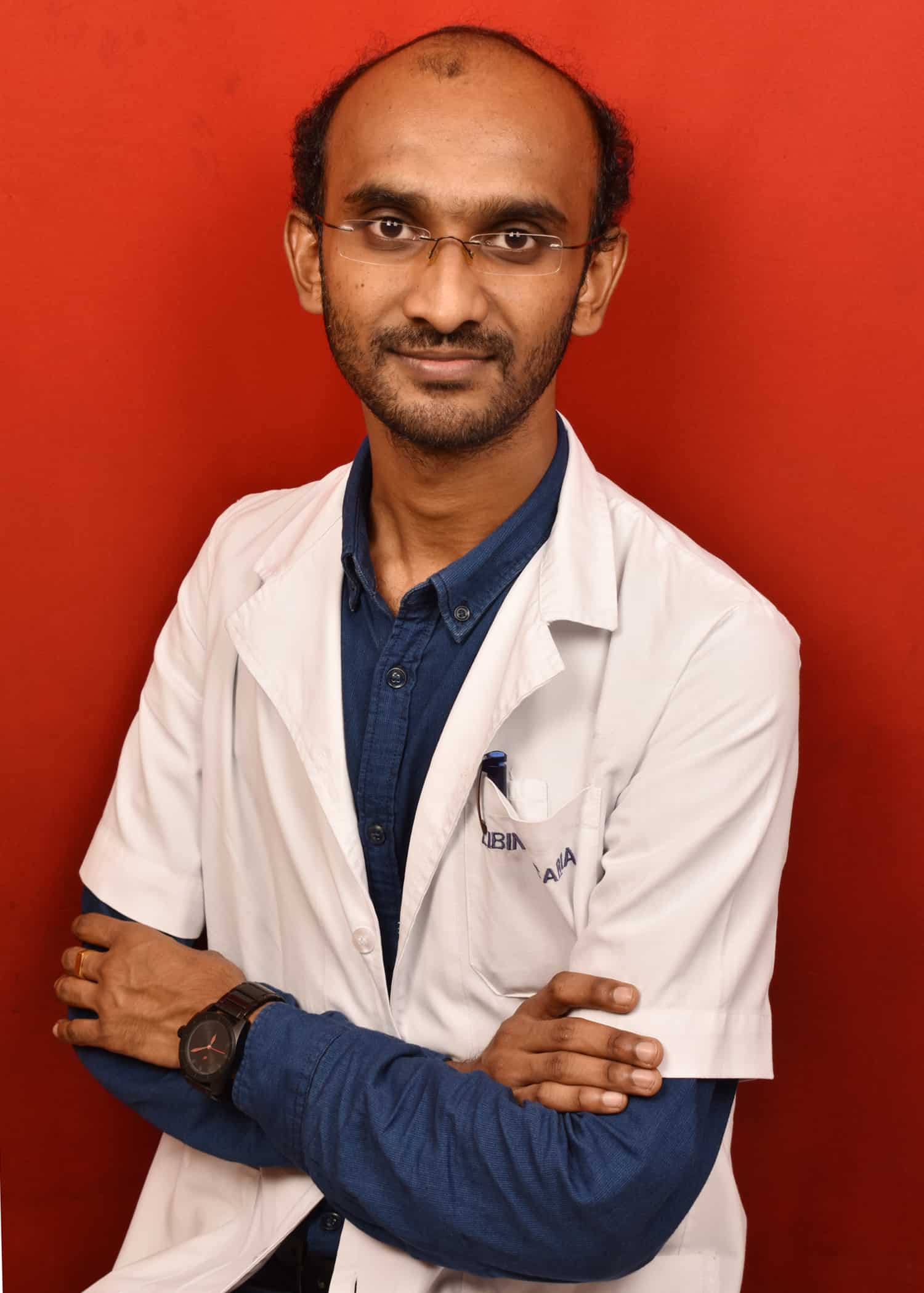
“The personal satisfaction you get by doing something with a direct social benefit is very high,” says Libin Scaria, who works as a clinical radiotherapy physicist at the Tata Memorial Centre in Mumbai, India. His bustling workplace is a national centre for cancer treatment, research and prevention, funded by the Department of Atomic Energy.
Scaria helps cancer patients directly in his work. Every morning he tests the linacs used to treat patients, making sure they deliver high-energy X-rays safely and accurately. Specializing in breast cancer and brain and spinal cord tumours, he also designs treatment plans – a key responsibility of physicists in India.
He is never bored. “That’s the thing that I like, because every patient is a different challenge.” The rest of Scaria’s time is divided between teaching radiation therapists and medics, and carrying out project work, such as the introduction of new treatments. He enjoys the resulting variety in his day. “Its a wide area. There is so much to do.”
Following one of two possible paths in India, Scaria began his job after a one-year internship at the Indo American Cancer Centre in Hyderabad. As many physicists work in remote areas with a shortage of experienced colleagues, the experience is essential, says Scaria. “It enables junior physicists to practise independently from the beginning of their career.”
As part of his training, Scaria also completed a one-year diploma in Radiological Physics at the Homi Bhabha National Institute, also in Mumbai, following a bachelors and masters in straight physics. The government-run diploma is highly regarded. Though competition for places is fierce, Scaria recommends it over the alternative, a medical physics masters, where the quality of training is variable.
Heather Williams was just 15 when her interest in nuclear medicine was sparked during a high-school physics class, learning how images of radioisotopes injected into patients could be used to diagnose diseases. “I was just fascinated at this idea of putting radioactive stuff inside people, which sounds like a pretty bad idea on the surface of things.” Today, she is a principal physicist at the Christie Hospital in Manchester, UK, a specialist cancer care and research centre.
In fact, nuclear medicine is a vital and safe set of techniques for diagnosing and treating a variety of diseases, including cancer. Williams’ work combines clinical duties with research and teaching, with a heavy focus on PET. “It’s a really nice mix for me.” Her qualifications include a master’s in medical physics and a PhD in PET imaging. She also completed the NHS training scheme and obtained state registration, allowing her to practice as a clinical scientist, a legally protected title.
Much of her clinical work revolves around image quality and radiation safety, as for a short time following injection, patients become walking radioactive sources. “We need to have diagnostic-quality images that we can pass on to the doctors, so they can make decisions about what’s going on inside the patient.” She has to be mindful of any factors that could degrade the images, and must often come up with solutions on the spot. During a scan, for instance, a patient might not be physically able to hold a particular position that gives the best quality images.
Williams finds her clinical duties especially rewarding as she knows she is directly helping the patient sitting in front of her. “I really like coming to work and thinking that I’m going to make a difference today.”
A brush-shaped structure called a pappus creates vortex rings that help keep dandelion seeds aloft, according to a comprehensive study by researchers at the University of Edinburgh in the UK. The team was also able to created artificial structures that mimicked the seeds.
Dandelions are yellow wildflowers that grow in temperate regions. Widespread and prolific, the plant is often considered a weed but is also edible and has a number of culinary uses.
The dandelion flower – actually a collection of multiple flowers – matures into white blowball full of single-seed fruits attached to a bundle of bristle filaments called a “pappus” (see figure). Resembling a chimney brush, the pappus helps the windblown seed to stay aloft over incredible distances. Although most dandelion seeds travel less than 2 m from their flowers, seeds can easily travel over 100 km in warm, dry air.
Fluid mechanics
A pappus has 100–110 filaments, which together have a large surface compared to the area of the dandelion seed it bears. Scientists had thought that dandelion seed can fly thanks the large drag force (or resistance to moving air) of individual filaments. By combining fluid mechanics, plant biology and microfabrication, the Edinburgh team has discovered that the effect of drag alone cannot explain why the seeds are so good at remaining airborne.
“We had reasons to believe that the lift enhancement from an individual filament would not be enough to provide sufficient force for the dandelion to remain aloft for so long. Therefore, we decided to investigate whether there was an interaction between the filaments that was strong enough to generate a flow feature that would operate up the sides of the entire dandelion seed instead of beside the individual filament,” explains Ignazio Maria Viola, who led the fluid mechanics aspects of the research.
Smoke and light
To gain a better understanding of the object’s aerodynamic properties, a dandelion pappus was studied using X-ray computed microtomography to assess its features and porosity – the latter being a measure of how easily air can flow through the structure. Then, researchers built a vertical wind tunnel that blew air upwards to keep a seed to hovering at a fixed height. The airflow in the tunnel was traced by illuminating smoke with a laser and recording its motion with a high-speed camera.
The wind tunnel experiment showed that a “separated vortex ring” forms in the air just above the dandelion filaments. “The existence of the vortex had been considered from the theoretical point of view in the past, but it was argued that it would be too unstable to occur in nature,” explains Viola.
As it turns out, the vortex is stabilized by the airflow through the filaments. As the air flows around each filament, it also interacts with the flow of its neighbouring filament, thus creating a so-called “wall effect”. The flow through the filaments is considerably reduced due to this interaction, which, in turn, buoys-up the dandelion seed.
Once the separated vortex is stabilized, it increases the ability of the seed to fly, because it confers an upward force on the seed. What is more, the pappus structure is four times more efficient at keeping the seed aloft than a parachute-shaped structure of similar mass.
Slotted disks
To further understand the process, the team used microfabrication techniques to make pappus-inspired structures from tiny disks of silicon. These disks had radial slots cut into them to allow air to flow through, in the same way that air can flow between the filaments of a pappus. They found that disks with no slots did not create separated vortex rings, whereas slotted disks with porosities similar to a pappus did create separated vortex rings.
The team also explored whether dandelions from different parts of the world had similar pappus structures. After obtaining seed samples from all over the world, they found little variation in the number of filaments in the pappus – suggesting that the separated vortex observed in Edinburgh dandelions was not a local fluke.
The dandelion has somehow magically understood the perfect number of filaments necessary for the vortex stabilization
Ignazio Maria Viola
Even samples collected from distant locations such as Japan or China have very similar number of filaments. “The dandelion has somehow magically understood the perfect number of filaments necessary for the vortex stabilization,” says Viola.
The team believes that there are many more examples of structures with tiny filaments and hairs with interesting fluid mechanics functions across both animal and plant kingdoms. Apart from flying, these structures may be used in swimming, feeding or even cleaning.
The biologist Camilla Pandolfi from the University of Florence told Physics World “Flying seeds evolved to maximize their dispersal distance by optimizing the shapes of their little parachutes, and the [Edinburgh team] found a new interesting way they are doing it”. She adds, “I am very glad to see that other scientists are paying attention to the many different seed dispersal strategies, as they are a huge unexplored source of inspiration for future technologies”.
Umklapp electron-electron scattering in so-called moiré superlattices made from aligned layers of 2D materials could degrade the high-temperature mobility of charge carriers. That’s the conclusion of a new study by researchers in the UK, US and Japan, who say the effect would limit the potential applications of these technologically important materials in high-mobility devices operating a room temperature. The problem could be overcome, however, by misaligning, or twisting, the 2D layers with respect to each other.
Umklapp electron-electron (Uee) scattering is the process that gives pure metals an electrical resistance, explain the research team, which was led by Vladimir Fal’ko and Andre Geim of the National Graphene Institute at the University of Manchester in the UK. It is the only intrinsic mechanism that allows electrons to transfer momentum to the crystal lattice, but it is difficult to measure in an experiment because it is often masked by other dissipation phenomena.
Fal’ko and colleagues have now shown that Uee scattering dominates the transport properties of superlattices that are made by placing layers of graphene on top of hexagonal boron nitride (hBN). Uee processes in these heterostructures lead to giant excess resistivity that rapidly grows as the period of the superlattice increases, causing their room-temperature mobility to plummet by more than an order of magnitude when compared to ordinary, non-superlattice graphene devices.
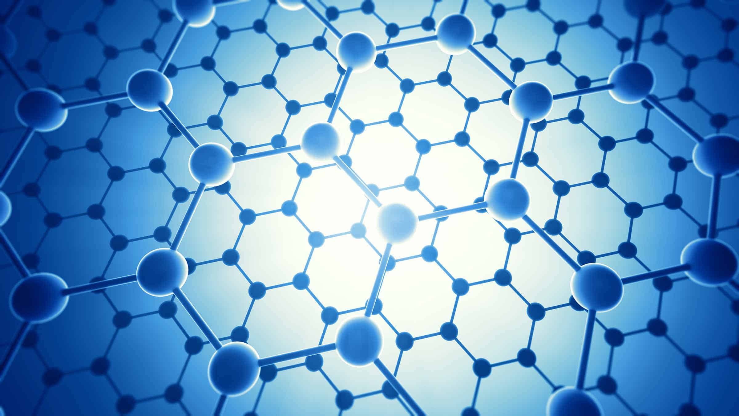
The researchers engineered their superlattices by aligning graphene with an hBN substrate. This process produces a moiré pattern as a result of the small lattice mismatch (of 1.8%) between the two materials. The moiré lattice produces a potential with a period of around 15 nm when the two materials are perfectly aligned, and this periodic potential dramatically alters the material’s electronic properties – transforming it from a “zero-gap” semiconductor to one that does have a band gap.
In a zero-gap semiconductor, the electron valence and conduction bands just touch each other at the so-called Dirac point, while conventional semiconductors have an energy gap between the bands. At the Dirac point, the relationship between the energy and momentum of the electrons can be described by the Dirac equation and resembles that of a photon, with the electrons moving at a very high, relativistic, speed.
One of the most important things to happen in a moiré superlattice is the creation of a mini Brillouin zone around the Dirac point. The size of this zone depends on the misalignment angle between the graphene and hBN and the resulting moiré period. Since the Brillouin zone is small compared to that in normal metals, Uee scattering is the dominant effect in graphene/hBN superlattices, say the researchers, who obtained their result by measuring the resistivity of different superlattice devices and a “reference” device in which the graphene and hBN were intentionally misaligned by more than 15°. This device had a moiré period of less than 3 nm.
“Uee scattering in long-period moiré superlattices degrades the intrinsic high-temperature mobility of graphene’s charge carriers,” they report in Nature Physics 10.1038/s41567-018-0278-6. “This limits the potential applications of epitaxially-grown graphene/hBN heterostructures, which are inherently aligned, for room-temperature high-mobility devices.”
The researchers advise that the 2D crystals that form the heterostructure should be misaligned, or twisted, to achieve high carrier mobility at room temperature.
Commercial systems that harness the power of light are allowing researchers to probe everything from novel materials to fundamental physical systems. These recent highlights show how light-based tools can be applied to carbon nanomaterials, biomolecular structures, and the quantum and atomic worlds.
Raman measurements extended to “challenging” inorganic materials
Raman spectroscopy has become a powerful tool for probing the structure and properties of graphene, carbon nanotubes and other thin-film structures. That’s why Princeton Instruments, a US-based manufacturer of high-performance cameras and spectroscopy equipment, has now added 532 nm Raman capabilities to its popular FERGIE spectrograph.
(Courtesy: Shutterstock/Mopic)
Raman measurements at 532 nm offer better sensitivity and higher resolution than those at longer wavelengths, allowing researchers to use FERGIE to characterize challenging inorganic materials such as graphene and other nanostructures. “FERGIE provides a carefully conceived ecosystem that not only allows researchers to design an experiment quickly but also to switch between different experiments in minimal time,” comments Peng Zou, FERGIE product manager at Princeton Instruments.
The FERGIE system includes modular accessories that allow users to perform high-precision measurements using several complementary characterization techniques, and it is now fully equipped for both 532 nm and 785 nm Raman applications.
Molecular interactions brought into view
Optical tweezers have become an essential tool for manipulating single cells and molecules, so much so that its invention in 1986 won Arthur Ashkin the 2018 Nobel Prize for Physics. Scientists and engineers have continued to refine the technique in the intervening years, and Lumicks is the first company to offer a commercial system that combines optical tweezers with fluorescence microscopy. This system, called the C-Trap, allows molecular interactions to be visualized and manipulated at the same time.
The C-Trap in action
Researchers in the Netherlands have recently used the C-Trap to devise a new technique to quantify the molecular tension in DNA and other biomolecular structures (Nano Lett.18 2274). The technique, which measures the fluorescence from dyes intercalated in the DNA, achieves a resolution of 1–3 pN over a range of at least 0.5–65 pN.
Laser lights up atomic and quantum research
A tunable continuous-wave laser from HÜBNER Photonics has been specifically designed for demanding applications in atomic physics and quantum optics. The high-precision C-WAVE laser delivers single-frequency radiation in the 450–650 nm and 900–1300 nm ranges, with output powers reaching 200 mW at visible wavelengths and 400 mW in the near infrared.
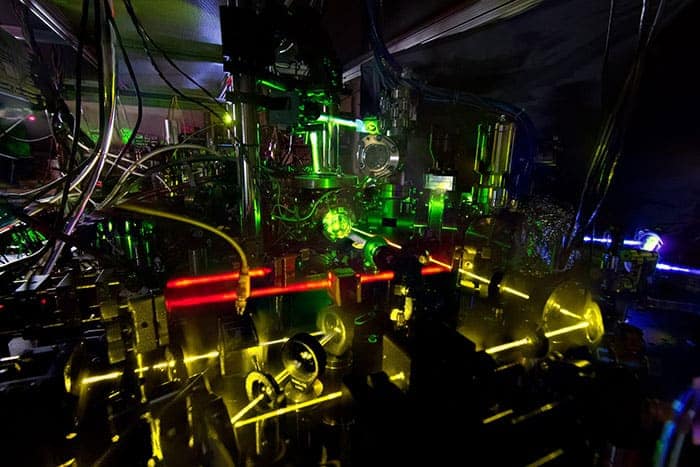
Lasers for atomic physics at NIST
A recent innovation is the addition of precise wavelength control, called AbsoluteLambda, which allows the wavelength to be selected automatically with high accuracy (set point ± 1 MHz) and drift-free wavelength stabilization (±1 MHz). Full product information is available on the HÜBNER Photonics website.
Visit the Physics World Buyers Guide to find the best technology and supplier for your next scientific project.
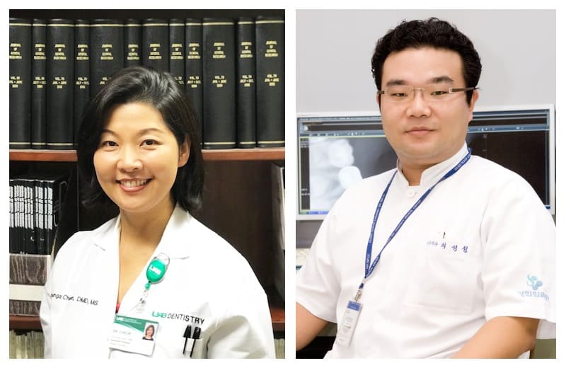
Authors Kyounga Cheon (left) and Sung Chul Choi (right) and their colleagues designed a novel gel to fill teeth, enabling a better root canal treatment.
Root canal treatment, a common dental surgery, currently relies on clotted blood to fill the empty tooth canal. Most times it goes well. But having a reliable, biodegradable filling that promotes healing and regeneration of the tooth would be a great advance. Sung Chul Choi from Kyung Hee University, Kyounga Cheon from the University of Alabama at Birmingham and their colleagues have developed a gel filling for root canal treatment that releases nitric oxide and antibiotics inside the treated tooth. The pilot study shows that this new canal filling material has potential for boosting regeneration of the impaired tooth (Plos One 10.1371/journal.pone.0205534).
When the pulp of a tooth, its blood vessels and nerves, is infected, root canal treatment becomes necessary. This can happen when the pulp is exposed following damage caused by caries or mechanical injury. In such cases, the infected tissue inside the tooth is removed, the area is disinfected, medicine and canal filling material are placed in the cavity and the crown is restored.
Currently, blood clots are used as a canal filling material. The composition of these blood clots cannot be controlled and the procedure, while mostly successful, can have negative effects like discolouration or fractures of the tooth. Also, the procedure can damage the tooth’s stem cells, which are needed for regeneration. The team from Seoul consequently set out to develop a better canal filling material.
The new gel is made from peptide amphiphiles, molecules that self-assemble into gel-like structures based on their charge. To add the nitric oxide, the researchers reacted the amphiphiles with poly-lysine as a nitric oxide donor before heat-induced polymerization. The antibiotics ciprofloxacin and metronidazole were encapsulated in the gel during polymerization.
Schematic of the treatment with peptide amphiphile gel (PAYK-NO gel). The infected tissue is removed, gel and a new crown placed in and on the tooth, and the tooth regenerates, including nerve and blood vessel growth. (Courtesy: C-Y Moon et al. Plos One 10.1371/journal.pone.0205534)
Blood pressure, Viagra and now teeth
The discovery that nitric oxide is a signalling molecule in the body has allowed the development of drugs such as Viagra and drugs to treat high blood pressure. But it seems that nitric oxide signalling, along with its antibacterial effects, can be beneficial for pulp regeneration in teeth too.
The researchers found that nitric oxide-releasing gel had antibacterial effects. Also, nitric oxide helps wound healing and blood vessel growth by preventing death of blood vessel cells (vascular endothelial cells) and by regulating vascular endothelial growth factor. This allows the interior of the treated tooth to regenerate.
Avoiding antibiotic resistance
The local application of antibiotics through a gel inside the tooth is preferable to systemic application to fight antibiotic resistance. Local release exposes only a small number of bacteria to antibiotics, compared with the large number of bacteria in the whole body. It is thought that antibiotic resistance is only beneficial to bacteria that are exposed to antibiotics, as it requires a lot of energy to maintain. Releasing antibiotics directly in the tooth may therefore help to limit the spread of antibiotic resistance.
The antibacterial effect of nitric oxide that the authors observed in their study might actually allow them to leave out the conventional antibiotics completely in the future. This would be even better for preventing antibiotic resistance.
For optimal tooth regeneration three components are required: a biodegradable scaffold; dental mesenchymal stem cells (tooth stem cells); and growth factors. The new gel provides the biodegradable scaffold, while the use of nitric oxide and the applied concentrations of antibiotics seem to have no adverse effect on the stem cells.
In the future, the authors want to supplement the gel with growth factors, to support the growth factors naturally present in the treated tooth.
Medical physicists. What do they do, exactly? It’s not a well-known field and, as a former medical physicist myself, I’m no stranger to the puzzled looks and confusion – I was once mistaken for a physiotherapist. Perhaps one reason for the mix-up is that medical physics is a dizzyingly diverse field. When it comes to a career, though, that’s good news, as there is plenty of choice.
“It’s a microcosm of physics all in one field,” says Simon Cherry, whose lab develops optical and positron emission tomography (PET) imaging technology at the University of California Davis. PET, for example, is a medical-imaging technique that exploits both nuclear and particle physics: positron-emitting radioisotopes are mapped in the body using gamma rays emitted on annihilation of the positrons. Physicists in magnetic resonance imaging (MRI), meanwhile, use electromagnetic theory, while those in ultrasound imaging apply acoustics.
Health care, academia and industry are the big three sectors where medical physicists work, and research jobs can be found in all three. In hospitals, clinical medical physicists also provide essential services, such as safe, accurate cancer treatment, and introduce new technology so that patients receive the most up-to-date care possible. For example, medical physicists are playing a major role in the arrival of proton therapy in the UK this year.
In industry, physicists support increasingly sophisticated medical technology, and they deliver services such as radiation protection. Companies range from global firms – like the vendors of accelerators used for radiotherapy – to smaller, more niche enterprises that make device components. Many of these businesses recruit graduates.
Contact with patients varies between jobs. “There are [areas] where you’re mainly concerned with the images or equipment and you very rarely see a patient at all,” says Heather Williams, principal physicist in nuclear medicine at the Christie Hospital in Manchester. “But that motivation to put the patient at the centre of everything that you do is common across all medical physicists.” And indeed, when you ask medical physicists what they love about their jobs, making a difference is a recurring theme.
Talking to medical physicists about their jobs, making a difference is a recurring theme
Now retired, Mark Tooley, president of the UK’s Institute of Physics and Engineering in Medicine (IPEM), personifies the hard-to-pin-down nature of medical physics. In his career, physics and engineering blurred into one another. He held several roles as a physicist, though he was an engineer by training. Fascinated by electronics and amateur radio, Tooley’s first role following his clinical training was as a physicist at Bart’s Hospital in London in the mid-1980s, applying signal processing to extract useful information from signals from the body. “Communications and signals were always an interest of mine,” he says. Back then, he used an IBM XT personal computer. “It cost £6000 and had a 10 MB hard disk, and I thought this was absolutely amazing.”
In his first big project, which was also his PhD, he worked with a cardiologist. Together, they measured electrical signals inside the heart to distinguish abnormally high heart rates caused by disease, from those due to other causes. Their research ultimately led to a patent. Such interdisciplinary collaborations are common and essential, in clinical practice and research. “That’s when ideas happen,” says Tooley.
Echoing the point, Cherry recommends prospective PhD students should find labs that work closely with clinicians, for a good start in research. “The best that you can do is immerse yourself in an environment where you’re close to a hospital and you’re talking to physicians.” Working on the front line, they have an intimate understanding of clinical problems and whether new solutions can work in everyday practice.
As data sets grow larger and analysis becomes more sophisticated, solid computing and programming skills are vital too. “Now things such as machine learning have become so important, not to mention lots of other advanced statistical methods that we use, students do need to be prepared to do some computational work,” says Cherry.
Translating research into the hospital can be a long-term endeavour. Some 13 years in the making, and a defining project in his career, Cherry’s lab is developing the first total-body PET scanner. With the prospect of scanning its first patients within the next year, the system promises 40 times the sensitivity of those currently in hospitals.
Clinical physics can offer more immediate rewards – physicists can see the patients they are helping day-to-day. Especially in smaller clinics, a physicist’s day can also be really varied. “You may be doing some [treatment] planning in the morning and by the afternoon you’re on a [treatment] machine sorting out a problem,” says radiotherapy physicist Robert Farley, head of medical physics in the South Tees Hospitals NHS Foundation Trust in Middlesbrough. “Then you have to come back and advise a consultant on a specific patient treatment.”
Where do I start?
Clinical training programmes are available globally, including in the UK, the US and India, all with varying entry requirements and structures, though fierce competition for places is common. Alternative routes are also possible for those researchers who may be looking to change careers, as are jobs as a medical physics technologist.
Before going further, Farley recommends talking to people in the field, visiting departments and getting work experience to get a good feel for the discipline. Keeping an open mind is important, he advises. Switching from a research career in chemistry using magnetic resonance techniques, he assumed MRI would suit him best. But after a tour of a local radiotherapy department, he was captivated. “I was [also] told by a friend that they couldn’t imagine me working with patients,” he says. “So don’t rule things out because, actually, you might surprise yourself.”
Healthy move
Thinking of a career in medical physics, but unsure which of the many routes are for you? To find out more about the day-to-day of working in a diagnostic, clinical or industrial role, Jude Dineley talks to eight physicists who’ve made the plunge, to find out what inspires and drives them:
Being both data-driven and a vital element of discovery research, radiomics is poised to be a pivotal process for the development of new diagnostic imaging strategies and therapies, delegates learned at the French national radiology congress JFR 2018, held in Paris from 12 to 15 October.
Rapid progress is being made, but as those in the field of radiomics seek to stratify which imaging parameters are useful for answering certain questions, they are facing a number of challenges, according to Laure Fournier, professor of radiology at Hôpital Européen Georges Pompidou in Paris.
“Traditionally, physicians were suspicious of data-driven research, but because of recent progress making its statistics sturdier, it is happening in all areas, including medicine, and imaging has opened up to it. There has been a shift in paradigm from mistrust to interest, particularly in its potential in discovery research,” Fournier told AuntMinnieEurope.com ahead of her JFR 2018 presentation in the session “AI for Idiots”.
In this image, the tumour is segmented to define a region-of-interest in which radiomics parameters are calculated. (Courtesy: Laure Fournier)
“Radiomics is increasing efficiency in research using high throughput techniques and that’s why we should do it. But we must understand the physics of it,” she noted.
In theory, radiomics uses big developments in data science technology and understanding to better sift through parameters to find the promising ones, and make research more efficient. For example, doctors interested in survival as an outcome, or in tumour response to specific therapies, need to understand which imaging parameters are associated with them.
Fournier explained, however, that looking at parameters often generates a black box effect, whereby radiologists don’t understand what they are seeing, or the physical significance of it. The radiological image is merely a representation of a physical wavelength and its interaction with tissue, and there are a lot of technical elements during the processing of that image that will influence the imaging parameters.
She pointed to the texture parameters as a key example. These parameters reveal the heterogeneity of an image, yielding additional information that can’t be seen with the naked eye. This quantification takes place at pixel level but radiologists need to understand what influences this quantification. Furthermore, the parameter might be used in a predictive manner, which helps doctors to make a diagnosis or select appropriate therapy. However, if the CT scanner or protocol changes, then will the parameter still be useful?
Laure Fournier
“You have to understand how a parameter is produced and what the measurement means. If not, this will lead to errors that will impact diagnosis and therapy,” Fournier noted.
The solution is that radiomics researchers can’t stop at parameter discovery. She illustrated the point through an example from her own radiology department. The group has discovered an interesting parameter (elongation) for the evaluation of HPV infection in head-and-neck cancers. Now the department’s radiologists are working with the mathematicians and physicists to see what it means and to understand its sensitivity to different technical acquisition techniques. The team is also testing different calculation techniques to make the parameter more robust.
Like artificial intelligence (AI), radiomics is based on data-driven research, but whereas AI is a tool, radiomics is a process that one can partly control, according to Fournier. If one can make mistakes in radiomics due to acquisition technique sensitivity, this is even more true in AI, but in the latter, there seems to be less focus on this factor, she said.
“Another difficulty encountered in radiomics and AI is the problem of validation in data-driven research: There is not enough data and what exists is too similar. For example, when teaching algorithms you have to cover every single possibility or the algorithms might not recognize the exceptional situations and deal with them correctly when they crop up,” Fournier pointed out. “No one knows how many patients it takes to train an algorithm whether for the purpose of radiomics or AI, but with radiomics one can vaguely perceive the issues of a lack of diversity, while in AI it is less apparent.”
As well as improving research efficiency through traditional use, radiomics can open the door to new unsupervised discoveries. For example, instead of having a predetermined question, and looking for associated parameters, one can simply feed information using a radiomics program and explore if there are different groups of patients that emerge from the data.
This heatmap represents parameter clustering from which new data can emerge when radiologists use radiomics in an “unsupervised” and discovery-driven manner. (Courtesy: Laure Fournier)
This clustering can be represented visually by heatmaps that colour-code the number values of a parameter to yield a synthetic view of massive data that is understandable to the naked eye.
“This unsupervised use could lead us to understanding there are different patient profiles that previously have not been identified,” Fournier said. “For example, if we notice that some patients don’t respond to therapy and this imaging profile never responds, we can seek to understand the biological differences expressed in the image as to why they don’t respond, and this helps to stratify different patient subgroups to different treatments.”
Therefore radiomics acts as an important source of new medical discovery, she continued. Discerning new biological profiles will lead to the development of new therapies and drugs.
“Instead of imaging being downstream of drug development, it will be upstream,” she said.
One challenge is this new field does not benefit yet from standardization. Each research group has their own software and parameters. Fournier pointed to one group of physicists that has made strides in this area with its Imaging Biomarker Standardization Initiative (IBSI), a pdf of standardized guidelines including recommendations and vocabulary, so that results published in the field can be reproducible. IBSI is free and updated regularly, but is not yet peer reviewed.
Once issues of standardization are resolved, Fournier is positive about the future of radiomics’ role in harnessing big imaging data and mining it in the coming years.
“Radiomics will allow new discoveries and understanding of images and what they can contribute to patient care,” she said.
Along the US east coast, offshore wind power would have the greatest market value off New York, Connecticut, Rhode Island and Massachusetts. That’s according to US researchers who developed a method to assess the economic value of potential offshore wind sites.
The technique shows that the market value of offshore wind could vary from $40 to more than $110 per megawatt-hour. It included the influence of factors such as renewable energy credits
“What we are trying to provide is relevant information to policy makers and to the public and to developers,” says Dev Millstein of Lawrence Berkeley National Laboratory. “The idea is that this information will help each region make informed decisions about policies related to offshore wind development.”
The US lags behind Europe and China in its uptake of offshore wind, hosting just a single 30 MW offshore wind farm off the coast of Rhode Island. According to Andrew Mills, Millstein and colleagues at Lawrence Berkeley, the reasons for this could include stiff competition from natural gas, onshore wind and solar, as well as the presence of complex regulations and a lack of existing infrastructure. This is despite the fact that there are strong winds and shallow waters off heavily-populated areas of the US east coast – factors that generally favour offshore wind power.
To see the economic case with better clarity, the researchers estimated the market value of offshore wind at some 7,000 potential offshore wind sites using data going back to 2007. For each site, and for each hour between 2007 and 2016, they estimated the wind speed and the potential for energy generation. They then matched each site to a connection point on the electricity grid, and for each of these points calculated the varying energy price and energy value.
The researchers included the possibility of wind farms selling renewable energy credits – additional revenue gained by utility companies that better their portfolios of renewables. They also estimated the value or benefit of additional effects of offshore wind farms, such as the lowering of gas and electricity prices and the reduction of air pollution and greenhouse gases.
The result was a range of market values that each region off the coast of the eastern US could see for offshore wind. “This provides information about what type of cost structures would be profitable in each region, and what other value streams might be important for local policy makers, and residents, to consider when developing policies related to offshore wind,” says Millstein.
According to Millstein and colleagues, energy consumers would receive some value, possibly temporary, from a reduction in gas and electricity prices, and emissions would be significantly reduced. In addition, the group found that offshore wind was more valuable per megawatt hour than onshore wind. “This was due to offshore wind being located closer to population centres, and also due to offshore winds having closer correlation than onshore wind to high-price hours,” explains Millstein.
The researchers believe ongoing work should explore how the value streams of offshore wind will change in the future as a result of, for example, sensitivity to natural gas prices.