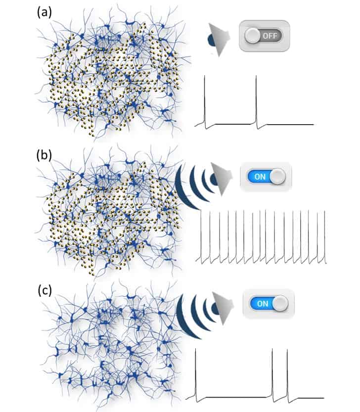
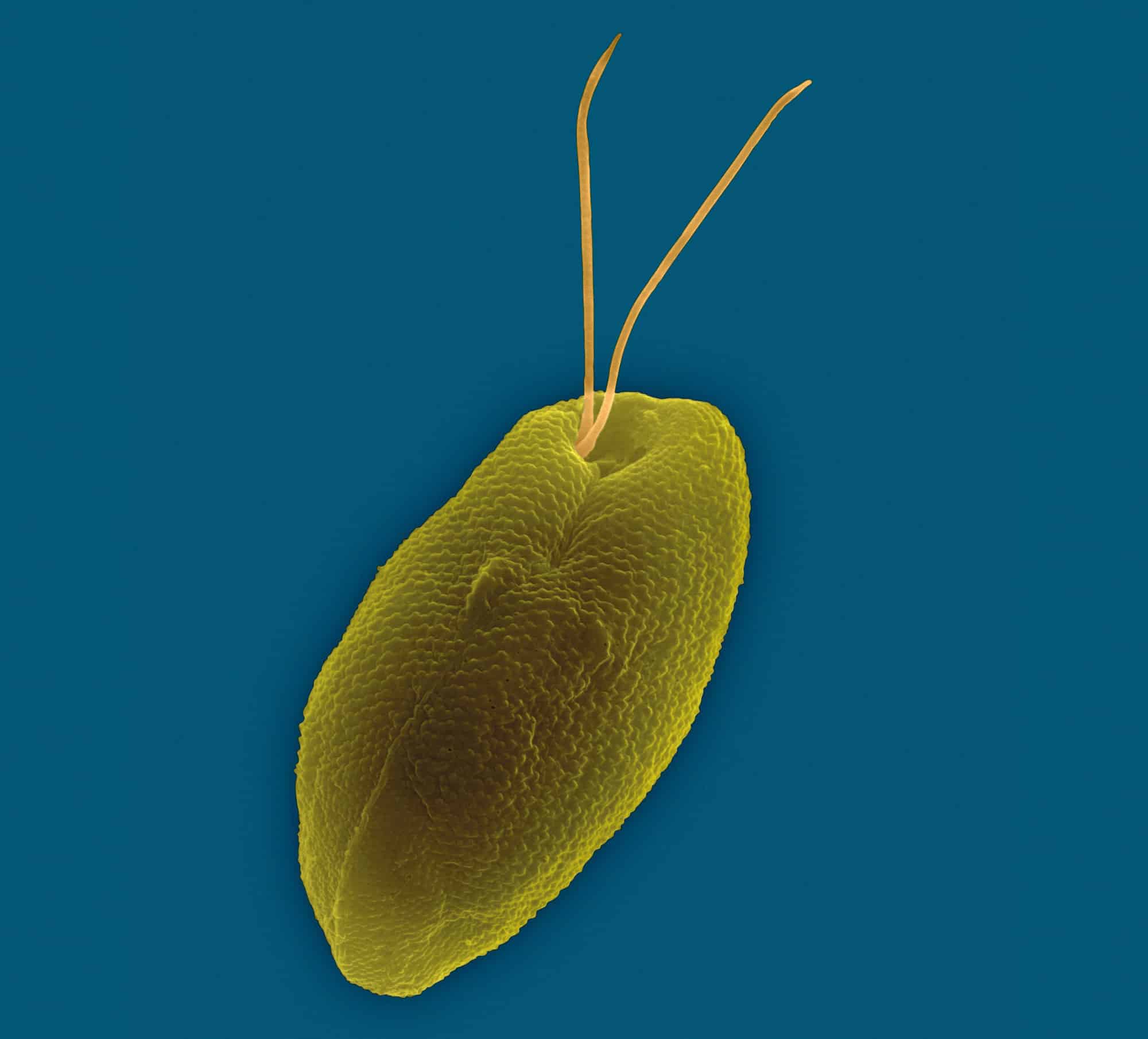
The understanding and treatment of brain function and neurological disorders took a step forward with findings from a study of piezoelectric nanoparticles stimulated by low-intensity pulsed ultrasound. Italian researchers showed that piezoelectric barium titanate nanoparticles (BTNPs) adsorbed on neuronal membranes induced a response to a gentle mechanical stimulation. This offers the potential to use nanoparticles and ultrasound to perform non-invasive and highly selective modulation of the activity of central nervous systems (J. Neural Eng.15 036016).
The researchers created cultures of hippocampal and cortical neurons taken from the brains of rat embryos and coupled them with micro-electrode arrays (MEAs). BTNPs were prepared, stabilized and incubated within the cell cultures for a few hours.
For the experiments, a planar disc ultrasound transducer was placed on top of the MEA chamber, at 8 mm from the neuronal culture. The stimulation protocol consisted of a series of ultrasound pulses with durations ranging from 0.5 to 1.5 s, delivered at 0.5 Hz. The researchers used each electrode of the MEA to record electrophysiological activity of the neurons. They recorded spontaneous and induced action potentials before, during and after ultrasound stimulation.
The presence and distribution of BTNPs were acquired from fluorescence microscopy images taken on the neuronal cultures upon completion of the experiments. The team evaluated and statistically analysed spike detection, mean firing rate and post-stimulus time histograms.
Lead author Camilo Rojas from the University of Genova and co-authors reported that when neuronal networks incubated with piezoelectric BTNPs were exposed to ultrasound, there was an evident increase in the recorded network activity (spiking and bursting) with respect to spontaneous activity before and after stimulation. This occurred with both cortical and hippocampal cultures.
Neurostimulation using ultrasound and piezoelectric nanoparticles.
The authors repeated the stimulation three times on the same network in 5 min intervals. When the ultrasound source was turned off, the neuronal network returned to original baseline levels, demonstrating the reversibility of the induced effects. Experiments repeated using different neuronal preparations from five different animals exhibited reproducible results. The team treated a total of 13 cultures with piezoelectric BTNPs.
The researchers also conducted experiments with a control group of neuronal networks incubated either with non-piezoelectric BTNPs of the same size or without nanoparticles at all. The authors reported that in both cases they never observed a significant response to ultrasound, although the same cultures responded to the electrical stimulation as expected.
Network response
To determine whether the observed firing activity upon ultrasound stimulation was due to the collection of independent single neuron responses or a collective network response, the authors applied a synaptic receptor antagonist “cocktail”. Their objective was to isolate neurons from a network context.
They determined that treatment with blockers of synaptic transmission significantly decreased responses to piezoelectric ultrasound stimulation and that after 10 min, the network did not show any response to electrical stimulation. The authors suggested that this is “an indication that, even if the mechano-electric transduction performed by the piezoelectric BTNPs works at the subcellular level, the measurable effect is at the network level”. Additional experiments aimed at controlling the level of network overall excitability would help better understand the (sub)-cellular mechanisms underlying the obtained network response.
Because the ultrasound-induced responses of the neuronal cultures were reversible, the researchers were able to perform repeated experiments on the same cell culture, such as varying the intensity or pulse duration of ultrasound stimulation. This enabled them to determine the clear dependence and linear relationship between the relative increase in the mean firing rate of the network and the pressure field intensity. “This finding suggests that a possible steering of the network activity can be implemented by fine-tuning the ultrasound amplitude,” they wrote.
The authors suggest that acoustic-electric stimulation enabled by piezo-nanomaterials without genetic intervention is a promising strategy to generate physiologically relevant responses in a minimally invasive way. “We foresee the possibility of highly selective and efficient neural stimulation by providing suitable nanoparticle functionalization to address specific groups of neurons in the central nervous system with virtually single cell resolution, together with the use of focused ultrasound,” they concluded. “This would represent a valuable innovative tool for the generation of new non-invasive neural interfaces.”
Peter Grünberg, the German condensed-matter physicist who shared the 2007 Nobel Prize for Physics with Albert Fert, has died at the age of 78. Grünberg and Fert independently – and at roughly the same time – discovered the effect of giant magnetoresistance (GMR). It not only created a new field of research known as spintronics, which uses the spin of the electron to store and transport information, but also led to a dramatic rise in the amount of data that can be stored on computer hard-disk drives.
Grünberg was born in 1939 in Pilsen (now in the Czech Republic) and gained his PhD in physics from the Technische Universität Darmstadt in Germany at the age of 29. After spending three years as a postdoc at the University of Ottawa in Canada, he moved to the Forschungzentrum Jülich in 1972, where he remained for the rest of his career, retiring in 2004.
It was at Jülich where Grünberg co-discovered GMR. A consequence of the quantum-mechanical nature of electron spin, GMR is the substantial change of resistance that occurs in wafer-thin sandwiches of magnetic and non-magnetic metals when they are exposed to a magnetic field.
In 1988, Grünberg’s group used the simplest system of a single layer of chromium sandwiched between two layers of iron. The team noticed that when the relative orientation of the magnetic fields of the magnetized layers were reversed, the resistance changed by about 10%.
Working independently at the same time, Fert and his group at the Université Paris-Sud in France created some 30 alternating layers of iron and chromium finding a resistance change of 50%. For their discovery, the duo shared the 2007 Nobel Prize for Physics.
‘An outstanding scientist’
As well as creating the new field of spintronics, both groups realized the potential technological importance of their discovery. It has since led to a dramatic effect on both science and technology allowing hard-disk read heads to detect ever smaller regions of magnetic data and is exploited in virtually all of today’s personal desktop computers and laptops as well as some digital music players. Indeed, in the years following GMR’s commercialization in the late 1990s, the annual rate of increase of hard-disk storage capacity trebled.
As well as winning a Nobel prize, Grünberg also received many other honours. He won the German Future Prize for Technology and Innovation in 1998 and the Wolf Prize in 2006. He was also given the freedom of the city of Jülich in 2008.
“We lose an outstanding scientist who has set standards worldwide in the field of solid state research,” noted Wolfgang Marquardt, chief executive of Forschungszentrum Jülich. “[Grünberg] was not only an excellent researcher, he was above all a valued and popular colleague.”
As the world’s population grows so does the amount of food required to feed it. But what’s the most efficient way to produce that food? Weight-based feed conversion ratios – the amount of feed administered over the lifetime of an animal divided by weight gained – show that beef is more resource intensive to produce than chicken, but this approach may be missing important details, as researchers in the US have now shown.
New results generated by scientists from Johns Hopkins University together with a colleague at Stanford University indicate that the meat production efficiency of some species of fish can be compared to pigs or even cattle. Only farmed Atlantic salmon was found to be on a par with chicken, one of the most efficient to produce forms of animal protein.
The study, published in Environmental Research Letters (ERL), looks at how feed conversion results change when additional factors such as the edible portion of the animal and the nutritional density of the feed are taken into account.
Aquaculture – or farmed seafood – is a fast-growing sector and viewed by many as a promising route to satisfying increased global demand for meat. Seafood has lower weight-based feed conversion figures compared with terrestrial animals, suggesting greater production efficiency. Water plays a role as buoyancy allows fish to expend less energy in moving and staying upright.
But what happens when the analysis is expanded to rank species by protein retention and calorie retention? In other words, the ratio of protein (or calories) in the edible portion of the animal divided by the amount of protein (or calories) in the feed.
The nine species – giant tiger prawn, whiteleg shrimp, common carp, grass carp, pangas catfish, channel catfish, tilapia, rainbow trout and Atlantic salmon – considered in the study represent over half of the global production of fed aquaculture. For land animals, the team focused on the top three livestock groups produced for meat worldwide – chicken, pigs and beef cattle.
It took time for the scientists to track down all the data they needed to calculate protein and calorie retention for all 12 species, but their search was worthwhile. Feeding the information into the equations showed that chicken performed best for both protein and calorie retention, followed by Atlantic salmon.
“A major takeaway from our work is the importance of using multiple measures when determining the efficiency of animal production,” said Jillian Fry from the Johns Hopkins Center for a Livable Future. “I consider the results of this study to be most relevant for NGOs, academics, and other stakeholders focused on creating sustainable, resilient food systems.”
During the FC Barcelona vs Paris Saint-Germain match in March 2017, Barcelona scored the vital sixth goal the team needed during the final minute of the game. The home crowd celebrations shook the ground enough to record a large signal on the seismometer installed by Jordi Diaz in the basement of the Institute of Earth Sciences Jaume Almera-CSIC building, some 500 m away from the Camp Nou stadium.
Initally Diaz installed the kit as a fun way to communicate about seismology to visitors. When he realized that the equipment could have serious uses too he added a second system, as he revealed at a press conference at the European Geosciences Union general assembly in Vienna.
Not only does the seismometer pick up signals from football crowds as they enter and leave the stadium and jump up and down after goals, in 2016 it even distinguished between songs at a Bruce Springsteen rock concert based on the ground movements as 65,000 people danced to the different rhythms. Because the music fans’ movements were more co-ordinated than the football fans, the signal the seismometer detected was clearly distinct to that from goal celebrations.
As well as “footquakes”, the kit also detects vibrations from traffic with enough detail to show when traffic lights on Diagonal Avenue – some 100 m away – turn red, from the subway system, from fireworks, marathon runners, storms and oceanic waves. The 2.8 Hz signals created by the runners revealed they were running at a pace of 170 steps per minute.
Apart from monitoring football matches, dancers and runners, the seismometer could provide additional information to engineers by revealing whether buildings respond similarly to different types of vibrations. It could also monitor traffic from a distance.
I grew up on a farm in southern Indiana and, to be honest, I must have been born a physicist because I didn’t really fit in with the farmers. It wasn’t until I arrived at Massachusetts Institute of Technology (MIT) as an undergraduate that I found my people. After I received my bachelor’s degree I spent a couple of years designing weapons for the US Navy, and then I went to graduate school to study plasma physics because I was really interested in fusion research. However, oil was cheap at the time, so the Department of Energy cut our funding. One day, my PhD adviser pulled me aside and told me that I could start drinking a lot of coffee and get in at 7 a.m. every day to finish my PhD before the money ran out, or I could get a Master’s degree. I chose the former, which somewhat ruined my health. However, the output from that work led to what really got my career started.
It turns out I was a better inventor than I was a physicist – inventing is part physics, part engineering and part business, all mixed together – and my biggest invention came as I was trying to finish graduate school. I was feeling a little depressed because I had thought I was going to be a fusion researcher for the rest of my life, but then I happened to walk into a Walmart and as I was looking at their hammers I realized every hammer that had ever been made had been designed incorrectly. If you hammer things all day long with an ordinary hammer, the shock and vibration will eventually give you lateral epicondylitis, or tennis elbow. Manufacturers knew this and designed the shape of their hammer heads to minimize this effect, but that leaves you with a hammer with reduced momentum transfer. I figured out that if you put air into the hammer’s grip in certain strategic locations, you can eliminate most of the shock and vibration, and that allows you to change the shape of the hammer to give it greater momentum transfer. Now, almost all hammers sold in the US have this technology in them. The invention has sold about $1.5bn in total, and I ended up applying a lot of the skills I learned in developing the hammer to my work at NovaCentrix (which was then called Nanotechnologies, Inc. – we’ve gone through about four different names).
How did you become involved in NovaCentrix?
I was hired by the founder of our company because I was an inventor, not because I had a plasma physics background – although the latter did help. Our original device began as a weapon during the Strategic Defense Initiative in the 1980s. We changed it around to make nanoparticles instead. The current device uses an intense, pulsed arc discharge (~100 kA) – a synthetic lightning bolt, if you will – to ablate electrodes, and make silver and aluminium nanoparticles. These particles have several interesting applications, and we still sell them industrially, but it’s hard to make a living and pay the bills when you’re just selling particles. By 2004, the company was in the process of going bankrupt, so I decided to start experimenting. I knew I was going to lose my job anyway, so I thought I would at least have some fun. That’s what led us into printed electronics and the photonic curing technology we use now.
How did that invention develop?
At the time, I had not really heard of printed electronics. I just thought “Wouldn’t it be interesting if you could use an ordinary printer to print an electronic circuit onto plastic or paper?” So I was approaching it from a fresh perspective, and we had these silver nanoparticles. I thought, hmm, I could take these particles, make a dispersion out of them, put it in an inkjet printer cartridge and use the printer to print silver traces. I was able to print the traces, but the bigger issue was that in order to make wires, you have to sinter the particles together. This is a little problematic as silver melts at 962 °C, but paper famously combusts at Fahrenheit 451 (233 °C). You don’t need 962 °C to sinter silver nanoparticles, as chemical and other techniques somewhat work, but in order to be effective and do it quickly, you still need a much higher temperature than paper or plastic can take.
Now, recall we were already using a pulsed-plasma device to make nanoparticles. It makes a lot of light, and as I had a background in radiation, I theorized that I could use a flash of light from a flashlamp to heat these silver traces and sinter them in less than a millisecond, without damaging the plastic or paper substrate. It worked! As we already knew how to make reliable, intense arc discharges, scaling this new process up by building an industrial flashlamp system was within our capabilities. To use a very relevant analogy, if all you have is a hammer, everything looks like a nail, and that’s how we developed the process we call photonic curing.
As a start-up, your team is not your most important asset. Neither is your technology – despite what your technical people might think
What are the advantages of that process?
If you really want to make circuits cheaply, you probably need to print them, and if you want to make a lot of them, you need to do so on a big roll of paper or plastic. The printing can be done fairly quickly, but if the ink takes 10 minutes to cure, and you’re running the roll through an oven at, say, 100 m per minute, you’re going to need a kilometre-long oven. That isn’t practical, and although you can squeeze it down a little bit, you will still end up with an oven the size of a large building. We’ve replaced that with a device called a PulseForge® that is less than 1 m long and uses a fraction of the energy.
How did you develop the market for that device?
Printing electronics is still considered a new field, and we also had a new technology. That combination gives you a lot of opportunities – for success, yes, but also for failure. Consequently, we realized that to sell a product in this environment, we had to do a lot of vertical integration. Back when we were just making nanoparticles that meant asking our customers what they wanted to do. One of the responses was “Can you make a dispersion?”, so we learned how to do dispersions and then we made inks and then we learned how to print them properly, and finally we processed the printed circuits with our photonic curing technology and made a machine that did it all. And because this is still a new field, and our livelihood is at stake, we still take a lot of input from our customers and put that (as well as the profit from the sale of the equipment) back into developing new equipment and new enabling technologies around it. The net result is that we end up having the best technology and our customers end up being evangelists for it as well.
How did you get funding for NovaCentrix?
Early on, we had venture capital funding. This was during the late 1990s and very early 2000s, when the dotcom boom meant there were a lot of people around who didn’t have a lot of experience, but because they had made a lot of money, they thought they were geniuses. They weren’t, of course, but the person with the money is generally the person in charge. So in the early days of the company we had what you might call a “seasoned management team” because that’s what the venture capitalists wanted to see. At one point, we had more vice-presidents than we had technicians. However, it turns out that as a start-up, your team is not your most important asset. Neither is your technology – despite what your technical people might think. I mean, technology is nice, and we had a lot of exciting patents. But a patent alone won’t even buy you a pint. In reality, the greatest asset of any company isn’t the team, or the technology, or even the product: it’s your customers, and they are what saved us back in 2004.
The way it happened was that I was playing with this photonic curing technology, and although the technical folks in the company were getting excited about it, our business folks were more worried about their next job – because as I said, we were in the process of becoming bankrupt. Then my boss and I went on a trip to the South Dakota School of Mines and Technology, where a lab director told us about the printing they were doing with nanosilver-based dispersions on low-temperature plastics. They needed a higher conductivity on these low-temperature materials. So when he passed around a sample printed on plastic, I asked him to show me the resistance. Immediately, I pulled out a $7 disposable flash camera I had bought and flashed his sample, which instantly reduced the resistance by a factor of two. My boss gave me a look and was getting ready to let me have it, but then the lab director became very animated and started saying things like “I want one of these machines right now. Will you build me one? I’ll get you the money!” That, in addition to a similar incident with Sun Chemical, caught the interest of one of our early investors, the Munson family. They had been minority investors, but Charlie Munson, who’s our current CEO, saw what was happening and they bought the company. So, it does involve a little bit of faith on both the technical and business end.
What do you know now that you wish you’d known when you were getting started?
People at a new company think they can do anything, and they tend to chase flowers in the wind – a tiny source of revenue here, something else there – instead of focusing on what their product is going to be. You sometimes hear “Don’t put all your eggs into one basket,” but the only people who truly deliver are the people who, to some extent, do just that. We should have said, “This is our core competency; this is what we can do.” Instead, we’d have eight projects going on even though we only had the resources for two. That approach guaranteed failure for all of them. It’s very important to identify the one or two projects that have the highest possibility of success, and worry about the other ones later if you get to it. That is very tough for a researcher to learn, and it’s also difficult for a business person because they’re afraid they might miss an opportunity. But doing so hurt us quite a bit. One example is that in early 2001 we secured a large amount of money to expand our nanoparticle business. The money came so quickly and so easily that our management became a little greedy and they started talking to other venture capitalist firms to find a better offer. But then the September 11th attacks happened, the American economy tanked and a lot of that money was retracted. We ended up laying off a lot of people and nearly went under – we nearly went under several times, in fact. I think being too greedy and not focusing was part of that.
Any advice for somebody who’s just starting a nanotechnology company now?
It’s important to know your customer. I see many inventors, physicists and engineers who like to build stuff. They get excited about doing that. But they don’t really think about, okay, when you build this, what are you going to do with it?
I also think it’s very important to go through the mental exercise of “what if?” It’s almost an adage that a start-up will always need more money than its founders expect, it will always take longer than they think to finish and there will be resources they need that they didn’t think about. There is a cartoon I love that shows some dogs shipwrecked on a life raft, and one of the dogs says: “Okay, we have enough food for two weeks. Who votes we eat it all right now?” And of course all the dogs raise their paws, because that’s what dogs do. But in some ways start-up founders are similar in that they’re often too optimistic. They use so many of their resources early on towards activities that are not important to achieving success. That’s what you need to avoid.
One final thought regarding the choice of key personnel. Usually, when early-stage companies build their team they look for specialists. This is a mistake as invariably the job they were originally hired for will change or even go away. Specialists generally can’t pivot. Instead, we chose to hire expert generalists. This paid off well, as when the company pivoted, they could follow in unison. Later on, as the company grew, specialists became a better choice.
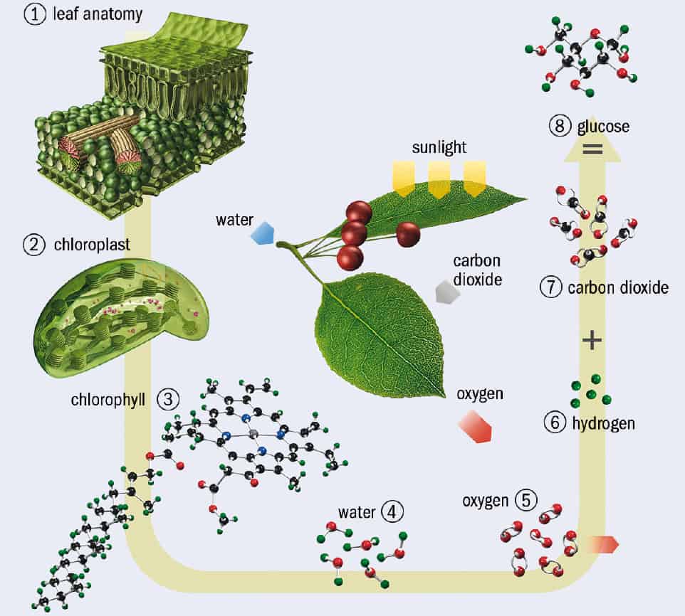
The quirks of quantum physics are something you might expect to find under exotic conditions in a laboratory, but not in a meadow. Yet in recent years, a blossoming idea called quantum biology proposes that life’s molecular mechanisms deploy some of those notoriously counterintuitive behaviours. Ten years ago, researchers reported evidence that photosynthesis – the process by which green plants and some bacteria turn sunlight into chemical energy – gains light-harvesting efficiency by exploiting the phenomenon of “quantum coherence”. This involves the superpositions of electronic quantum states, which seem able to explore many energy-transmitting pathways at once. If so, quantum mechanics is assisting the fundamental energetic process that drives all life on the surface of the Earth.
It was a remarkable claim. But was it true? The question has been hotly debated over the past decade. Early talk of “quantum coherence” in photosynthesis being akin to that in quantum computers, where it underpins the faster and more efficient computation that those devices achieve, has now largely evaporated in favour of a more nuanced picture. And some researchers insist that such coherence plays no useful role in photosynthesis at all.
On the one hand, Greg Engel of the University of Chicago, who was involved in the initial work suggesting quantum coherence as a new design principle of nature, says that “the general notion that the language and mathematics of quantum information, including coherence, can be used to understand photosynthetic dynamics in ultrafast spectroscopy experiments seems to be growing in acceptance”. But biophysical chemist Sebastian Westenhoff of the University of Gothenburg in Sweden says that ever more scientists in the field regard the earlier work as a misinterpretation, and that “there is no such thing as quantum coherence in [natural] photosynthesis”. So what’s the argument all about?
Get the green light
Let there be light: In photosynthesis, sunlight is converted into chemical energy in the form of sugars (glucose). In plants, photosynthesis occurs in the chloroplasts of the plant cells. Chlorophyll within the chloroplasts is used to synthesise glucose from water (H2O) and carbon dioxide (CO2) using the energy from sunlight, with oxygen (O2) released as a by-product. (Courtesy: Carlos Clarivan/Science Photo Library)
Both plants and photosynthetic bacteria capture solar energy in much the same way (figure 1). Photons of sunlight are absorbed by the chlorophyll molecule and other light-absorbing “chromophores” in a large assembly of proteins and other molecular structures, which together make up a “photosystem” embedded in the so-called thylakoid cell membrane. This energy creates an electronic excited state of the chromophores, and the energy must be channelled towards other molecules in the “photosynthetic reaction centre” – an enzyme that is the site of the light reactions of photosynthesis. Containing the pigments such as chlorophyll, it is surrounded by light-harvesting complexes that enhance the absorption of photons. There, the energy separates positive and negative ions by some significant distance, rendering them unable to neutralize one another by charge recombination. This separation of charge sets up an electrochemical potential that is ultimately tapped to drive enzyme-assisted reactions for making the energy-storage molecule adenosine triphosphate (ATP) – the main reservoir of chemical energy for powering biochemical metabolic reactions in cells (figure 2).
Bright crop: The light-harvesting complex (orange) found on the thylakoid membrane (blue) in the chloroplasts of plants. This is the location of chlorophyll and carotenoid pigment molecules (orange) that take part in photosynthesis. They absorb incoming photons, and the excitation energy is transferred to the “reaction centre”, which here comprises a pair of chlorophyll molecules. The energy is transferred out through an electron acceptor (red, upper centre) and used in further reactions elsewhere on the membrane. (Courtesy: Science Photo Library)
But this transfer of solar energy to the reaction centre is extremely efficient: almost every photon of the captured solar energy is used by the cell. The mystery is how photosynthetic organisms achieve such efficiency in a warm, wet, disorderly environment.
Random hops or straight walks?
Until quantum coherence – the synchronous phase relation between different states of a quantum system – entered the picture, energy transport in photosynthesis was thought to involve interactions between the electronic states of chromophores. The idea was that the energy is passed on to a neighbouring chromophore with a slightly lower energy level. But there was thought to be no coherence – no in-phase resonance – in this coupling. Instead, the energy transport was pictured as a random (incoherent) hopping process between chromophores, guided by an overall energy gradient, like a drunken sailor staggering downhill.
But even as far back as the 1930s it had been suggested that the efficiency of photosynthetic energy transfer might come from some kind of synchronization of the wavelike electronic excitations – known as excitons – between chromophores. In 2007 physical chemist Graham Fleming of the University of California at Berkeley and his co-workers claimed to find evidence that this is indeed what happens. If there really was coherence among excitons, they reasoned, it should be possible to see interference between the different pathways: so-called quantum beats, like the acoustic beats audible when sound waves of similar frequency interfere. Such coherence would allow the excitation to find the optimal route to the reaction centre, without wasting energy through random hopping.
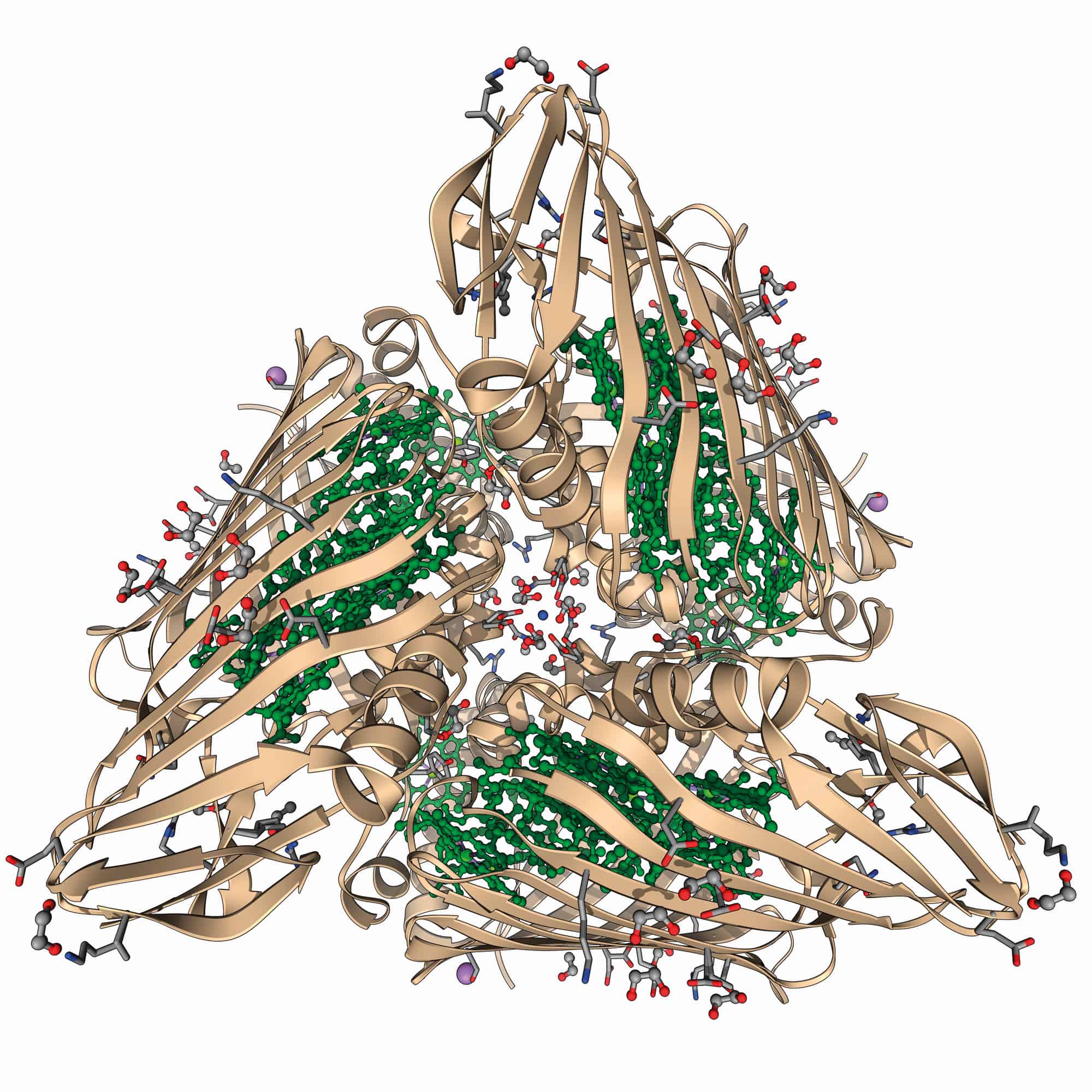
Fleming, with postdoc Engel and others, used a technique called photon-echo spectroscopy to study the energy-transfer process in the so-called Fenna–Matthews–Olson (FMO) pigment–protein complex – the part of the photosynthetic apparatus that mediates energy transfer in the thermophilic bacterium Chlorobium tepidum. This method involved exciting coherent excitons using an ultrashort “pump” pulse from a laser, and then probing the way the excitation evolved over time using subsequent ultrashort laser pulses to look for signs of beating.
At the same time Fleming and his collaborators Hohjai Lee and Yuan-Chung Cheng, also at Berkeley, studied energy transfer between two types of pigments in the photosynthetic reaction centre of a different kind of photosynthesizing bacterium. Although the researchers saw the beats that they had anticipated in both cases (suggesting that coherence was involved) the studies were conducted at temperatures much colder than the physiological conditions under which such organisms operate in nature.
Reaction centre: A molecular model of the Fenna–Matthews–Olson pigment–protein complex, as found in the green sulphur bacterium Prosthecochloris aestuarii. At the centre of each of the three proteins is a bacteriochlorophyll molecule. (Courtesy: Laguna Design/Science Photo Library)
But in 2010 Engel, now in Chicago, reported that quantum coherence in the C. tepidum FMO complex survived even at room temperature. Meanwhile, another of Fleming’s former postdocs, photochemist Gregory Scholes at the University of Toronto in Canada, collaborated with chemist Elisabetta Collini and co-workers to look for such effects under ambient conditions in yet another type of organism: photosynthetic cryptophyte algae, which can harvest light efficiently enough to carry out photosynthesis even in dim conditions. They too saw beats lasting for longer than a tenth of a picosecond at room temperature. This putative electronic coherence, they said, was an order of magnitude longer-lived than had been previously thought – and long enough for the energy to get transferred from the chromophores to the photosynthetic reaction centre.
Natural computing?
This seemed remarkable. Quantum coherence between quantum bits (qubits) placed in entangled states is what physicists are seeking to exploit in quantum computers. But qubits are typically cooled to within a fraction of a degree of absolute zero, and even then, coherence survives only for brief instants, among just a handful of bits, before decoherence sets in due to disturbance from the surrounding environment. So intuition seemed to insist that a quantum effect like this would never survive the warm, wet conditions of living cells. How could photosynthetic organisms sustain it?
But the closer researchers have looked, the more complications and nuances have arisen. Did the early work really detect electronic quantum beating at all? What was actually measured was an oscillating spectroscopic signal; the question is how to interpret it. In 2013 David Jonas of the University of Colorado at Boulder and co-workers argued that the “beats” were in fact caused purely by molecular vibrations – the process of Raman scattering – and not by quantum coherence among excited electronic states (PNAS110 1203).
Engel acknowledges that it was impossible in the original experiments to distinguish definitively between the two possibilities. One way to tell them apart is to make synthetic light-absorbing chemical systems tuned to avoid any coincidence in the energies of excitonic and molecular vibrational energy states – then those two types of state are less prone to coupling. Engel and his co-workers did just that, making dimers of two different molecules that mimicked the light-absorbing groups of photosystems rigidly linked together (Science340 1431). In 2013 they reported that this system also shows quantum beats that did not appear in the vibrational Raman signals.
But when the same idea was tested by Dwayne Miller of the Max Planck Institute for the Structure and Dynamics of Matter in Hamburg, Germany, and his team using dye molecules joined together in pairs by a more flexible linker, they came to a different conclusion (Nature Chem.6 196). They argued that the previously observed coherence was too small in amplitude to originate in excitons, and was indeed a kind of classical resonance effect involving molecular vibrations. Besides, they said, any electronic coherence decays too rapidly to have any meaningful biological consequences.
Miller has also now re-examined the behaviour of the FMO complex itself (PNAS114 8493). “We went back and redid the original work at room temperature and actual physiological conditions, and it is absolute certain there are no long-lived electronic coherences that direct energy transfer”, he says. “The beats are comparable in amplitude, frequency and decay rate to trivial Raman vibrations of the electronic ground state excited in the process. It is Raman that they saw, not long-lived electronic coherence.”
Mixed vibes
Quantum theorist Michael Thorwart of the University of Hamburg agrees. In 2011 he reported calculations of the electronic and vibrational states in the FMO protein which, he says, implied that long-lived coherence wasn’t feasible (Phys. Rev. E84 041926). “At that time, these theoretical calculations were the best ones available”, says Thorwart. He collaborated with Miller to interpret the recent experimental measurements of excitations of FMO, and he says that the findings match those earlier predictions. Any coherence is tiny and comes from vibrational coupling of the electronic states, somewhat akin to the famous synchronization of wall clocks observed by Christiaan Huygens in the 17th century due to vibrational coupling through the wooden board on which they were mounted.
Light lover: Coloured scanning electron micrograph of Rhodomonas salina – a marine cryptophyte algae. Most of these single-celled organisms contain chloroplasts and so can carry out photosynthesis in dimly lit surroundings. (Courtesy: Dennis Kunkel Microscopy/Science Photo Library)
Increasingly the consensus seems to be that molecular vibrations lie at the core of the disputed observations. And yet Elisabet Romero of the Free University of Amsterdam and her co-workers have carried out spectroscopic experiments that they interpret as evidence that vibrations in the reaction centre of the photosystem from spinach support quantum coherence between excitonic states, by strengthening the interactions between them, and promoting coherent energy transfer (Nature Phys.10 676).
Very recently, Scholes (now at Princeton University) and co-workers, including veteran photosynthesis expert Robert Blankenship of Washington University in St Louis, Missouri, have told a slightly different tale. They conducted pump-probe spectroscopy on tailor-made mutant versions of the FMO protein complex (Nature Chem.10.1038/nchem.2910). These variants have different electronic states from the native form, and so if excitons are involved in the spectroscopically observed beats – the characteristic sign of coherence – then these beats should have a shifted frequency. But they don’t: the signal stays just the same. Scholes says this indicates that the oscillations comes purely from vibrations of the electronic ground state (the lowest-energy state). Miller says that this is consistent with earlier work from several other groups, and that “there now seems to be consensus that the original beats were vibrations, not excitons”.
So, while Romero and co-workers’ findings are similar to what Engel originally claimed, Miller and Scholes’ teams are certain that it’s vibronic coupling that is at play. While the beats are real enough, it’s how to interpret them that’s the problem.
Quantum(ish)
Yet Scholes concedes that his new results do support the original contention that “the molecules in the FMO protein are coupled in a special way and this may aid energy transport by directing it or making it quicker”. According to Romero, this tuning of molecular vibrations to the right frequencies for transferring energy makes the photosystem what she calls a “quantum-designed light trap”. When you look at photosynthetic reaction centres for a range of organisms, she says, “there is only one design that is conserved, which suggests that nature has found a design able to perform efficient charge separation and has maintained it”. In other words, she says, natural selection seems to have favoured this quantum-optimized process.
Until quantum coherence entered the picture, energy transport in photosynthesis was pictured as a random hopping process, guided by an overall energy gradient, like a drunken sailor staggering downhill
But Miller argues that the strength of the vibrational coupling is far too low to enhance energy transport. He sees an imprint of evolved optimality in the very absence of quantum coherence – in the fact that it is very rapidly lost after photo absorption through decoherence. “It turns out that nature has evolved to not beat decoherence but exploit it,” he says. It’s precisely because decoherence causes the dissipation of energy that the energy transfer can find its way gradually downhill along the most energy-efficient path, guided by how electronic properties vary from place to place in the molecular environment. “It’s a bit like laying paving stones down a hillside to direct hikers to stay on the trail and not explore the full landscape,” he says.
Full circle
So then, is photosynthesis “quantum” or not? “The observations show that there is correlation between the wavefunctions of the states involved in energy or electron transfer,” says Romero. “But these effects are not considered by some scientists as truly quantum coherence in the sense that entangled states of quantum computing are understood.” And Engel agrees that to compare the two is to invoke “the wrong language”.
Maybe so – or maybe these correlations and coherences, mediated by vibrations, are in any case too weak to have any biological relevance, and we still have to think of exciton states in the photosystem as being more or less localized to particular molecular groups, with incoherent transfer of energy between them. That’s what Miller and Thorwart think. “We have come full circle,” Miller says, “and it seems that the early picture of energy transport as a largely incoherent process has withstood the challenge.”
Romero says that arguing whether energy transport is a true quantum effect or not isn’t terribly productive anyway. “In my view, the relevant issue is to understand how plants or other photosynthetic organisms are able to transfer energy and electrons on an ultrafast timescale in the right direction with high quantum efficiency,” she says. If we understand that, we might be able to do something useful with it. “This knowledge holds crucial lessons about how to engineer human-made systems with the capacity to convert sunlight energy to electrochemical energy to produce electricity or, even better, to produce solar fuels catalytically,” Romero asserts. That’s an idea everyone can get behind – but first we need to know how nature does it.
Superconductivity has been observed for the first time in a quasicrystal – a solid material with atoms that are arranged in an ordered pattern that does not have translational symmetry. Keisuke Kamiya and Noriaki Sato at Nagoya University in Japan and colleagues created the quasicrystal by altering the ratio of elements in a specialized metal alloy – and found that it is a superconductor a temperatures lower than 0.05 K. The discovery could lead to the creation of new materials that display fractal superconductivity.
Conventional superconductivity arises when electrons interact with atoms in a crystalline lattice, causing lattice deformations called phonons that propagate through the crystal. These deformations contain pockets of excess positive charge that tend to attract pairs of correlated electrons called “Cooper pairs”. Unlike single electrons, which are fermions, Cooper pairs are bosons and can therefore condense at low temperatures to form a superconductor that flows without encountering resistance.
Long-range order
The atoms in a quasicrystal have long-range order but the pattern of atoms does not repeat itself periodically in space – and therefore a quasicrystal is not a crystalline lattice. As a result, the theory of conventional superconductivity does not describe quasicrystals.
To look for superconductivity in a quasicrystal, the team experimented with an alloy of aluminium, zinc and magnesium that has quasicrystal and crystalline structural phases – depending on the relative abundances of the three metals.
The team began with the alloy in an “approximant crystal” phase that bears some resemblance to a quasicrystal but actually has a lattice that repeats in space. They reduced the aluminium content of the alloy while keeping the magnesium content almost constant and found that the critical temperature marking the onset of superconductivity decreased gradually from about to 0.8 K to about 0.2 K. “However, at 15% aluminium, the alloy transformed into a quasicrystal, and the critical temperature plummeted to about 0.05 K,” says Kamiya.
Dramatic jump
At 0.05 K, the specific heat of the quasicrystal alloy jumped dramatically, and magnetic flux inside the material was almost entirely blocked. These are both important signs that a transition to a superconducting phase had occurred.
The team describe the critical temperature as “extraordinarily low” and say that it explains why it had previously been difficult to observe superconductivity in quasicrystals. Closer inspection of the properties of the quasicrystal superconductor suggest that the formation of Cooper pairs arises from the weak-coupling of electrons. Although relatively uncommon, weak coupling is seen in other materials including the approximant crystal phase of the alloy used in the study.
Dirty superconductivity
According to Sato, this similarity could mean that the observed superconductivity is not related to the quasicrystalline nature of the alloy – but is rather “dirty superconductivity” that occurs in imperfect crystals.
“However, the theory of quasicrystals also predicts another form of superconductivity, based on fractal geometry in quasicrystals. We believe there is a strong possibility that fractal superconductivity makes at least some contribution, and we would be excited to finally measure it.”
The team is examining the interplay between this fractal geometry and the weak coupling electron pairs to explore a new area of superconductivity.
Researchers have produced fine, thread-like “yarn” batteries using a scalable carbon nanotube (CNT) based roll manufacturing processes. The batteries have specific capacities competitive with Li-ion technology, can be knitted into fabric, folded, stretched and washed, offering promising power storage for low-profile applications.
Lithium-ion batteries offer power densities far in excess of previous rechargeable battery types, like nickel-metal hydride and nickel-cadmium, allowing portable electronics to become ever more sophisticated and power-hungry. Their power density however comes at the cost of manufacturing ease and safety, so they are not appropriate for all applications. The development of new battery types is accelerating with the aim of diversifying the power storage options available for technological applications with different needs.
For a long time primary (non-rechargeable) batteries have relied on the electrochemistry of Zn anodes and MnO2 cathodes, but experts had considered it impossible to recharge these cells. However, through studies of the cathode structure and choice of electrolyte researchers have recently achieved high reversible capacities with Zn and MnO2. Using these recent developments researchers in Hong Kong have developed a scalable method to produce highly durable, waterproof, high-capacity cells in the form of knittable yarn, which may have extensive applications in flexible, transparent, or wearable electronics.
Spinning an electric yarn
The process begins with CNT fibres for high strength current collectors. On these the Hong Kong research team deposits cathode and anode materials using roll-dipping and electrodeposition processes respectively: The active cathode material consists of MnO2 nanorods precipitated onto the surfaces of the multiwalled CNTs, and Zn metal is electroplated onto the anode. They then wound the ~100 μm diameter cathode and anode fibres around an elastic thread and coated them with a polymer hydrogel electrolyte – a highly porous cross-linked polyacrylamide impregnated with an aqueous ZnSO4 and MnSO4 solution. They then encapsulate the assembly in a silicone sheath to keep it all together and watertight.
In their report Chunyi Zhi and co-authors demonstrated the scalability of their process by manufacturing a 1.1 m long cell. They were able to demonstrate stable electrochemical cycling while bending, knotting, and stretching the cells. In addition, under rapid cycling conditions (10 min dis/charge) the cells retained 98.5% of their specific capacity after 500 cycles.
The physical robustness and pliability of these cells makes them suited to unique niche applications, such as medical implants, and flexible electronic devices. Their low toxicity, low cost and good safety characteristics may also make them excellent candidates for power sources for ubiquitous wearable electronic devices.
A new material made from hydrogels etched with nanocrystal patterns and rat heart cells can change colour as the heart cells expand and contract. Inspired by skin colour changes in chameleons, the “heart-on-a-chip” platform might be used to investigate the fundamental mechanisms involved in disease aetiology and organogenesis as well as to test drugs for heart disease in an alternative to animal testing.
Photonic crystals are nanostructured materials in which the periodic change of the refractive index on the length scale of visible light produces a photonic bandgap through which certain light wavelengths can pass through while light in other ranges is reflected. This means that the colour reflected by the crystals can be tuned by changing the bandgap. In chameleons, this gap is the distance between nonclose-packed guanine nanocrystals and it can be varied by deforming the surrounding elastic matrix. This allows the animal to change its colour over the entire visible spectrum, but it is usually from yellow to green.
Synchronous shifts in the photonic band gaps
“Inspired by the structural colour-shift mechanisms in these animals, we constructed flexible inverse-opal hydrogel films etched with nanocrystals patterns assembled with engineered rat cardiomyocyte tissue,” explains lead author of this study Fanfan Fu of the School of Biological Science and Medical Engineering, Southeast University in Nanjing, China. “As the heart cells beat, they contract and elongate and this causes the substrate hydrogel to do the same. This movement appears as synchronous shifts in the photonic band gaps, causing light of different wavelengths to be reflected from the gel nanostructure.”
The researchers, led by Yuanjin Zhao, integrated this material into a microfluidics system to make a heart-on-a-chip device in which the colour changing properties allowed them to measure the beat frequency of heart cells – for example, after administering isoproterenol (a drug that lowers heart rate). The colour shifts on the chip corresponded with measurements performed on live organisms that had also been given the drug.
Drug-testing applications
“This chip could be used to test different types of cardiomyocyte drugs and also provides an ideal platform for studying the growth and differentiation of heart cells,” Fu tells nanotechweb.org. “We can use the chip to study how induced pluripotent stem cells develop, for example, or how other stem cells differentiate into cardiomyocytes.”
And that was not all: Fu and colleagues also organized the cardiomyocytes on the hydrogel film surfaces in an anisotropic way, so as to better mimic the conditions in a real heart. To do this, they used silicon wafers containing micro-groove patterns that allowed the hydrogel film to self-assemble into a particular structure – which has the same shape as a butterfly.
Intelligent robot actuators
When the heart cells beat, the hydrogel expands and contracts and the thrust from this expansion and contraction changes the bending angles of the film. “The ‘bionic butterfly’ thus has a specific structural fingerprint at each thrust, which makes it appear as though it is flapping its wings as the colour shifts from the outer edge of the structure and spreads to the inside of the wings,” explains Fu. “We believe that this type of structure could be useful for making intelligent robot actuators in the future.”
The team, reporting its work in Science Robotics DOI: 10.1126/scirobotics.aar8580, says that it would now like to make such devices from its material. “We also plan to further optimize the biohybrid structural hydrogel so that that we can detect single cardiomyocytes on our heart-on-a-chip platform,” adds Fu.
Monitoring forests provides important clues to the health of our planet. Comparing sequences of satellite images can reveal change across large geographic areas over time. Field measurements add to this by highlighting forest growth or decline from the ground up. But what’s the most efficient way of linking these data together to complete the picture?
The group’s output shows that human activities control biomass removal in most years. It also highlights that fire can shift forest systems strongly into carbon loss.
“Our goal here was to connect the measurements on the ground with the measurements from space in a way that leverages the strengths of both approaches,” said Robert Kennedy of Oregon State University.
Landsat satellites have captured images of our planet from space for decades, providing researchers with views of the forest canopy from above. But these data only tell part of the story, particularly when it comes to quantifying the amount of biomass.
“Forests grow up, and generally the taller the forest, the more carbon is there, especially in the kinds of evergreen needleleaf forests that dominate much of the study area we examined,” said Kennedy.
It’s difficult to monitor this vertical dimension from above, which is why it’s still necessary to go into the forest and measure trees. But performing such a forest inventory and analysis (FIA) is time-consuming and expensive, so it’s only conducted at sample locations rather than across the whole landscape.
To enrich the data, the team used a gradient nearest neighbour method, which begins by converting satellite imagery into values that represent the environmental conditions of the landscape. “Once we have defined the numbers that describe what the trees experience, we can link those numbers to the FIA plots with rich information that field crews have painstakingly measured on the ground,” said Kennedy.
Put simply, it means that the group can meaningfully apply the field data – a gold standard – across the entire landscape to generate much more detailed biomass estimates. The downside is that each satellite pixel, which represents an area of around 30 m2, could be assigned information from an FIA plot that may be far away in real life. It’s a balance that the researchers are dialing into their analysis.
“Somewhere in between the pixel and the whole study area is a sweet spot of accuracy and usability, and we continue to work on the best ways to find that sweet spot,” added Kennedy. “It’s not perfect, but the statistical methods we used seemed to go a long way toward painting a good picture of what’s happening on the ground.”
Encouraged by the system’s success in tracking higher biomass forests, the researchers are extending their approach. “We’re keen to see how these stories evolve as we expand to the rest of Washington, Oregon, and California,” said Kennedy. “The extension promises to be interesting because we’ll be including many more forests that are dry and affected by fire and insects, so we’re curious to see whether the relative impacts will change.”
A full description of the monitoring framework, including a flow-chart of the analysis pathways, can be found in Environmental Research Letters (ERL).