A new technique to synthesize radioactive upconverting nanocrystals that was previously used to extract uranium from ore could be used to make efficient radiotracers for biomedical imaging, say researchers at the University of Pennsylvania in the US. The nanophosphors, which emit beta-particles, could be ideal in theranostic applications and even targeted in vivo imaging when combined with CT scanning.
Upconverting materials emit light at a wavelength that is shorter than the wavelength of light they have been photoexcited with and are promising for applications in biomedical imaging. The so-called anti-Stokes shift in these materials limits the autofluorescence of nearby molecules within a sample. This significantly reduces background signals, allowing for better target detection.
Rare-earth compounds for upconversion
Although there are several materials and molecules capable of upconversion, rare-earth compounds are particularly good at converting near-infrared (NIR) light to visible light. These materials transfer the energy from the absorbed NIR photons in the form of excitons (excited electron-hole pairs) so that they are emitted with a higher energy (or shorter wavelength).
Another form of imaging commonly employed in nuclear medicine relies on unstable isotopes within radiolabelled molecules or nanoparticles (known as radiotracers) to produce a signal around the target. Radiotracers do not need to be externally excited, which allows the signals coming from them (beta-emission, for example) to penetrate deeper into tissue than optically-stimulated nanoparticles.
Beta-emission and upconversion in one system
A team led by Christopher Murray made the nanophosphor sodium yttrium fluoride (NaFY) doped with the rare-earths erbium (Er) and ytterbium (Yb), and radiolabelled with 90-Y. This nanocrystal upconverts near-infrared light to the visible thanks to the rare-earth dopants and emits beta-particles thanks to the 90-Y.
The presence of both beta-emission and upconversion in one system is very rare in nature, explains team member and lead author this study Stan Najmr, and allows for deep tissue imaging because the two modalities are involved.
Hydroxide metathesis method
The researchers made their nanophosphors using a hydroxide metathesis method that was previously used to extract uranium from ore. “This method allows us to exchange one anion (for example, chloride) with another (trifluoroacetic acid), which is crucial for homogenously incorporating the radioactive element into the host matrix of the nanophosphor,” says Najmr.
The nanophosphors are produced through the rapid thermal decomposition of the rare-earth trifluoroacetates. “Once they have been synthesised, we silica coat them using the ‘Stober technique’. This allows us to disperse them in water, which is required for biomedical applications. Since these nanocrystals are fluorides, they are fairly inert to their environment.”
Extending the technique to other radioactive rare-earths
“This particle system is an excellent candidate for biomedical trials, especially in theranostics,” Najmr tells nanotechweb.org. “Combined with CT scanning and surface functionalisation, these nanocrystals could be used for targeted in vivo imaging.”
The researchers hope that their work will encourage other groups to consider this synthesis route for developing rare-earth nanocrystals and silica architectures. “For our part, we are eager to extend this technique to other radioactive rare-earths, such as lutetium (Lu) and samarium (Sm),” says Najmr. “These elements will provide new signals like gamma radiation that might lead to additional pathways for multimodal imaging.”
I was always interested in taking things apart to understand how they worked. I think my parents found it a bit stressful at times, so it was cool that physics was a class at school that was all about figuring out how stuff works. It probably helped that getting good grades in physics was easier for me than learning a foreign language. During my final year of my schooling in Germany, we covered special relativity and had a whole week of project work preparing a presentation on the topic for the rest of the school. I think this really cemented my interest in physics and that I wanted to study it at university. I thought that if physics could explain space–time, then what couldn’t it explain? I did both undergraduate and postgraduate physics degrees at the University of Manchester, UK, including a summer studentship at CERN in Switzerland and two years based at Fermilab in the US.
How did you get interested in software development and machine learning?
When I was 13 I saved up my birthday money to buy a computer – I started writing small programs (and playing games). Then I got interested in building websites, and ever since, I have been fascinated with the wide variety of things you can do if you start plugging bits of software together, discover how they work on the inside and then modify them to do something new. It is kind of like gaining a superpower, one that you can learn! My first experience with machine-learning was during my PhD at Fermilab. The task was to identify electrons in the D0 experiment with high purity and efficiency. With youthful naivety I thought we could simply combine all existing discriminative features in an ensemble of decision trees. When that turned out to not outperform existing methods my curiosity grew. After my PhD I got involved with an open-source project called “Scikit-learn”. It is the most widely used software package for machine-learning in the Python programming language. The realization that I, or anyone really, could work with some of the world’s experts on this topic was a great discovery for me.
Did you ever consider a permanent career in experimental particle physics at CERN?
For sure. After finishing my PhD, I moved to CERN to work as a research fellow on the LHCb experiment. Being able to work in that environment for a long time was certainly very attractive. However, my perception was that the high level of competition leads to a large amount of luck being involved when it comes to who, out of a large pool of very good candidates, eventually lands that dream role. With my skills and contacts, I felt I could make a more direct impact on scientific progress by focusing on creating the tools that researchers need and training them in their use. That’s why I decided to leave my postdoctoral position at the Federal Institute of Technology in Lausanne in Switzerland after two years and founded Wild Tree Tech.
What are some of the challenges of moving from academia to setting up Wild Tree Tech?
The biggest challenge was having the self-confidence to take that step into the dark. I was fortunate to already have a good number of contacts all around the world from my open-source work. However, I didn’t know anybody in Zurich, where I had just moved to. So I spent the first few months doing nothing but meeting new people and telling them what I now do, while not being quite sure yet what that was exactly – combine that with trying to land my first contracts and wondering “Am I doing this right?”. Having a network of friends and more experienced entrepreneurs who I could turn to was very important. Navigating the administrative jungle was stressful but less difficult than I thought it would be. The biggest lesson I learnt is that you can just ask people for help. Almost everyone is happy to help you, or recommend someone that can. Most people remember how it was when they started out.
The biggest lesson I learnt is that you can just ask people for help
Tim Head
What are some of the projects you are currently working on at Wild Tree Tech, especially in the field of open-source data?
Wild Tree Tech is a small consultancy that creates and customizes open-source software tools for data scientists and researchers. Our aim is to improve the user experience of analysing data and our motto is “data done better”. We are currently working on Binder – an open-source project makes it much easier to share your software with others. By clicking on a single link, a user can try out someone else’s project from the comfort of their web browser. This is great for sharing research code with colleagues, teaching material with students or even using whole books that contain programming exercises with your readers. Researchers at CERN are also interested in using it for reproducible research purposes, open-data initiatives or outreach events. It is nice to still have a link with my former life. Another favourite project of mine is setting up data-analysis infrastructure for the Open Humans Foundation to make it easier for its users to analyse the data shared via the platform. I also teach hands-on courses for data scientists, programmers and academics who want to learn the ins and outs of machine-learning. It is a great feeling being able to, in just a few days, instil the idea that this is something anyone can learn and understand how powerful these tools are. I feel like I am passing on my computer-given superpowers to others.
How has your physics background been helpful in your work?
Being able to look at very complex problems, and break them down into small chunks that I know how to solve is invaluable. Years of research have given me the confidence that handling unforeseen and as-yet-unsolved challenges on a daily basis is not something that I need to be worried about. After all, that is the definition of research! As a consultant, people come to me for expert opinions and advice. Truth be told, sometimes you just need to say, “I don’t know, but I am sure I can find out for you.” That confidence comes from having done research for several years.
Any advice for today’s students?
Keep learning new things. By trying out new things you will meet new people who share your values and ideas about life. Work with them on (crazy) projects, because like the click-bait headlines on the Internet say “You won’t believe what happened next”. With all your skills in physics, maths, thinking-like-a-scientist and hands-on hardware experience you are pretty much unique, on a global scale. Be confident of the skills you have and humble about the things you don’t know. Also, if you aren’t enjoying what you are doing now, consider switching to something new. I have yet to meet someone who regretted actively making a change, but I know many who never tried and wonder “what if”. Very often the only way to find out if something will work is to try doing it.
Some of the research discussed in this article is described in a paper in the journal Nature. That paper has since been retracted.
The strongest experimental evidence yet for the existence of Majorana quasiparticles in solids has been found by two independent groups of physicists. The research could lead to the creation of topologically-protected quantum computers that are robust to the harmful effects of environmental noise.
In 1937 the Italian physicist Ettore Majorana predicted a fermion that would behave as its own antiparticle. While no such particle has ever been found, it turns out that Majorana’s mathematics also describes “Majorana zero modes” in hybrid semiconductor/superconductor systems. This has led to the prediction of Majorana quasiparticles that can be both an electron and a positive hole at the same time.
In 2010, two groups of physicists showed theoretically that, in certain electric and magnetic fields, a semiconductor nanowire coupled to a superconductor should also produce Majorana quasiparticles. This is because an electron tunnelling into the superconductor would be reflected as a positive hole and result in a constant current at zero voltage.
Peak piques interest
Two years later, Leo Kouwenhoven of Delft University of Technology and colleagues observed a current peak at zero voltage, but it was only about 5% of the predicted height. Moreover, they were unable to verify that the height of the peak is independent of parameters such as the length of the nanowire.
“One of the big worries…is that there’s some other zero-energy bound state localized at the end of the wire,” says theoretician Felix von Oppen of the University of Berlin, who was involved in the 2010 research. He adds, “That would also lead to such a zero bias peak, but one would not expect it to occur as robustly at zero bias – one should be able to move it around – and most certainly one would not expect the height to be fixed”. Testing these properties has therefore been high on experimentalists’ wish lists.
Physicists who accept the 2012 results explained the disparity between the theoretical and experimental values by noting that the theory requires a clean interface between semiconductor and superconductor, and this was not the case experimentally. “You basically put a superconductor on top of a semiconductor,” explains Hao Zhang of Kouwenhoven’s Delft group. “The interface was very rough, and you had a lot of disorder and dirt, so when you measured the Majorana signature you also measured this dirt effect and the Majorana signature became very weak.”
Steady improvement
Since then, the Delft researchers have steadily improved the interface, and in the new research, they present results from a single indium antimonide nanowire partially covered with superconducting aluminium. “Now it’s basically atomically flat,” says Zhang: “There’s no defects, no dirt at all at the interface.”
Independently, Charles Marcus and colleagues at the University of Copenhagen arrived at similar results using slightly different techniques and nanowires made from indium arsenide antimonide rather than indium antimonide. Both groups confirmed that the zero-bias peak appears to be quantized at the fixed value theoretically predicted over a range of various different parameters.
These ultra-clean observations of Majorana zero modes have two consequences. Firstly, Zhang predicts that they will “convince most people in the community” of the existence of Majorana quasiparticles. Secondly, by showing that Majorana modes can be carefully manipulated like this, the results could lead to the use of Majorana zero modes for robust storage of quantum states in quantum computing.
Quantum braiding
“In our nanowire, we split one electron into two and put it on the wire’s two ends to create two Majorana fermions,” explains Zhang. “Because the quantum information is stored non-locally, local fluctuations don’t destroy it.” The researchers are now working towards “braiding” quantum information into topologically protected forms by moving Majorana fermions around each other: “The information in a knot doesn’t change if you just shake it – you have to take a pair of scissors and cut the fibres to destroy the information. That’s where the topological protection comes from,” explains Zhang.
Von Oppen reflects on the results, “On the one hand it’s very gratifying and promising to see that these things actually come out,” he says, “and on the other hand it’s really a tribute to fact that these samples have gotten a lot better over the past five or six years and the interface between the superconductor and the semiconductor has improved a lot.”
The Kouwenhoven group describes its work in Nature and the Marcus group’s research is described on arXiv.
Awake small-animal PET provides a powerful tool to gain unique insight into mechanisms underlying cognition and behaviour. Various methods have been developed that allow brain function imaging on conscious and freely moving rodents, with motion tracking and motion compensation algorithms used to realign the measured lines-of-response.
Uncertainty introduced by noise and jitter, however, and brief periods of fast animal motion with insufficient sampling rate, can reduce the resolution of the motion-corrected images compared with their stationary counterparts. An Australian research team has now devised a method to experimentally measure this residual blurring and use the estimated motion-dependent blurring kernel to improve contrast in the motion-corrected images (Biomed. Phys. Eng. Express4 035032).
The researchers characterized residual blurring by measuring the point spread function (PSF) in image space using a point source attached to the animal’s optical tracking marker. They hypothesized that the reconstructed PSF should contain information about the animal’s motion, which could then be used to construct a blurring kernel for image deconvolution.
“Point sources have been previously used to measure and mitigate resolution degradation due to physical effects such as parallax errors and positron range,” explained first author Georgios Angelis from the University of Sydney. “Since insufficient motion tracking is yet another factor that degrades image resolution, even when employing motion compensation, we expected that a point source would be appropriate to measure the overall reduction in resolution in motion-affected scenarios.”
Moving phantoms
Angelis and colleagues evaluated their proposed approach by using the microPET Focus220 scanner to image moving phantoms. They constructed point sources from 18F-containing molecular sieves embedded in a cube of tissue-equivalent material. The sources were placed on hot-rod phantoms, which were robotically controlled to perform simple lateral movement, star-like motion and realistic rat head motion. For each pattern, they examined three speed settings.
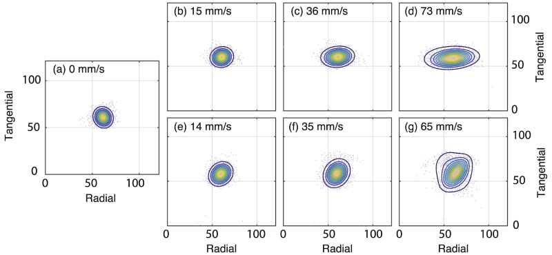
The researchers reconstructed the emission data using an iterative motion-compensation algorithm. They then manually isolated the point source from the image, modelled it as a mixture of two Gaussian distributions, and extracted the motion-dependent PSF. They observed that the shape of the PSF was dependent on the motion pattern, with lateral motion leading to elongated PSFs and star-like motion giving more rounded PSFs. For both, the width broadened as phantom speed increased.
Isocontours of the fitted motion-dependent PSFs. The isocontours correspond to different motion speeds during data acquisition; lateral-only motion is shown on the top row and star-like motion on the bottom row.
For PET scans with lateral motion at 15 mm/s – a speed typically encountered during an awake rat study – motion-corrected reconstructed images had slightly lower resolution than an image from a stationary acquisition. For moderate (35 mm/s) and fast (70 mm/s) motion, residual motion blurring was clearly visible. Similar findings were seen for the scans with star-like motion.
The team then used the PSF within an iterative Lucy-Richardson deconvolution algorithm to mitigate residual blurring in the motion-corrected images. The deblurred hot-rod images appeared highly similar, in terms of resolution and contrast, to their stationary counterparts. In particular, the three larger sets of rods (4.2, 4.0 and 3.2 mm) in the deblurred images appeared more circular compared with the unprocessed images.
To quantify differences in contrast before and after deblurring, the researchers examined line profiles through some of the rods. The profiles showed a clear difference in contrast (peak-to-valley ratio) between the stationary and motion-corrected images. After deblurring, the reconstructed line profiles for stationary and motion-corrected images were highly similar.
Realistic rat motion
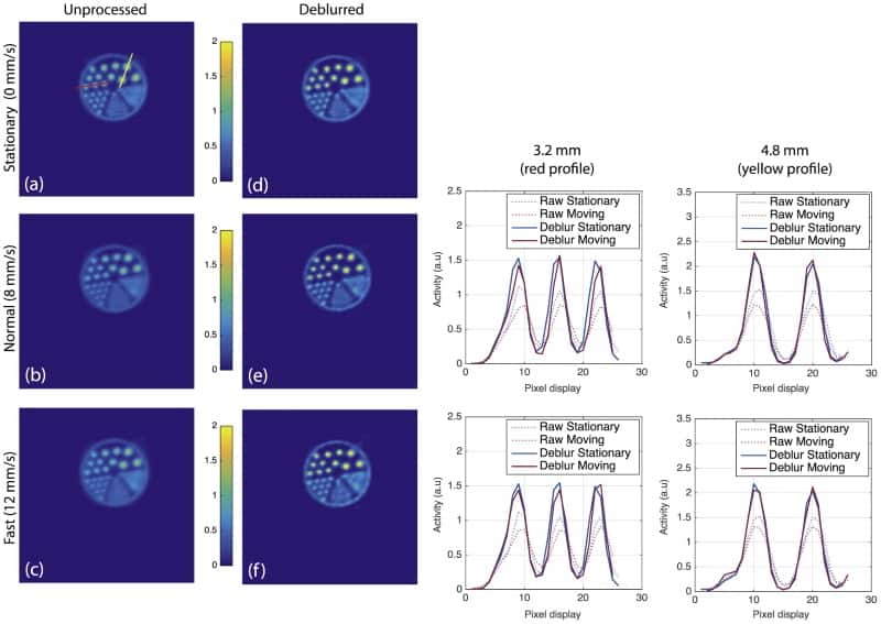
The researchers also applied realistic motion (taken from head movements of an awake tube-bound rat) to the phantom using a high-precision robot. Motion parameters included translations and rotations with an average speed of 8 mm/s and maximum speeds of 60-80 mm/s across all three axes. They also emulated a more agitated animal with an average head speed of 12 mm/s.
Motion-corrected hot-rod phantom images of realistic rat head motion without post-reconstruction processing (first column) and after de-blurring using the estimated motion-dependent PSF (second column). The third and fourth columns show line profiles through the 3.2 mm and 4.8 mm rods, respectively, for unprocessed (dotted lines) and deblurred images (solid lines), compared with their stationary counterpart.
As before, the motion-corrected images were slightly degraded compared with the stationary acquisition, with resolution deteriorating more for the fast-moving case. Comparing unprocessed and deblurred images demonstrated the improvements in contrast recovery offered by the post processing.
The authors note that although they didn’t show results from a moving animal, it should be fairly straightforward to transfer this technique to their routine freely moving animal experiments. “We have previously performed pilot experiments, where the point source was attached to the optical marker glued on the animal’s forehead, which was well tolerated by the animal,” Angelis noted.
In future work, they plan to incorporate the proposed methodology during image reconstruction (rather than post-reconstruction) using a nested deconvolution approach, which should further improve image quality, primarily due to noise reduction.
“Noise reduction is particularly important for other approaches we are working on, such as direct 4D reconstruction algorithms and deep learning neural networks to derive reliable physiological parameters from the brains of conscious animals,” Angelis explained. “Our overall aim is to improve image quality and parameter estimation for awake and freely moving animal scans, and this approach may provide a straightforward means to overcome basic hardware limitations.”
Jim Bridenstine, a Republican Congressman from Oklahoma, has been sworn in as NASA administrator – the political appointee who leads the space agency. The appointment comes 15 months after former astronaut Charles Bolden resigned as administrator. Brindenstine is 42 and was approved for the job by the US Senate by a single vote margin last week.
“I look forward to working with the outstanding team at NASA to achieve the president’s vision for American leadership in space,” Bridenstine said in a statement. But as he sets out on that mission, the former US Navy pilot faces a series of high-flying opportunities and potential difficulties, including the fate of of manned flight beyond Earth orbit and of advanced observatories and the burgeoning commercialization of spaceflight.
The unprecedented closeness of the Senate vote, which came more than seven months after the Trump administration nominated Bridenstine, indicated the degree of discontent that surrounded his candidacy. Critics in and outside the Senate complained that Bridenstine lacks the experience as an astronaut or manager of space missions that most of his predecessors possessed. They also expressed fears that he will take a partisan approach to managing the traditionally non-political space agency. And they pointed to his less than enthusiastic acceptance, before and during his Senate testimony, of the reality of human-induced climate change, an important factor in NASA’s Earth exploration research.
“Politically divisive”
“The NASA administrator should be a consummate space professional,” said Florida Democratic senator Bill Nelson during Senate debate, noting that Bridenstine’ only direct connection to space issues was as executive director of the Tulsa Air and Space Museum and Planetarium from 2008 to 2010. “What’s not right for NASA is an administrator who is politically divisive and who is not prepared to be the last in line to make that fateful decision of go or no go for launch,” Nelson added. Despite his criticisms, however, Nelson has indicated that he would work with Bridenstine if he were confirmed.
Critics also remain unconvinced by Bridenstine’s moderation of his initially sceptical view of human-caused global warming. “I am aware of his dismissive statements about climate change,” Pennsylvania State University climatologist Michael Mann told Physics World. “I believe that he has the wrong stuff when it comes to the sort of leadership NASA needs in the 21st century.”
Bridenstine’s supporters point out that neither James Webb, NASA’s administrator during the build-up to the Apollo Moon-landing programme, nor Sean O’Keefe, who led the agency from 2001 to 2004, had experience in space projects before their appointment.
Not naïve
Bridenstine demonstrated an interest in space issues in 2016, when he sponsored a bill called the American Space Renaissance Act in Congress. “It’s a very thoughtful look at needed reform,” says John Logsdon, professor emeritus in George Washington University’s Space Policy Institute. “He’s not coming in naïve.”
G Scott Hubbard, former director of NASA’s Ames Research Center and overseer of the agency’s first Mars programme who is now at Stanford University, agrees. “He has exhibited a significant interest in the policy,” he says. “He wasn’t just grabbed from the hallway.”
Bridenstine certainly inherits a tough set of issues from Robert Lightfoot, who leaves NASA next week after service as acting administrator since Charles Bolden’s departure. NASA must rely on Russian and commercial spacecraft to launch its astronauts until it develops its Space Launch System and Orion spacecraft. It must work with Congress on deciding whether or not to pull out of the International Space Station. It must deal with delays in its two major observatory projects – the James Webb Space Telescope and the WFIRST.
Back to the Moon
But perhaps NASA’s biggest challenge will be to carry out the Trump administration’s pivot to the Moon as a gateway on the path towards manned missions to Mars. In those efforts, “Bridenstine will put even more reliance on the commercial sector,” Logsdon says. “There’s no going back, nor should there be.” Meanwhile a new chief scientist, the physicist and former director of NASA’s Planetary Science Division Jim Green, will join Bridenstine in the agency’s headquarters.
As observers see it, the new administrator needs another critical individual on board: a deputy administrator schooled in the culture of NASA and space science. Such an appointment “would tend to quieten the concern about being a political hack,” Hubbard says. So far, no potential nominees have emerged for that post. “We’re all very interested to see what sort of person the administration needs to support Bridenstine,” says Logsdon.
“Our results warn that if we allow climate change to continue unchallenged, European cities are likely to suffer more frequently the devastating effects of high winds, including storm surges in coastal areas,” said Oscar Martinez-Alvarado from the UK’s National Centre for Atmospheric Science. “However, an important detail about our study is that we assumed the most extreme climate change scenario in which the concentration of greenhouse gases keeps increasing throughout the 21st century.”
The team hopes that actions to tackle rising global surface temperatures such as targets on reducing greenhouse gas emissions will mean better news for Europe’s weather.
Advances in computing power allow researchers to explore weather systems – such as the extra-tropical cyclones that dominate autumn and winter weather over Western Europe – in more detail than ever before. But sting jets are particularly challenging for climate modellers to capture.
“They are relatively small-scale features, only a few hundreds of kilometres wide, and therefore can only start to be realistically resolved in models with a horizontal grid spacing of around 12 km,” said Martinez-Alvarado. “However, typical climate models work with horizontal grid spacings of around 60 km.”
To reduce the computational burden, the team inferred the likelihood of sting-jet occurrence indirectly, using precursors.
“In previous work, we showed that sting jets are associated with the occurrence of a certain type of instability in the atmosphere, which would manifest itself as the downward motion of air forming (or enhancing) a sting jet,” said Martinez-Alvarado.
Applying their proven sting-jet diagnostic to present day results, the scientists found that 32% of cyclones exhibited the precursor. When they modelled climate behaviour out to the year 2100, the figure rose to 45%.
“We believe that this increase is due to the enhanced availability of moisture in a warmer climate, but more work is needed to confirm this hypothesis,” said Martinez-Alvarado.
In addition, the group, including scientists based at the University of Reading, the University of Oxford and the UK’s Met Office, is keen to know more about the nature of the energy transport process. In other words, by what mechanisms the energy of sting jets, which are present 1 to 2 km above sea level, reaches the ground.
When I cycle through Hyde Park in London on my way to work, pausing as I cross the Serpentine to admire the view of the Houses of Parliament, I rarely think about what is going on beneath my tyres. There are just too many above-ground distractions to spend much time wondering about the subterranean network of plant roots weaving their way through the dark and mysterious soil below. And anyway, what have roots got to do with physics?
Roots are complex branched systems whose topology and structure determine the entire physiology of the plants above them. The tip of each branched root has distinct regions where different cell activities take place. At the very end there is the “meristem” where cell divisions occur. Moving up the root, an “elongation zone” comes next where the cells stop dividing and instead elongate. It is followed by a “differentiation zone” where the cells stop elongating and start differentiating.
The direction and rate of root tip growth are influenced by the signals they perceive from the environment, and the organization of the internal tissue within each root tip plays a big role in this root-soil interaction. As root tips explore, the signals help them seek out the most efficient paths to discover water and nutrients. The composition of the soil will influence the architecture of the root system, which ultimately determines the efficacy of the exploration. It seems that evolution has placed enormous pressure on root tips to understand their environments – and it has taken until now for us to start asking why.
The physics of the soil–root interactions that optimize subterranean structures is crucial for plant survival. The roots sense what is going on around them with remarkable detail at the appropriate length scale. “We are not talking about the scale of quantum mechanics, but interactions described by classical physics,” says Giovanni Sena, a development biologist at Imperial College London in the UK, who has a strong background in theoretical physics.
Roots are constantly exploring their space, steering through natural force fields – gravitational and electromagnetic fields as well as magnetostatic and electrostatic – and experiencing the mechanical and chemical properties of the soil they live in. Gradients in light, chemicals, temperature and oxygen provide positional information that allows the root to navigate and forage efficiently. These kinds of root responses are known as a “tropisms”.
Charged insides
The impact of mechanical forces on roots is relatively intuitive – stress and strain can change the shape, number and organization of growing tissue – and the alignment of root tips with the gravitational field is also relatively well understood. However, there is still no model that can effectively describe the much more subtle interaction between an external electric field and the internal electric field of biological tissue.
Keeping a plant alive without damaging it during measurements is no mean feat, and near-on impossible without understanding instrumentation and optics
We know that living cells actively maintain a negatively charged cytoplasm – the material inside a cell excluding the nucleus – and this establishes a non-zero membrane potential. Oscillations in the membrane potential create a temporal pattern, while a spatial pattern is produced by the differences in potential between cells. Yet there are still many unanswered questions. Could these patterns, for example, be crucial to tissue organization and morphogenesis, the process that causes an organism to develop its shape? Is spatial variability just stochastic noise? Or is it an actual pattern that is used as “positional information” during morphogenesis? Until now, how bioelectricity in plant tissue impacts their shape and function has never been defined.
One of Sena’s collaborators, Michael Levin at Tufts University in the US, is studying the bioelectrical cues of membrane potentials and using them to describe cell behaviour. Levin argues that cracking “the bioelectrical code” – understanding how patterns are encoded into biological networks – would allow us to programme cell activity and transform synthetic bioengineering and cancer treatment (Mol. Biol. Cell25 3835). Sena’s group is collaborating with Levin and partially funded by the Allen Discovery Center at Tufts University to develop models of bioelectric circuits within roots that store patterning information during morphogenesis.
Cut to regenerate
For a physicist, Arabidopsis – a small flowering plant also known as thale cress – is an ideal subject to observe and model because it has transparent, slow-growing cylindrical roots. It is now well established that if you cut the end off its root tip, it will regenerate and reform with no external influences, as shown in figure 1 (Nature457 1150). However, like a lot of developmental biology, a comprehensive understanding is still missing.
1. Root regeneration: Optical sections of regenerating Arabidopsis roots, taken with confocal microscopy on consecutive days after tip excision. Scale bar: 50 µm. (Courtesy: Giovanni Sena)
Electrotropism – the alignment of a root with an electric field – has also been observed for decades, but not explained, and Sena’s lab further identified that if you place the cut root tip in a weak electric field, it is more likely to regenerate (Regeneration3 156). The field impacts some hormones more than others but cause no obvious changes in tissue patterning. These are fascinating results that science still can’t explain: why does an external electric field make tissue regenerate faster? And why does a plant root align with an electric field in the first place?
Understanding the regeneration of damaged tissues would be a game changer in controlling morphogenesis. For roots it can be modelled as a self-organizing 3D system of interacting and coupled cells, which divide with a certain probability – a complicated mathematical model. Instead of just being a cylinder, the root becomes a 3D complex system of interacting units, where the units respond to an electric field. Despite advances in the computational models for developmental biology, there is still no effective way to model the increased regeneration.
Unlike animal cells that can move and change shape because they do not have cell walls, plant cells have a rigid extracellular cellulose coat that restricts their ability to migrate when embedded in a tissue. The driving force to shape plant organs is instead cell division – exactly where and when cells divide will strongly influence the overall tissue shape. The dynamics of cell division events are entirely non-trivial during morphogenesis, and at present very little is known about their spatial and temporal correlations.
Physics tells us that if you have a complex system undergoing a transition there is a sudden change of the correlation length. Sena wants to identify how the spatial and temporal correlation of cell divisions change during regeneration, and how regeneration is influenced by the physical forces that the root tip is growing in.
Experiments with tiny plants
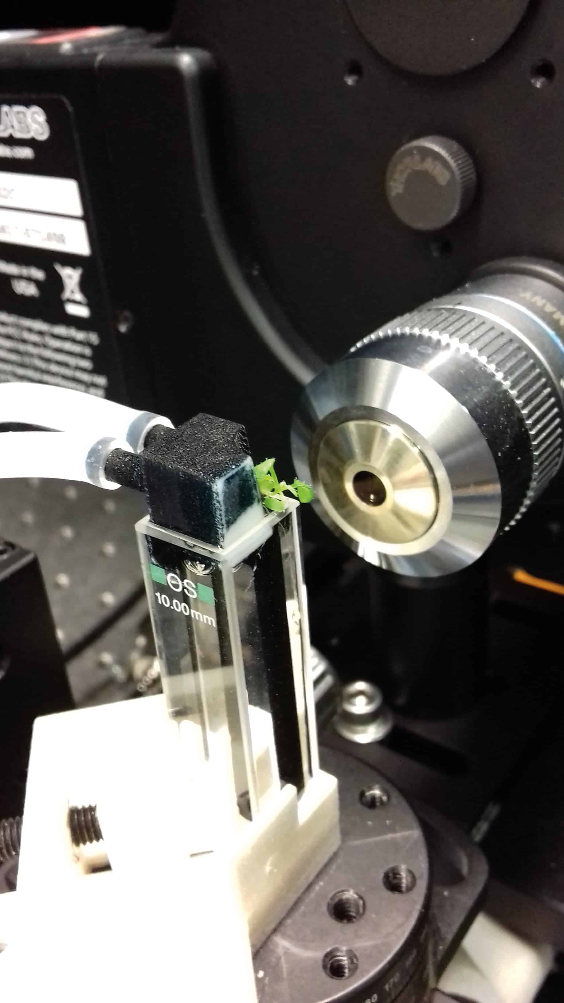
To investigate dynamic developmental processes, scientists need to image living tissues at cellular resolution regularly over an extended period of time. Keeping a plant alive without damaging it during measurements is no mean feat, and near-on impossible without an understanding of instrumentation and optics. To overcome these challenges, building on his previous experience as postdoc at Rockefeller University in New York (PLOS ONE6 e21303), Sena’s team created its own automated light sheet fluorescence microscope (LSFM), combining optical sectioning with a miniaturized plant growth chamber.
A physicist’s favourite: Thale cress has ideal roots for studying regeneration. (Sena Giovanni)
At any given time, the root is illuminated by a thin laser sheet (about 4 µm across), reducing the incident photo-energy and acquisition time by avoiding the need for lateral scans. The plant is held within a cuvette that is continuously illuminated and held at a constant temperature, with oxygen and an actively circulated liquid nutrient solution to keep it growing. Cell divisions are identified by tagging a protein – that appears just before and is destroyed just after cell division – with a fluorescent marker.
Because plants are so sensitive to gravitational fields, Sena had to play with the geometry of the microscope, with the fluorescent signal collected by a long-working distance lens normal to the excitation light-sheet (generated by a laser beam through a cylindrical lens) and the cuvette positioned vertical in the microscope. The stage is automatically repositioned to keep the root tip within the focus of the objective. As even slow-growing Arabidopsis can extend by a few millimetres per day, Sena and his group wrote an algorithm to compare the most recent image with the one before and automatically reposition the stage in 3D.
Before Sena’s dynamic LSFM, confocal microscopy only let researchers take daily images of regenerating root tips. If you wanted to know whether the root reforms or not, confocal microscopy was fine, but if you wanted to investigate regeneration at high temporal resolution, it was not enough. The LSFM allows Sena to grow a root and observe in 3D every cell division event every 10 minutes for at least one week at a time – and by measuring both cut and uncut roots, his team can compare the dynamics of regeneration against normal growth.
A glowing worm
On a computer, the process looks like a glowing worm wiggling across the screen, but it’s really the meristem of a root tagged by fluorescent proteins being tracked by Sena’s automated microscope stage. Whenever and wherever the root blinks, a cell division event has occurred. The video represents a 2D projection of a week of LSFM data collected every 10 minutes. For a non-expert, watching cell division is breathtakingly beautiful, but for Sena, it is just more intriguing physics and biology. Alongside the sped-up video, he can track the number of cell divisions, analyse their temporal and spatial distributions, characterize the bursts of proliferation activity and the more quiet periods, and in general compare normal and regenerating growth. The data are messy – well, we are dealing with biology, not electrons – but the implications are profound.
There is no doubt that physics contributes significantly to Sena’s team’s understanding of plant morphogenesis. In his lab almost all the researchers are experimental physicists – one even used to build particle detectors at CERN – while the others are biologists who have seen the light. If you gave their data to pure biologists, they’d jump immediately to genetics, and start a quest for “mutants” – genetic defects sufficient to modify the observed cell behaviour. Conventional plant science is familiar with visualizing root tips as static slices, but Sena argues that the dynamics are much more important – abnormal processes might look perfectly normal when viewed as static images. Plant morphogenesis, influenced by cell division, is an outcome of evolution – so how and why natural selection has resulted in these division dynamics is an underpinning question for biosciences. And the answers could provide a whole new perspective of those subterranean networks that I cycle above every day.
A single molecule has been created by combining individual atoms of sodium and caesium, using optical tweezers to guide them into place. The technique, devised by Lee Liu and colleagues at Harvard University and Harvard-MIT Health Sciences and Technology, could help chemists to study chemical reactions far more precisely by giving them control over the individual atomic and molecular collisions. The team hopes that their method will be used in a variety of fields to create diverse, complex molecules, allowing for discoveries of previously unforeseen molecular properties.
Conventional studies of chemical reactions involve observing the macroscopic results of large numbers of collisions of atoms and molecules – rather than studying individual collisions. Currently, chemists need to compare experimental reaction rates with theoretical models to calculate the probabilities of individual collisions taking place – a process that is fundamental to the understanding of chemistry. An alternative, and more precise, technique is to study interactions between individual atoms and molecules – something that requires great experimental dexterity.
To begin their interaction process, Liu and colleagues use magneto-optical traps to prepare reservoirs of stationary atoms of sodium and caesium at just a few hundred microkelvin. “Cooling and controlling atoms and molecules to temperatures where they are standing still allows for easier manipulations of their properties, interactions and reactions,” explains team leader Kang-Kuen Ni.
Polarizing forces
The chilly temperature means that individual atoms can be easily loaded onto optical tweezers – specialized, highly-focused laser beams that can trap dielectric particles including atoms. To confirm that the tweezers had been loaded successfully, the researchers use spectroscopic techniques to check for the presence of each atom.
One optical tweezer – based on 700 nm-wavelength light – holds the sodium atom, while another tweezer at 976 nm holds the caesium atom. To bring the atoms close together, the team move the tweezer containing the sodium atom so that it overlaps with the tweezer containing the caesium atom. Then, the 700 nm light is switched off. The 976 nm laser is capable of holding both atoms, so the sodium and caesium are trapped together.
The team found that simply bringing the atoms together is not enough to form a molecule – a pulse of light is required. “The atoms couldn’t turn into a molecule just through an isolated collision, as they had to simultaneously conserve energy and momentum – something very difficult to achieve in experimental conditions,” Ni explains. “To solve this, we shone another laser tuned to the resonance wavelength of the molecule, driving them to form the molecule.” The excited molecule which formed quickly decayed into a stable ground state, allowing for observations of its properties. “This was the first time that a molecule has been assembled starting from exactly two atoms, and establishing individual control over each of them,” said Ni.
New and unexpected
Even for such a simple molecule, the team observed new and unexpected properties in their creation. “We saw new molecular spectroscopic lines, and that contributed to a deeper understanding of the overall picture,” explains Ni. The researchers believe their technique could soon be expanded to study complex chemical processes.
“We are now thinking of working with many optical tweezers and many molecules simultaneously, allowing for studies of their interactions or reactions,” Ni says. With this technology available, it could soon be possible for chemists to synthesize their own designer molecules, with applications ranging from new medicines to qubits in quantum computers.
Researchers have challenged one of the few certainties of earth and climate history: the link between erosion and the drawdown of carbon dioxide from the atmosphere.
For decades, it has been a given that heavy rainfall on steep mountain slopes is likely to chemically weather the exposed rock and precipitate a chemical reaction that ends with carbonate minerals on the ground and with less of the greenhouse gas carbon dioxide in the atmosphere.
So geochemistry and the weather between them help moderate the planet’s climate.
But geologists and oceanographers who took another and closer look at the process in action – in the central mountains of Taiwan, hammered by three or more major typhoons each year – say they are not so sure.
They report in the journal Science that the same erosion process could be a source of carbon dioxide, releasing it into the atmosphere far faster that it can be absorbed by the newly exposed rock.
And the agency at work in this unexpected process could be biology: the researchers found evidence that tiny microbes in the mountain soils were consuming sources of organic carbon trapped in the rock, and releasing CO2 into the atmosphere.
On the face of it, the process may not be severe enough to upset the global calculations that add up to what climate scientists call the carbon budget – the annual traffic of carbon in the form of greenhouse gases from atmosphere to living things and then into the rocks and oceans – but it is yet another reminder that the climate machinery is still incompletely understood.
“This goes against a long-standing hypothesis that more mountains mean more erosion and weathering which means an added reduction of CO2. It turns out it’s much more complicated than that,” said Jordon Hemingway, of Harvard University, who led the study.
The Earth’s crust, powered by heat from the mantle below, is permanently in a state of levelling and reconstruction: powerful subterranean forces build mountains and the steady attrition of wind and rain immediately begin the process of wearing them down.
Complicated picture
But mountains are part of the climate machine. There is a theory that the rising of the Himalayas and the Tibetan plateau provided the barrier that made the South Asian monsoons possible, and a secondary theory that the increased rainfall on the freshly raised mountain slopes weathered so much rock that the planet’s levels of atmospheric carbon dioxide took a dive, to precipitate 30 million years of Ice Ages.
A closer study of the soil, bedrock and river sediments in Taiwan revealed a more complicated picture. The scientists found that almost 70% of the organic carbon initially present in the weathered bedrock had been oxidised by soil microbes, to put, for every square kilometre they measured, somewhere between six and 18 tonnes of carbon back into the atmosphere.
This is not enough to set alarm bells ringing. But it does suggest that the intricate details of the carbon budget depend not just on what happens on the planet’s surface, but also in the teeming life beneath – and sometimes far beneath – the surface.
This is basic research at a down-to-earth level: climate science can’t make sense of what is happening now without a better understanding of what has always happened, and of the swings in planetary temperatures over the past 4.5 billion years.
Clear understanding
Researchers are confident that they understand the cycle of Ice Ages, and they also have a clear idea that the biosphere plays a hand in keeping the planet at liveable temperatures, but they also know that the high altitudes are more than usually affected by climate change driven by ever-higher ratios of greenhouse gases released by the combustion of fossil fuels by seven billion humans.
Their fears extend to Alpine economies and the plants and animals that live in the mountains. Now it seems clear that some long-term questions require explanation at the microscopic level.
“Looking backwards, we’re most interested in how these processes managed to keep the levels of CO2 in the atmosphere more or less stable over millions of years. It allowed Earth to have the climate and conditions it’s had – one that has promoted the development of complex life forms,” Dr Hemingway said.
“Throughout our Earth’s history, CO2 has wobbled over time, but has remained in that stable zone. This is just an update of the mechanism of geological processes that allows that to happen.” – Climate News Network
Stress is a major modulator of neuronal networks and its consequences can be transmitted to others. With this in mind, a team from University of Calgary embarked on a journey to decipher whether transmitted stress has the same consequences on synapses as authentic stress (Nature Neuroscience21 393). Their findings in mice suggest that the lasting effects on synapses are the same for both authentic and transmitted stress, with implications for other stressful events.
Stress induces metaplasticity
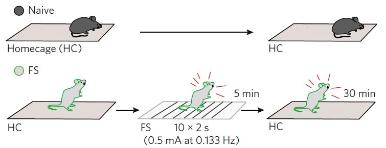
In the experiments, the authors subjected mice to footshock stress for 5 min and, upon returning them to their homecage, to high-frequency electrical stimulation for 30 min. They discovered that acute stress resulted in persistent changes at glutamate synapses (glutamate is the major excitatory neurotransmitter in the nervous system) on corticotropin-releasing hormone (CRH) neurons. This enables the induction of metaplasticity, activity-dependent changes in neural functions that modulate subsequent synaptic plasticity. The CRH neurons are found in the paraventricular nucleus of the hypothalamus, one of the most important autonomic control centres in the brain, and their activation is necessary for the rise of the endocrine response to stress.
Naive control mice were left undisturbed in the homecage. Footshock mice were subjected to 0.5 mA for 2 s, every 30 s for 5 min and then returned to the homecage for 30 min. Reprinted by permission from Nature Neuroscience21 393 Social transmission and buffering of synaptic changes after stress, Sterley et al., 2018
Pheromones help transmit stress
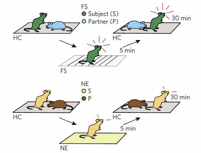
To study the effect of social interactions on metaplasticity, mice were subjected to footshock or a novel environment for 5 min and returned to a same-sex control partner in the homecage for the next 30 min. Interestingly, stress is transmitted by the release of alarm pheromones, predominantly from glands in the anogenital area, which are sensed by the partner during investigative behaviour. The triggering of this type of behaviour and synaptic priming require the activity of CRH neurons (which the authors determined using whole-cell recordings from CRH neurons in hypothalamic slices).
This experiment suggested that transmitted stress primes glutamate synapses in male and female subjects with different sensitivity and, importantly, that the effects of authentic stress in females are reduced by the presence of a partner.
Same sex littermates were housed in pairs. In the experimental pair, the subject was removed, exposed to footshock and returned to the partner in the homecage for 30 min. In another pair, the subject was taken to a novel environment for 5 min but not exposed to footshock, and then returned to the partner in the homecage for 30 min. Reprinted by permission from Nature Neuroscience21 393 Social transmission and buffering of synaptic changes after stress, Sterley et al., 2018
In another experiment, the team showed that the synaptic priming induced by transmitted stress can also be propagated by a partner mouse to a tertiary group member, with similar effects (the same synaptic load).
The findings of this study indicate the importance of mitigating the effects of stress and how the information extracted from the experience of a distressed subject can have adaptive benefits. Moreover, the impacted neuronal circuits can this way prepare the individuals for subsequent challenges.