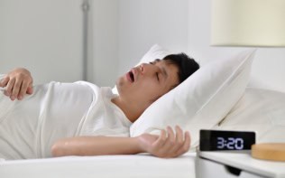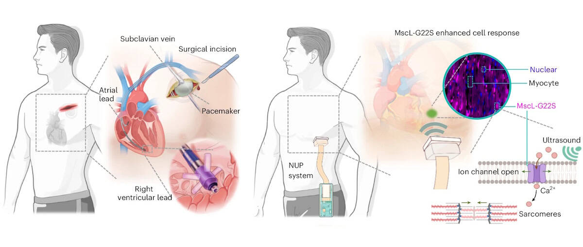
Cardiac arrhythmia – a disorder in which the heart beats too fast, too slowly or irregularly – affects millions of people worldwide. Currently, arrhythmias are treated using pacemakers to regulate the heart’s rhythm, but traditional pacemakers require invasive surgical implantation and intracardiac leads that risk infection and tissue injury. As an alternative, a research collaboration headed up at Massachusetts Institute of Technology (MIT) and the University of Southern California (USC) has developed a non-invasive pacemaker that’s worn like a sticker on the chest and stimulates the heart using ultrasound.
“Ultrasound offers a unique combination of deep tissue penetration, spatial focusing and non-invasive delivery,” explains first author Chen Gong from USC. Previous approaches for ultrasound-based cardiac pacing, however, lacked reliability or required the use of microbubbles. In this new study, Gong and collaborators employed a technique called sonogenetics, in which cardiac cells are genetically altered to increase their sensitivity to ultrasound. Once exposed to ultrasound waves, the engineered cells trigger the opening of ion channels that let in calcium, which signals the cells to squeeze and beat.
“Our central motivation was to address the limitations of conventional pacemakers, while preserving precise control of cardiac rhythm,” says Gong.
The team first examined whether ultrasound could modulate cardiac cells in vitro, using sonogenetically engineered human heart muscle cells. Ultrasound stimulation caused almost three-quarters of the engineered cells to beat in synchrony with the ultrasound waves. In contrast, unaltered cells did not exhibit such behaviour. Importantly, ultrasound exposure did not impact the cells’ expression of biomarkers for inflammation, myocardial injury or oxidative stress, demonstrating the safety of this approach.
The non-invasive ultrasound pacemaker
The researchers designed a sonogenetics-based non-invasive ultrasound pacemaker (NUP) capable of real-time imaging and steerable stimulation, describing the device in Nature Biomedical Engineering. The postage stamp-sized device, which sticks to the skin via a bioadhesive interface, incorporates a 64-channel phased-array transducer, as well as data acquisition, wireless transmission and power modules.
To assess whether this prototype NUP could achieve non-invasive cardiac pacing in sonogenetically engineered rats, they attached the device onto the rats’ chests and applied ultrasound stimulation through the chest wall to the heart.
At an acoustic pressure of 2 MPa and a 40 ms pulse duration, the NUP increased the rats’ heart rates from 240 to 360–540 bpm, with a higher pulse repetition frequency leading to increased heart rate. Upon stopping the ultrasound stimulation, the heart returned to its natural rhythm. In control rats without the genetic modification, NUP could not control the heart rate effectively.
The team demonstrated that the NUP could electrically steer the ultrasound beam focus with a spatial precision of less than 1 mm, enabling targeting of different regions of the heart to induce chamber-specific pacing. NUP also successfully treated cardiac arrhythmias induced in the engineered rats, returning their heart rates to normal levels. In contrast, ultrasound stimulation of control rats did not effectively treat their arrythmias.
“A major objective of this work was to move beyond proof-of-concept stimulation and demonstrate a wearable system that could realistically support daily use,” explains Qifa Zhou, group leader at USC. “The current prototype integrates imaging, stimulation, wireless communication and battery-powered operation into a wearable format.”
Clinical potential
For future clinical use, the NUP device needs to automatically detect the wearer’s heart rate, locate heart chambers and deliver stimuli. To achieve this, the researchers used a cloud-based, artificial intelligence (AI)-powered imaging feedback loop that determines the heart rate and stimulation coordinates, and steers the ultrasound beam to target sites.
To test its feasibility for human-scale applications, they assessed the acoustic energy penetration of the device. Using simulations and experiments on a pig heart beneath layers of tissue, they confirmed that, even after tissue attenuation, NUP could deliver sufficient ultrasound pressure (approximately 2 MPa) to a human heart to enable stimulation at clinically relevant pacemaker sites.
The team also assessed the biosafety of the NUP approach, finding that the delivered ultrasound energy remained below approved safety limits and generated minimal thermal effects during and after stimulation. They also confirmed that the sonogenetic engineering induced no off-target effects, immune responses or pathological changes in the rats.
“In the long term, we are optimistic about applying sonogenetics in humans,” explains co-author Gengxi Lu from MIT. “Sonogenetics is different from gene editing – it does not aim to rewrite a patient’s DNA sequence, but instead enables target cells to temporarily or controllably express ultrasound-responsive proteins. At the same time, extensive clinical validation will still be needed to establish safety and long-term effectiveness.”
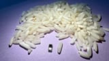
Light-activated pacemaker is smaller than a grain of rice
The team is now focusing on clinical translation and validation of the NUP technology, which includes improving gene delivery strategies, validating the system in larger animal models, and developing closed-loop control approaches that combine real-time physiological sensing with adaptive stimulation. There’s also much engineering work to be done, such as device miniaturization, and improving battery life, skin coupling and motion robustness.
“For cardiac pacing, we envisage that the final goal of NUP technology is to be a permanent alternative to a long-term implanted pacemaker,” Xuanhe Zhao from MIT tells Physics World. “More broadly, we are interested in expanding ultrasound-enabled bioelectronic medicine beyond cardiac pacing toward other organs and therapeutic applications where non-invasive, spatially precise modulation could have clinical impact.”