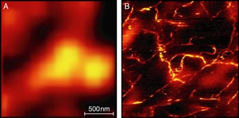
Scientists in the US have produced the highest resolution optical image to date – showing details of structures that are less than 30 nm across. Lukas Novotny from the University of Rochester and colleagues from Portland State University and the University of Harvard used a technique known as “near-field Raman microscopy” to look at carbon nanotubes (A Hartschuh et al. 2003 Phys. Rev. Lett. 90 095503)

Advances in nanotechnology rely on researchers being able to manipulate individual structures on the nanoscale. Many new methods have been introduced, such as scanning probe techniques, optical tweezers and atomic force microscopy. Although these ultra-high resolution imaging techniques can detect the presence of very small objects, they cannot actually “see” them.
Raman spectroscopy involves sending laser light through a sample and measuring how the light is scattered. The technique can provide much more detailed structural information about molecules than other imaging methods, because it measures unique vibrational modes of the material being studied.
Hartschuh and co-workers focused a laser beam against the side of a silver wire tip, measuring about 10 nm across. They then scanned the tip over a sample surface at a distance of around 1 nm. The interaction between the electromagnetic energy in the silver tip and the atoms in the sample created packets of light that the researchers collected, filtered and analyzed. This technique is known as “near-field” surface enhanced Raman spectroscopy and can boost the Raman signal intensity by factors of up to 1015 – which allows single molecules to be investigated.
The method can be used to identify the chemical composition of a material and can even detect whether a carbon nanotube is lying horizontally or vertically. This is an improvement on conventional “far-field” microscopy methods (see figure) which do not show such detail.
The researchers now hope to refine their system so that they can image structures – such as proteins – that measure only 5nm across. This means “sharpening” the silver tip or experimenting with different shaped points.


