Scientists at IBM have imaged the spin of an individual electron for the first time by combining magnetic resonance imaging with atomic force microscopy. The new microscope might eventually be able to produce three-dimensional images on the atomic scale and might also be used in read-out devices for spin-based quantum computers (D Rugar et al. 2004 Nature 430 329).
Although an atomic force microscope can image individual atoms, it can only be used to provide information about the surface of a sample. Magnetic resonance imaging (MRI), on the other hand, can probe deeply into an object and provide three-dimensional images. MRI measures the signal produced by the magnetic moments or “spins” of protons that have been aligned by a magnetic field. However, the spatial resolution of the technique is still limited to about a micron. The new technique is better than this by a factor of 40.
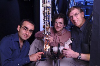
Dan Rugar and colleagues at the IBM Almaden Research Center have now combined both these techniques to make a magnetic resonance force microscope (MRFM) and have used it to detect the spin of a single electron some 100 nanometres below the surface of a sample made of vitreous silica. “An electron spin is easier to measure than a proton spin because the magnetic moment of the electron is some 600 times larger than the proton’s,” Rugar told PhysicsWeb. “We eventually hope to reach single proton sensitivity.”
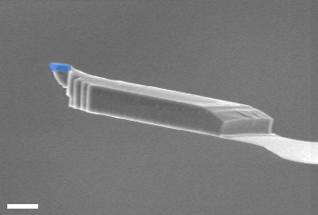
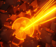
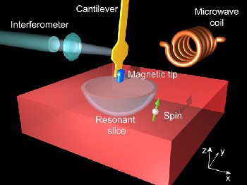
The IBM microscope consists of a nanometre-sized magnetic tip, made of samarium and cobalt, attached to a vibrating silicon cantilever that is 85 microns long and 100 nanometres thick. Rugar and co-workers positioned the cantilever about 125 nanometres above the silica sample and then applied a high-frequency magnetic field of 3 gigahertz to it. This excited electron spins inside the sample and flipped their direction (see figure 1).
The technique is able to distinguish between individual spins. The force produced by a single electron as it flips — which can be as small as 10-18 Newtons — switches from being attractive to repulsive and causes the cantilever frequency to change slightly. This change can be then be measured using a laser beam.
The scientists now hope to improve the sensitivity of the technique so that it is able to detect and image individual nuclear spins, which are much weaker than electron spins. “By coupling the powerful concepts of MRI with the exquisite sensitivity we have developed for detecting magnetic forces, we are hoping that MRFM can be developed into an atomic resolution instrument,” said Rugar.
