Graphene-based transistors that respond to changes in chemical solutions could be used to link electronic devices directly to the human nervous system. That is the claim of researchers in Germany who have built arrays of devices that respond to changes in the electrolytes surrounding living cells. The team hopes that its research could result in retinal implants that could help some visually impaired people see images.
The research centres on the small voltage that a neurone creates across its cell membrane when it fires, with the potential difference arising from sodium ions moving into the cell and potassium ions moving out into the surrounding solution. Since the 1970s, biophysicists have been trying to detect this sudden change in the electrolytic properties of the liquid surrounding a cell using a type of field-effect transistor (FET). These devices are called solution-gated FETs (SGFETs) and much of the initial research was done using silicon. But after graphene was isolated in 2004, some researchers realized that this material – a layer of carbon just one atom thick – could be used to create better SGFETs.
Clean, sensitive and flexible
According to Jose Garrido of the Technische Universität München, who has led the work, graphene offers several important advantages over silicon. First, the graphene surface remains clean – unlike silicon, which quickly forms a performance-degrading oxide layer when exposed to the electrolyte. Second, electrons in graphene have an extremely high mobility, which makes the device much more sensitive than silicon SGFETs. Finally, graphene is extremely flexible, which is good because any device implanted within the brain or similar tissue must be bendable.
A SGFET is a different take on a conventional graphene FET, in which the current flowing through its graphene channel can be controlled by changing the voltage applied to a nearby “gate” electrode. In a SGFET, in contrast, the gate voltage is kept constant and the graphene is exposed to the electrolytic environment of the cell. Any shift in the concentration of ions in the solution affects the electronic properties of the grapheme, thereby changing its conductivity and the current flowing in the graphene channel. The firing of a neurone is therefore detected as an electronic signal.
Brainy plans
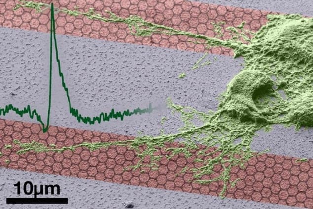
In their new work, Garrido and colleagues have created 8 × 8 arrays of SGFETs – with each individual transistor measuring about 10 μm across. These arrays were used to detect firing signals from neurone cells that were cultured on an artificial medium. The researchers have also shown that neurone cells are able to survive for long periods of time in close proximity to graphene layers. They now want to show that the SGFETs work in living tissue – rather than cell cultures – and that neuronal tissue is not adversely affected by the presence of the devices.
According to Garrido, an important application of the graphene SGFETs would be creating retinal implants that could improve the sight of visually impaired people. Indeed, he believes that an array containing about 1000 elements could provide the brain with enough information for a person to be able to perceive an image. Another important application could be as cortical implants to help people control artificial limbs.
Although creating a 1000-element array of graphene SGFETs is a straightforward process, Garrido says that integrating the technology within a person will require a great deal more work.
The technology is described in a preprint on the arXiv server.


