Researchers at Stanford University in the US have developed the first non-invasive imaging technique that can detect micron-sized structures within blood vessels in the brains of mice. The method involves detecting near-infrared fluorescent light from single-walled carbon nanotubes (SWCNTs) that are injected into the mice. The ability to monitor the structure of blood vessels – and the blood flow within them – is extremely important for treating conditions such as strokes, dementia and brain tumours.
Today, brain imaging mainly relies on techniques such as X-ray computed tomography and magnetic resonance angiography. However, these methods cannot image structures several microns in size. In addition, with these approaches it can take several minutes to acquire an image, which means that it is not possible to use them to monitor blood flow in real time.
Fluorescence-based brain imaging in the visible and near-infrared (NIR) regions of the electromagnetic spectrum (400–900 nm) is a good alternative but at the moment it requires skull-thinning or, worse still, craniotomy – where sections of the skull are removed and replaced with a transparent “window” – to work properly. This is because light at these wavelengths can only travel about 1 mm through the skull.
Window of opportunity
Now, a team led by Hongjie Dai and Calvin Kuo at Stanford has developed a new through-scalp and through-skull fluorescence imaging technique that goes a long way in overcoming these problems. The method makes use of the intrinsic fluorescence of SWCNTs in the 1.3–1.4 µm range. “We define this wavelength as the NIR-IIa window, and it represents just about the longest wavelengths for fluorescence imaging reported thus far,” explains Dai.
“Photons at these wavelengths are much less scattered than those in the 400–900 nm window when traversing biological tissues and are not absorbed significantly by water either,” says Dai. “All in all, this allows us to see deeper into the brain through intact scalp skin and bone than is possible with traditional fluorescence imaging, which is mostly done with <800 nm wavelength photons.”
“Compared with all other techniques for in vivo brain imaging (including MRI and CT), our technique affords higher spatial resolution”, he says. “It allows us to image single capillary blood vessels that are just microns across and as deep as 3 mm inside the brain.”
Need for speed
And that is not all: the technique is also fast, at 200 ms per frame or less, which allows blood flow to be monitored in real time. This is particularly important when treating stroke patients because in these cases blood flow in parts of the brain can be drastically reduced, causing serious damage.
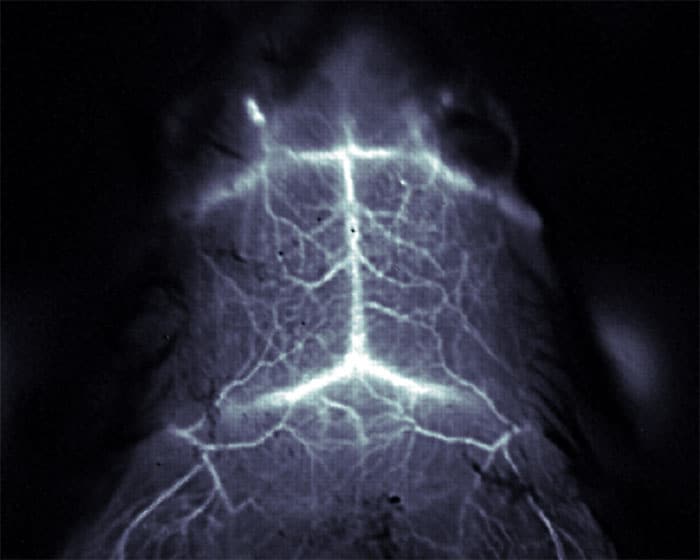
The team tested the technique by injecting mice with SWCNTs. The heads of the mice were shaved and illuminated with an infrared laser. The fluorescent light was detected using an array of photodiodes that created a 2D image of the brain. The researchers are now busy trying to image in 3D using their method. “We are also developing imaging agents for an even longer-wavelength window to further minimize photon scattering,” adds Dai. “And, we are looking at making NIR-IIa fluorophores that might potentially be used in human clinical trials.”
The imaging technique is described in Nature Photonics.
- This article first appeared on nanotechweb.org