A new technique for taking nanoscale images of the magnetic properties of materials has been unveiled by researchers in Switzerland. Unlike other nanoscale imaging methods, the technique can reach deep into a sample, and the team believes that it could provide new insights into the physics of magnetism and optimize magnets for a wide range of industrial applications including data storage.
Nanoscale images of magnetic structures can be obtained using low-energy “soft” X-rays or electrons. However, these can only penetrate 100–200 nm into a material and this restricts them to studying thin films and the surfaces of bulk materials. Neutrons can probe much deeper into materials, but their resolution is limited to 35–100 μm. Higher-resolution images of internal magnetic structure have been obtained only by techniques that destroy the material.
Higher-energy “hard” X-rays penetrate much deeper into materials and are used in tomography. However, magnetic interactions involving X-rays are very weak and magnetic tomography – which involves measuring three components of the magnetic field at every point inside an object – is fiendishly difficult.
Circular dichroism
Now, researchers at ETH Zürich and the Paul Scherrer Institute have developed a new imaging technique that allows researchers to do this using a phenomenon called X-ray magnetic circular dichroism.
“When you have circularly polarized light, and you are near the absorption edge of a magnetic material,” explains ETH Zürich’s Claire Donnelly, “you have preferential absorption of a certain spin for a certain polarization of light.”
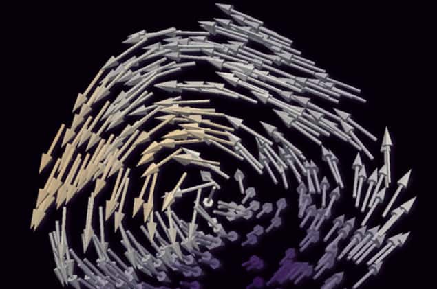
The researchers scanned a hard X-ray beam over the surface of a 5 μm-diameter micropillar of the intermetallic magnetic compound GdCO2. They recorded the change in the diffraction pattern as they rotated the pillar through 360°. The researchers then tilted the pillar through 30° about a different axis and repeated the process. Using a computer algorithm of their own design, the researchers used the data to reconstruct the magnetism at each point in the pillar with a spatial resolution of around 100 nm.
Bloch points
This allowed the team to make the first-ever direct experimental observations of Bloch points. These are monopole-like points where magnetic domains point in different directions. “These are not true Dirac monopoles of the kind everybody likes to make a fuss about,” says team member Sebastian Gliga, now at the University of Glasgow. “They are points of divergence of the magnetization within a material, which are fully allowed by Maxwell’s equations.”
The existence of Bloch points has been generally accepted for decades, and they were even used in a type of computer memory called bubble memory in the 1970s and 1980s. Until now, however, no technology has been able to image the magnetic domains around them. The team confirmed that the Bloch points are surrounded by circulating magnetic flux, which theoretical models predict is more stable than “hedgehog” Bloch points with all the domains pointing towards or away from one point. It also observed twisted “anti-Bloch” points for the first time.
The researchers believe the technique opens significant opportunity to explore magnetism: “Now we have the possibility to look inside these unconfined, bulk magnetic systems to see how they react to the application of a magnetic field, or to different temperatures,” says Donnelly. Her colleague Manuel Guizar-Sicairos of the Paul Scherrer Institute believes it also has significant industrial potential: “Once you can look into the internal structure and predict the bulk performance of the magnet, then you can tailor your production methods to create the properties that you want, such as greater efficiency, greater power or whatever.”
Speeding up
Peter Fischer of Lawrence Berkeley National Laboratory in California, who was not involved in the research, says it is “an achievement in demonstrating technique” and is well placed to take advantage of new fourth-generation coherent X-ray light sources: “This technique relies heavily on coherence,” he says. “The promise is that, whereas it took Claire Donnelly a day or two to collect the data, 10 years down the road it might be possible in a couple of minutes, because we have so much higher flux of coherent X-rays in the new facilities coming online as we speak. With this will come better spatial resolution.”
The technique is unveiled in Nature.


