Flash Physics is our daily pick of the latest need-to-know developments from the global physics community selected by Physics World‘s team of editors and reporters

Stellar family history
The family tree of nearby stars has been mapped by astronomers. Paula Jofré from the University of Cambridge and colleagues have applied biological principles to map how 22 stars are related to each other. In evolutionary biology, the DNA of living organisms allows scientists to map the route of evolution and determine the relationships between different species. While stars are very different to living creatures, they carry the chemical signature of the gas cloud they formed in, and will therefore share “chemical DNA” with other stars formed from the same cloud. Looking at 22 stars in our galactic neighbourhood including the Sun, the astronomers analysed chemical spectra taken by large telescopes in Chile, alongside data from the European Space Agency’s Earth orbiter, Hipparcos. Using computer algorithms, the team identified 3 groups of stars that share a common ancestor and six stars that were not statistically viable for any group. The researchers suggest that the thicker part of the Milky Way’s disc forms new stars more rapidly than other parts of the galaxy. Meanwhile, they also found that some stars may have originated from another galaxy that collided with the Milky Way in the distant past. The work, published in Monthly Notices of the Royal Astronomical Society, is a proof-of-concept study. Further datasets from Hipparcos’ replacement Gaia, more advanced telescopes and sky surveys, should allow astronomers to build a detailed stellar family history.
Electron camera images fragile metal–organic frameworks
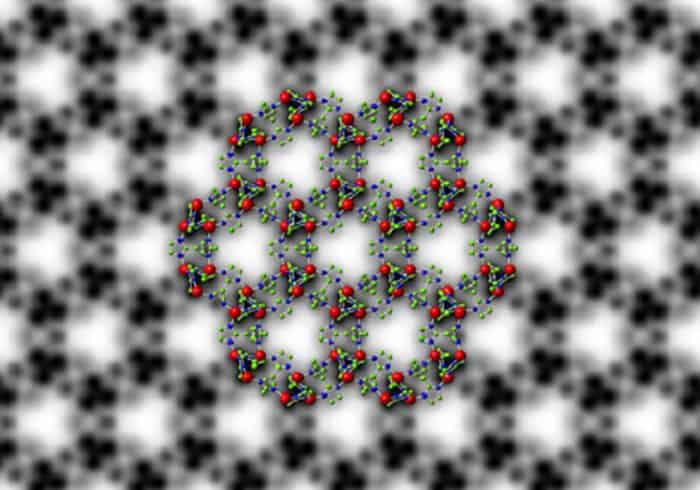
An extremely sensitive electron camera has been used to obtain transmission electron microscope images of fragile crystals called metal–organic frameworks (MOFs). Comprising metal atoms connected by organic molecules, MOFs are highly porous materials that can be engineered to store or transport specific gases. Determining the structure of a MOF is crucial to optimizing its performance, but doing so using electron microscopy is challenging because electron beams intense enough to create a useful image will destroy most MOFs. Now, an international team of researchers led by Yu Han at KAUST in Saudi Arabia have imaged a MOF using an electron-counting camera developed by US-based Gatan, which was also involved in the study. According to Gatan, the camera is able to detect individual electrons – making it possible to take microscope images under extremely low electron illumination. The team acquired images of ZIF-8, which is a MOF made of zinc atoms connected by 2-methylimidazole molecules. The images have a spatial resolution of 0.21 nm, which allowed the team to see individual zinc atoms in columns as well as the organic linking molecules. Han says that the research has already revealed new information about the porosity of the material, “which influences gas molecules transport in ZIF-8 crystals”. The research is described in Nature Materials.
Consortium will build full-body PET scanner

Researchers at the University of California, Davis, are leading a collaboration to build a prototype total-body positron-emission-tomography (PET) scanner. The EXPLORER consortium hopes to have a small-scale version ready for testing by July and the full-sized prototype by mid-2018. PET scanners use radioactive tracers attached to molecules of interest to show how organs and tissues are functioning. PET scans are mostly used to diagnose and monitor cancer, heart disease and forms of dementia. To gain multiple organ views of diseases such as measuring the spread of cancer, for example, it is necessary to scan the whole body because metastatic cancer can appear at many different sites. The detector in conventional PET scanners only covers a small part of the body, so imaging the whole body involves moving the patient through the scanner. The small detectors also mean that only about 1% of the available signal is actually collected. Another limitation of existing scanners is that they cannot collect dynamic data showing how the radio tracer distributes throughout the body. The new full-body PET scanner should provide a 40 fold gain in effective signal, compared with current scanners. This, say the researchers, will dramatically improve imaging capabilities and quality, revolutionizing the ability to study and diagnose disease as well as also reducing patient radiation exposure, which would allow more frequent use of PET scans. “Total-body PET is going to allow the interactions between different systems to be studied, providing a whole-body system view of disease that is not readily provided by other technologies,” says project leader Simon Cherry.
- You can find all our daily Flash Physics posts in the website’s news section, as well as on Twitter and Facebook using #FlashPhysics. Tune in to physicsworld.com later today to read today’s extensive news story on how to encourage teenagers to study physics.