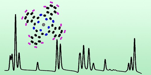
The sensitivity of a scanning-tunnelling microscope improves by up to a factor of 50 when the microscope’s usual tip is replaced by a superconducting one. The technique, developed by researchers at Christian-Albrechts-University in Kiel, Germany, could provide unprecedented levels of detailed data about molecules on the surface of a material. Such data could help scientists test and improve theoretical methods for understanding and even predicting a material’s properties.
Although vibrational spectroscopy is routinely employed to probe molecular properties and interactions, most techniques lack the spatial resolution and sensitivity to probe single molecules, explains team leader Richard Berndt. While inelastic tunnelling spectroscopy (IETS) with a scanning tunnelling microscope (STM) does not suffer from this problem, the small signal size of conventional IETS has so far limited the number of vibrational modes that can be observed in a molecule, with 1 or 2 modes out of 3N (where N is the number of atoms in the molecule) being a typical maximum.
Plenty of modes
“Our new technique increases the sensitivity of the STM, so far by factors up to 50, and as a result we see plenty of modes,” Berndt tells Physics World. “It simultaneously circumvents the resolution limit of conventional IETS, allowing us to provide detailed data on the vibrational modes of a molecule and how these modes change when they interact with their molecular environment.”
The researchers carried out their experiments in ultra-high vacuum with STMs operating at 2.3 and 4.2 K. For their sample material, they chose to study lead-phthalocyanine (PbPc) on a surface of superconducting lead. This sample provides a sharp feature known as a Yu-Shiba-Rusinov (YSR) resonance that arises when a localized spin, which the researchers prepared in their molecule, interacts with a superconductor – in this case, the lead substrate. Since the tip is also superconducting, it contributes an additional fairly sharp signal peak – the so-called coherence peak.
Electrons cross a “forbidden” region
When Berndt and colleagues applied a suitable voltage to the microscope, electrons from the peak in the tip inelastically tunnelled to the YSR peak on the sample. To do so, the electrons had to cross a so-called “forbidden” region as they tunnelled between the tip and the substrate, and they arrived with less energy than they started with. This energy difference comes from the excitation of vibrations of the PbPc molecule and it can be determined from changes in the conductance of the system. Using this technique, the researchers were able to enhance the signal (relative to tunnelling between two normal, non-superconducting surfaces) by a factor that is related to the product of the two peak heights.
Anyons could be spotted using scanning tunnelling microscopy
Since the experiments take place at cryogenic temperatures, the technique’s initial applications will be in basic science, says Berndt. “The technique will be able to provide detailed data on molecules at surfaces in an unprecedented fashion,” he explains. “It will also help us better understand the interactions between molecules, which are important for processes like self-assembly and properties like magnetism.”
The team is now trying to extend its method to other classes of molecules. “We will be attempting to understand the spectral intensities of the various vibrational molecules in these molecules,” Berndt says. “Currently, modelling can reproduce the mode energies fairly well, but the intensities hardly match the experimental data. We think that the time an electron spends on the molecule during the tunnelling process may play a role – but so far that is speculation. In any event, explaining the intensities will be a tantalizing nut to crack.”
The researchers report their work in Physical Review Letters.


