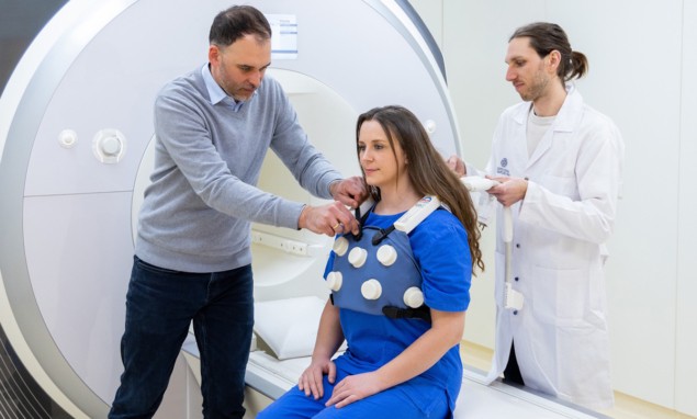
A new breast-imaging technique known as “panoramic breast MRI” could expand the use of breast MR imaging as a follow-up exam to investigate suspicious mammography findings. The approach is expected to improve image quality, simplify clinical workflow, be more cost-effective and aid image interpretation using a panoramic visualization of breast MR images.
In a step towards clinical adoption of this technology, researchers from the Medical University of Vienna have designed a prototype MRI coil that can be worn like a sports bra. The BraCoil, described in Investigative Radiology, is a vest-like receive-only coil array made of flexible coil elements for 3 T MRI. The design is suitable for a large range of body shapes and breast sizes, sufficiently covering both breasts and allowing for assessment of axillary lymph nodes. Importantly, the BraCoil enables MR imaging in both the supine (patient lying on their back) and prone (patient lying on their front) positions.
The adjustable BraCoil is designed to improve comfort, reduce preparation and acquisition time, and increase the signal-to-noise ratio (SNR) of the resulting images. The ability to perform supine imaging provides consistency in breast shape with other imaging and therapeutic modalities. To improve the display efficiency, the team proposes using the coil together with a panoramic reconstruction of the images, similar to that commonly employed in panoramic dental X-ray.
Principal investigator Elmar Laistler, from the university’s High Field MR Centre, and colleagues note that their BraCoil design, together with the high field strength of 3 T MRI, yields high SNR. This provides images with high resolution to better detect small lesions in the breast; it also has benefits for MR techniques such as diffusion-weighted imaging (DWI).
The BraCoil comprise a 28-channel, receive-only coil array, with an overall size of 55 x 25 cm. The array is organized into seven four-channel modules, enclosed by 3D-printed interface housings and textile layers. Each module contains four circular, single-gap, 8 cm-diameter coaxial coils made from thin and highly flexible coaxial cable. This design weighs less and is much easier to handle than commercially available breast coils, and does not require bulky positioning support.
Laistler and colleagues conducted a pilot study to assess the capabilities of the BraCoil and compare its performance with a 16-channel dedicated breast coil and a semi-flexible 18-channel multipurpose coil. Their specific objectives were to measure SNR and signal homogeneity in the breast volume, to assess acceleration possibilities and to determine whether panoramic reconstruction can reduce the number of slices to be read. The study included 12 healthy volunteers with variously sized breasts and one patient with suspected breast cancer.

The researchers found that the BraCoil produced an up to three-fold improvement in SNR compared with standard coils, with the highest performance seen for smaller-breasted patients. Parallel imaging techniques enabled acceleration factors of up to 6 × 4 to be employed. Panoramic visualization of supine breast images reduced the number of slices to be viewed by a factor of 2.1– 3.7 compared with axial images acquired by a standard coil in the prone position.
Because of the matching breast geometry between ultrasound and panoramic breast MRI with the BraCoil, a lesion originally not found in ultrasound could be localized in second-look ultrasound. This enabled ultrasound-guided biopsy (a much less costly option than MR-guided biopsy) to be successfully performed.
“We envision that panoramic breast MRI will be performed as a follow-up exam of suspicious findings on a mammogram. We do not expect it to replace mammography in general,” Laistler tells Physics World. “But in the long term, I do think that panoramic breast MRI can replace mammographic screening for certain subgroups, such as patients with dense breasts and/or young patients under 45 years at intermediate or high risk.”
Laistler says that the main reason breast MRI has not yet replaced X-ray mammography is its higher complexity, lack of accessibility and cost. “The conventional big and heavy breast coils are difficult for technologists to handle, and fail to deliver high-quality images in smaller breasts,” he explains. “Also, a contrast agent has to be injected to achieve sufficient sensitivity and specificity. DWI is the most promising candidate method to reliably detect cancer without contrast agent. But it is a technique that is demanding in terms of SNR and is susceptible to image artefacts from hardware imperfections.”

Deep learning improves multimodality imaging for breast cancer detection
“Our goal in developing the BraCoil is to make breast MRI more cost effective and robust by easier handling for the technologist, shorter set-up time, and faster image acquisition and interpretation,” says Laistler. “With future improved acquisition and reconstruction techniques, our vision is to even enable MRI breast cancer screening without the need for contrast agents.”
The researchers are currently conducting a study of about 60 patients with breast cancer to investigate the clinical performance of panoramic breast MRI. The next generation of the BraCoil will have additional coverage of the clavicular lymph nodes and an improved handling concept. Together with collaborators from Université de Lorraine, research is under way to develop motion detection and correction techniques to make supine panoramic breast MRI more robust against image blurring arising from breathing motion.


