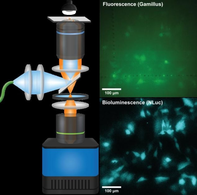
A new microscope inspired by the design of Keplerian telescopes produces much sharper images from luminescence from biological cells than was possible with previous devices. Dubbed the “QIScope” by its creators, the device’s highly sensitive camera can detect extremely low levels of light and could be used to observe delicate biostructures in greater detail and over longer periods of time without damaging them.
Many organisms naturally produce light via special enzymes in their cells. Although most such bioluminescent creatures are found in the ocean – think of anglerfish and firefly squid – there are also examples of terrestrial bioluminescent organisms, including bacteria and molluscs.
For researchers in life sciences, harnessing this light is an attractive alternative to imaging organisms using fluorescence. This is because it does not rely on strong external illumination, which can damage cells or interfere with the subtle signals they produce. The downside is that bioluminescence is feeble by comparison, so using it produces relatively low-resolution images.
Researchers led by Jian Cui of Helmholtz Munich and the Technical University of Munich, Germany, have now used a new detector technology called a quantum image sensor (QIS) to improve the resolution of bioluminescence imaging. By integrating this sensor into an unconventional optical microscope design, they increased the number of photons per pixel without sacrificing spatial resolution or field-of-view (FOV), as previous bioluminescence microscopes did.
“Telescope-within-a-microscope”
To avoid this restriction, which is known as vignetting, Cui explains that the team separated the two lenses and inserted a Keplerian telescope between them. “This ‘telescope-within-a-microscope’ reshapes the output of the objective lens to match the width of the tube lens’ back aperture,” he says.
The resulting “QIScope”, as the researchers call it, substantially reduces the size of the image while still capturing the full FOV. The result: an instrument with a higher signal-to-noise ratio and spatial resolution, leading to crisper images than was possible before.

MRI technique detects light-emitting molecules deep inside the brain
“New detector technologies are being developed all the time and some of them are very impressive,” Cui says. “However, we shouldn’t think about simply putting cameras on microscopes – sometimes you need to design the microscope around the properties of the camera. And this is what we have done.”
The researchers, who detail their work in Nature Methods, hope it will spur more interest in bioluminescence as an imaging tool. “There is a lot of untapped potential here and it could have advantages for certain applications such as studying photosensitive samples or low-abundance proteins,” Cui tells Physics World. “It could be used to study a range of biological systems – from single cells to organoids and tissue models. And since it can be used for long periods, it could reveal subtle and long-term changes in cell behaviour, so supporting progress in diverse research areas, including cell biology, disease modelling and drug discovery.”


