Researchers at Duke University have developed a novel way to improve the efficacy of radiation therapy, using a phototherapeutic agent activated by Cherenkov light produced by the therapeutic photon beam. The technique – radiotherapy enhanced with Cherenkov photo-activation (RECA) – shows promise for increasing both local control and tumour immunogenicity (Int. J. Radiat. Oncol. Biol. Phys. 100 794).
RECA works by using the clinical megavoltage (MV) photon beam to induce Cherenkov light throughout the irradiated tissue, while also delivering radiation dose to the tumour. This Cherenkov light – produced when high-speed secondary electrons travel faster than the phase velocity of light within the tissue – simultaneously activates light-sensitive psoralen within the target.
Psoralen is a drug that has anti-cancer effects when photo-activated by UV radiation. Cherenkov light generated from a MV treatment beam is particularly suited for psoralen activation as its emission is most intense in the short-UV region, with a peak around 300–320 nm that corresponds to the wavelengths where most psoralen photo-activation occurs.
“Radiation therapy is often considered a local therapy, effective at controlling local disease; but relapse may occur at distant sites,” explained lead author Mark Oldham. “RECA is an exciting approach because it has twofold potential: to increase the local efficacy of radiotherapy and amplify any systemic immunogenic response, which could help control distant disease as well. This would be an important therapeutic advance.”
Cell studies
First author Paul Yoon and colleagues investigated the basic mechanisms and effects of RECA in vitro using two murine cell lines: B16 melanoma and 4T1 breast cancer cells. They examined the cytotoxicity of RECA versus radiotherapy alone, as well as the expression of MHC I, which could indicate potential for immune response.
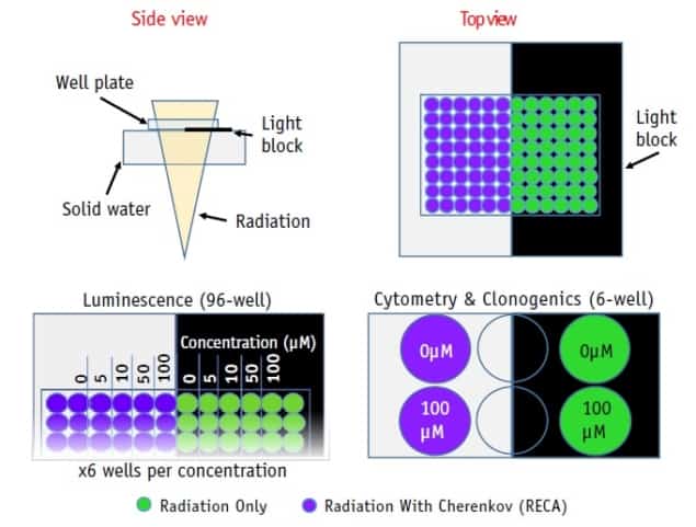
They placed well-plates of cultured cells on a solid water slab and irradiated from below using a beam from a TrueBeam linac. While all wells received the same radiation dose, a thin light block stopped the Cherenkov light from reaching half of the wells on each plate.
The researchers first irradiated arrays of both cell lines, incubated with varying concentrations of the psoralen derivative TMP, with 2 Gy of 6 MV radiation. They then performed luminescence assays to quantify the number of viable and metabolically active cells.
Cells exposed to full RECA treatment (including Cherenkov light) showed lower viability compared with cells that were only exposed to radiation. As psoralen exposure increased (from 0–100 μM TMP), the team observed a maximum differential at around 50 μM, where RECA increased cytotoxicity by 20% and 9.5% for 4T1 and B16 cells, respectively, compared with radiation and psoralen alone. They note that at low TMP concentrations (below 10 μM), cell viability was relatively constant, indicating that Cherenkov light alone does not enhance cytotoxicity.
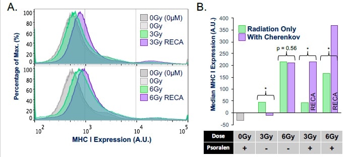
The researchers next performed flow cytometry to determine the effect of RECA on MHC I expression in B16 cells. They investigated pairs of wells treated with 3 or 6 Gy, with or without psoralen, and 0 Gy controls. One of each pair of wells received Cherenkov light and the other did not.

The flow cytometry data revealed a substantial increase in MHC I expression when Cherenkov light was present (about 450% and 250% at 3 and 6 Gy, respectively), compared with cells that received radiation and psoralen alone. Increasing MHC I expression has potential to increase tumour immunogenicity and may improve visibility to the immune system.
Finally, the team performed clonogenic survival assays on 4T1 cells 1–2 weeks after irradiation with or without exposure to Cherenkov light. At doses of 6 and 12 Gy, the assays revealed decreases in tumour cell viability of 7% and 36%, respectively, in RECA treated cells compared with identically treated cells with Cherenkov light blocked.
Boosting the light
To investigate whether the clinical radiation beam can be optimized to generate more Cherenkov light for the same radiation dose, the researchers irradiated a phantom with 6, 10 and 15 MV photons. The relative Cherenkov light intensity increased with increasing beam energy. The researchers also showed that adding a low-Z filter (for example, a block of polyurethane) to a flattening-filter-free 10 MV beam increased the relative Cherenkov light intensity per unit dose by 13% compared with the unfiltered beam.
The authors concluded that this work demonstrates how RECA, which is compatible with current standard-of-care radiation treatments, can increase cytotoxicity and potential immunogenicity over radiotherapy alone. Further work is required, however, to determine how these in vitro indications translate into an in vivo setting. As such, the team plans next to move on to in vivo small-animal studies.


